Clinical significance of OCT4 and SOX2 protein expression in cervical cancer
Bạn đang xem bản rút gọn của tài liệu. Xem và tải ngay bản đầy đủ của tài liệu tại đây (1.02 MB, 8 trang )
Kim et al. BMC Cancer (2015) 15:1015
DOI 10.1186/s12885-015-2015-1
RESEARCH ARTICLE
Open Access
Clinical significance of OCT4 and SOX2
protein expression in cervical cancer
Bo Wook Kim1,4, Hanbyoul Cho2, Chel Hun Choi1,3, Kris Ylaya1, Joon-Yong Chung1, Jae-Hoon Kim2*
and Stephen M. Hewitt1*
Abstracts
Background: Cancer stem cell markers have become a major research focus because of their relationship with
radiation or chemotherapy resistance in cancer therapy. Cancer stem cell markers including OCT4 and SOX2 have
been found in various solid tumors. Here, we investigate the expression and clinical significance of OCT4 and SOX2
in cervical cancer.
Methods: To define the clinical significance of OCT4 and SOX2 expression, we performed immunohistochemistry
for OCT4 and SOX2 on 305 normal cervical epithelium samples, 289 cervical intraepithelial neoplasia samples, and
161 cervical cancer cases and compared the data with clinicopathologic factors, including survival rates of patients
with cervical cancer.
Results: OCT4 and SOX2 expression was higher in cervical cancer than normal cervix (both p < 0.001). OCT4
overexpression was associated with lymphovascular space invasion (p = 0.045), whereas loss of SOX2 expression
was correlated with large tumor size (p = 0.015). Notably, OCT4 and SOX2 were significantly co-expressed in
premalignant cervical lesions, but not in malignant cervical tumor. OCT4 overexpression showed worse 5-year
disease-free and overall survival rates (p = 0.012 and p = 0.021, respectively) when compared to the low-expression group,
while SOX2 expression showed favorable overall survival (p = 0.025). Cox regression analysis showed that OCT4 was an
independent risk factor (hazard ratio = 11.23, 95 % CI, 1.31 - 95.6; p = 0.027) for overall survival while SOX2 overexpression
showed low hazard ratio for death (hazard ratio = 0.220, 95 % CI, 0.06–0.72; p = 0.013).
Conclusions: These results suggest that OCT4 overexpression and loss of SOX2 expression are strongly associated with
poor prognosis in patients with cervical cancer.
Keywords: Neoplastic stem cells, OCT4, SOX2, Prognosis, Survival, Uterine cervical neoplasms
Background
Cervical cancer is one of the most common gynecologic
malignancies worldwide and remains a leading cause of
cancer-related death for women in developing countries
[1]. Such high mortality rates are ascribed to disease recurrence despite cervical resection as well as ineffective
treatment options for advanced disease. Radiation therapy is widely employed for advanced cervical cancer, but
* Correspondence: ;
2
Department of Obstetrics and Gynecology, Gangnam Severance Hospital,
Yonsei University College of Medicine, 146-92 Dogok-Dong, Gangnam-Gu,
Seoul 135-720, South Korea
1
Experimental Pathology Lab, Laboratory of Pathology, Center for Cancer
Research, National Cancer Institute, National Institutes of Health, MSC 1500,
Bethesda, MD 20892, USA
Full list of author information is available at the end of the article
radiation resistance is an obstacle for cancer eradication.
Radiation resistance of cancer cells is acquired by intrinsic and extrinsic factors including tumor hypoxia, cell
cycle and DNA repair and radiation resistance in cancer
stem cells (CSCs) [2, 3].
CSCs are a small subpopulation of cancer cells that
have stem cell features such as self-renewal and the ability to differentiate into multiple cell types. Radiation
therapy or chemotherapy largely eliminates cancer cells,
including cervical cancer, but some tumor cells survive
and acquire radiation or chemotherapy resistance [4, 5].
These resistant cancer cells are difficult to eradicate, and
they show properties of CSCs.
Among CSC markers, octamer-binding transcription
factor 4 (OCT4) and sex determining region Y-box 2
© 2015 Kim et al. Open Access This article is distributed under the terms of the Creative Commons Attribution 4.0
International License ( which permits unrestricted use, distribution, and
reproduction in any medium, provided you give appropriate credit to the original author(s) and the source, provide a link to
the Creative Commons license, and indicate if changes were made. The Creative Commons Public Domain Dedication waiver
( applies to the data made available in this article, unless otherwise stated.
Kim et al. BMC Cancer (2015) 15:1015
(SOX2) are transcriptional factors involved in the regulation of several target genes including NANOG, Fgf4 and
Utf1, as well as OCT4 and SOX2 [6–10]. OCT4 belongs
to the POU (Pit-Oct-Unc) transcriptional factor family
and plays a key role in stem cell pluripotency and differentiation by determining the fate of embryonic stem
cells [11]. OCT4 expression in cancer stem-like cells is
associated with self-renewal and tumorigenesis via regulation of its target genes [12]. OCT4 expression has been
shown to be correlated with poor tumor differentiation
and metastasis, as well as poor prognosis in colon, pancreas and lung cancer [13–15]. SOX2, a member of the
SRY-related HMG-box (SOX) family of transcription factors, stimulates the reprogramming of adult cells into induced pluripotent stem cells and maintains stem cell-like
properties in cancer by complexing with other stem cell
markers such as NANOG and OCT4 [9]. SOX2 expression was reported to be correlated with tumorigenesis,
chemoresistance and maintenance of stem cell-like phenotype in cancer cells [16, 17]. In addition, SOX2 has been
shown to be highly expressed in premalignant lesions such
as squamous dysplasia and carcinoma in situ in lung [18].
Prior studies suggest OCT4 and SOX2 have a key role of
tumorigenesis and prognosis of cancer. However, the
prognostic significance of OCT4 and SOX2 is not clearly
defined in cervical premalignant and malignant lesion. In
this study, we investigated the clinical significance of
OCT4 and SOX2 expression in cervical neoplasia.
Methods
Patient selection
A total of 450 patients with cervical cancer and cervical
intraepithelial neoplasia (CIN) were collected from patients who enrolled at Gangnam Severance Hospital,
Yonsei University College of Medicine in Seoul, Korea
and the Korea Gynecologic Cancer Bank through Bio &
Medical Technology Development Program of the
Ministry of Education, Science and Technology, Korea
between 1996 and 2010. One hundred sixty-one
paraffin-embedded specimens of cervical cancer, 289
CIN and 305 matched normal tissues were included in
the study. Medical records were obtained to review patient data including age, cancer stage, tumor differentiation, cell type, tumor size, lymphovascular space
invasion (LVSI) and lymph node (LN) metastasis. Cervical cancer was staged according to the International
Federation of Gynecology and Obstetrics (FIGO) stage
and histologically classified and graded according to
World Health Organization (WHO) grade. Patients with
surgical indications underwent radical hysterectomy with
pelvic and aortic lymph node dissection. Concurrent
chemoradiation therapy was added in cases with risk factors such as LN metastasis, parametrial invasion and
positive resection margin. Inoperable patients underwent
Page 2 of 8
radiation or chemoradiation therapy. Tissue samples and
medical records were obtained with informed consent of
all patients and approval of the local research ethics
committee (approval no. 3-2010-0030; Seoul, South
Korea). This study was additionally approved by the Office of Human Subjects Research at the National Institute of Health.
Tissue microarray construction and immunohistochemistry
Tissue microarrays (TMAs) were constructed from 450 patients with primary invasive cervical cancer or CIN, as well
as 305 matched non-adjacent normal epithelial tissues.
After hematoxylin and eosin slides were reviewed by a
pathologist, areas containing each category were indicated
by marking them. Four 1-mm punches were then taken
from the corresponding regions of the paraffin blocks and
transplanted into a recipient paraffin block using a tissue
arrayer (Pathology Devices, Westminster, MD).
For immunohistochemical staining, all paraffinembedded sections were cut at 5-μm thickness followed
by deparaffinization through xylene and dehydration with
graded ethanols. Antigen recovery was performed in heatactivated antigen retrieval pH 6 (Dako, Carpinteria, CA)
for OCT4 and SOX2, and then specimens were incubated
with 3 % H2O2 for 10 min. Non-specific binding was
blocked with protein block (Dako) for 20 min at room
temperature. The sections were incubated with rabbit
polyclonal anti-OCT4 antibodies (Abcam, Cambridge,
MA; Cat. #ab19857) at 1:250 for 30 min or with rabbit
monoclonal anti-SOX2 antibodies (Cell Signaling,
Danvers, MA; Cat. #3579) at 1:500 for 2 h, respectively.
Subsequently, antigen-antibody reaction was detected
with EnVision + Dual Link System-HRP (Dako) and visualized with DAB+ (3, 3’-Diaminobenzidine; Dako). Tissue
sections were lightly counterstained with hematoxylin and
then examined by light microscopy. Negative controls
(substitution of primary antibody with TBS) were run simultaneously. Positive controls included testicular seminoma and lung squamous cell carcinoma for OCT4 and
SOX2 [19] antibodies, respectively.
Quantitative evaluation of immunostaining
Immunohistochemically stained slides were digitized at ×
20 magnification utilizing an Aperio Scanscope CS
(Aperio, Vista, CA, USA). Images were reviewed using
an online software application, Digital Image Hub (SlidePath, Dublin, Ireland). Once the areas were annotated,
they were sent for automated image analysis utilizing
TissueIA (SlidePath’s Tissue IA system, version 3.0,
Dublin, Ireland). Within Tissue IA, an algorithm was developed to quantify OCT4 and SOX2 expression levels.
The staining intensity of OCT4 and SOX2 was
categorized as 0 (no staining), 1+ (weak), 2+ (moderate)
and 3+ (strong). The overall immunohistochemical score
Kim et al. BMC Cancer (2015) 15:1015
(histoscore) was expressed as the percentage of positive
cells multiplied by their staining intensity (possible
range, 0–300) [20].
Page 3 of 8
Table 1 Patient clinicopathologic characteristics
Frequency
%
43.3a
Age
Diagnostic category
Statistical analysis
Histoscores were compared using one-way ANOVA test
and independent t-test. The immunohistochemical cutoff for high expression of tumor markers was determined through receiver operating characteristic (ROC)
curve analysis. The sensitivity and (1 - specificity) for
discrimination of dead and alive was determined for
each immunohistochemistry (IHC) score and plotted,
thus generating a ROC curve. The cut-off value was
established to be the point on the ROC curve where
sum of sensitivity and specificity was maximized.
Kaplan-Meier survival analysis was performed to determine the association of OCT4 and SOX2 expression
with survival, and the survival curves were compared between groups using log-rank tests. Multivariate analyses
of hazard ratio for death were performed using Cox proportional hazards regression. Chi-square test was used
to evaluate the association between OCT4 and SOX2.
Statistical analyses were performed using SPSS version
21.0 (SPSS Inc., Chicago, IL). A value of p < 0.05 was
considered statistically significant.
Normal
305
40.4
Low grade CIN
59
7.8
High grade CIN
230
30.5
Cancer
161
21.3
< IIA
118
73.3
> IIB
43
26.7
Well
2
1.3
Moderate
112
71.8
Poor
42
26.9
SCC
131
81.4
AD
16
9.9
Other
14
8.7
≤ 4 cm
112
69.6
> 4 cm
49
30.4
No
86
56.2
Yes
67
43.8
No
115
74.2
Yes
40
25.8
Negative
21
14.2
Positive
127
85.8
FIGO stage
Tumor differentiationb
Cell type
Tumor size
c
LVSI
Results
Clinicopathologic characteristics of cases
Table 1 presents the patients’ clinicopathologic characteristics. Of 161 patients with cervical cancer, 118 patients were stage IIA or less and 43 patients were stage
IIB or higher. The mean age was 43.3 years (range, 19–
83 years). The tumor sizes ranged from 0.2 to 12.0 cm
(mean, 2.8 cm). The histopathology included 131 squamous cell carcinoma, 16 adenocarcinoma, 7 adenosquamous and 7 other types (3 small cell carcinomas, 2
neuroendocrine and 2 mixed cell types). Patients with
cervical cancer were evaluated for survival analysis and
the mean follow-up time of surviving patients was
54.3 months (range, 1–179). Fifteen patients (9.3 %) died
during the follow-up period.
LN metastasisd
e
HPV test in CIN
CIN cervical intraepithelial neoplasia, FIGO International Federation of Gynecology
and Obstetrics, SCC squamous cell carcinoma, AD adenocarcinoma, LVSI
lymphovascular space invasion, LN lymph node, HPV human papillomavirus
a
mean value
b
calculated based on 156 cases with available tumor
differentiation information
c
calculated based on 153 cases with available LV invasion information
d
calculated based on 155 cases with available LN metastasis information
e
calculated based on only 148 cases of CIN with available HPV test data
OCT4 and SOX2 protein expression
Expression of OCT4 and SOX2 was evaluated by IHC
in cervical neoplasia and cancer specimens. Subsequently, we performed analysis of both markers using
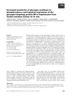
quantitative image analysis software. Representative
IHC images of OCT4 and SOX2 are presented in
Fig. 1. OCT4 expression was observed primarily in
the nucleus with limited cytoplasm expression, while
SOX2 was restricted to the nucleus (Fig. 1). Only nuclear staining was considered OCT4- and SOX2positive.
Of the cancer specimens, 92 of 161 cancers
(57.1 %) had high expression of OCT4 (histoscore >
200) and 125 of 161 cancers (77.6 %) had high expression of SOX2 (histoscore > 30). Association of
OCT4 and SOX2 expression with clinicopathologic
characteristics in cervical cancer is summarized in
Table 2. OCT4 and SOX2 expression was significantly
different depending on diagnostic category (p < 0.001).
OCT4 overexpression was associated with lymphovascular space invasion (p = 0.045), whereas loss of SOX2
Kim et al. BMC Cancer (2015) 15:1015
Page 4 of 8
a
b
c
d
Fig. 1 OCT4 and SOX2 expression in formalin-fixed, paraffin-embedded cervical cancer tissues. Representative immunohistochemical image of
OCT4 negative (a) and positive (b), SOX2 negative (c) and positive (d). Insets show high magnification of areas indicated with boxes. Scale bar: 100 μm
expression was correlated with large tumor size (p = 0.015).
There were no other correlations between OCT and SOX2
expression and clinicopathologic characteristics.
We next examined the association between OCT4 and
SOX2 expression, Chi-squared distribution was used in
malignant and premalignant lesions. In premalignant
cervical lesions, SOX2 expression presented a significant
correlation with OCT4 (p = 0.004), while there was no
association between OCT4 and SOX2 in malignant
tumors (p = 0.543; Table 3).
Prognostic significance of OCT4 and SOX2 expression
Five-year disease-free and overall survival rates were analyzed through the Kaplan-Meier plots as shown in
Fig. 2. In survival analysis with OCT4 expression, 22 recurrences and 13 deaths occurred in 83 cases of OCT4
high expression, while 7 recurrences and 3 deaths were
observed in 65 cases of low expression. The 5-year
disease-free and overall survival rates were 89.2 and
95.4 % in low OCT4 expression and 73.5 and 84.3 % in
high OCT4 expression. OCT4 overexpression was associated with shorter disease-free and overall survival than
the low expression group (p = 0.012 and p = 0.021, respectively) (Fig. 2a and d). In survival analysis of SOX2,
there were 20 recurrences and 8 deaths in 125 highexpression patients, while 8 recurrences and 7 deaths
occurred in 36 low-expression patients during the 5-year
follow-up period. The 5-year disease-free and overall
survival rates were 77.8 and 80.6 % in cases with low
SOX2 expression and 84.0 and 93.6 % in case with high
SOX2 expression cases. High expression of SOX2 was associated with better overall survival than low expression
(p = 0.025) (Fig. 2e). When survival of patients with expression of high OCT4/low SOX2 was compared with
survival of patients with low OCT4/high SOX2, KaplanMeier analysis revealed a significant difference in diseasefree and overall survival (p = 0.016 and p < 0.001, respectively; Fig. 2c and f).
Cox proportional multivariate analysis of relationships
between prognostic variables and survival are shown in
Table 4. FIGO stage was an independent survival factor for
both disease-free and overall survival analysis (p < 0.001
and p = 0.041, respectively). OCT4 overexpression showed
independent poor overall survival with a hazard ratio of
11.23 (p = 0.027), while high expression of SOX2 presented
better disease-free and overall survival compared to low
expression, as shown in Table 4 (p = 0.019 and p = 0.013,
respectively).
Discussion
OCT4 and SOX2 are important transcriptional factors
involved in maintenance of pluripotency and selfrenewal in cancer stem cells, aberrant expression of
OCT4 and SOX2 might contribute to carcinogenesis in
various cancers [15, 21, 22]. Radioresistance is important
in the treatment and prognosis of cervical cancer and it
is known to be associated with cancer stem cells [3].
This study examined the clinical correlation and prognostic significance of stemness-related OCT4 and SOX2
protein expression assessed by IHC in premalignant and
malignant cervical tumors. The results demonstrate that
OCT4 and SOX2 protein expression is elevated in premalignant and malignant cervical tumors compared to
normal cervix and this finding is consistent with a
Kim et al. BMC Cancer (2015) 15:1015
Page 5 of 8
Table 2 Association between clinicopathologic characteristics and OCT4 or SOX2 expression
OCT4
SOX2
p value
Mean Histoscore (95 % CI)
Diagnostic category
Mean Histoscore (95 % CI)
<0.001
<0.001
Normal
113.3 (105.1–121.4)
36.5 (32.3–40.8)
Low-grade CIN
197.6 (177.4–217.7)
40.0 (28.8–51.2)
High-grade CIN
219.0 (211.3–226.8)
91.7 (79.9–103.5)
Cancer
208.5 (196.7–220.3)
105.4 (91.8–119.1)
FIGO stage
0.498
< IIA
205.7 (191.6–219.7)
> IIB
215.1 (192.6–237.6)
0.529
108.1 (92.1–124.2)
98.3 (71.5–125.1)
Tumor differentiation
0.438
0.112
Well + moderate
203.8 (187.7–219.8)
112.8 (94.4–131.1)
Poor
213.4 (195.3–231.4)
90.1 (69.0–111.3)
Cell type
0.450
SCC
205.8 (192.4–219.2)
Other
217.2 (190.8–243.6)
0.060
111.7 (96.4–127.0)
78.3 (69.0–111.3)
Tumor size
0.868
0.015
≤ 4 cm
208.7 (194.2–223.2)
116.5 (100.1–132.8)
> 4 cm
206.5 (185.5–227.6)
80.3 (56.3–104.2)
LVSI
0.045
No
195.3 (175.8–214.8)
Yes
220.1 (205.3–234.9)
p value
0.106
115.5 (95.4–135.6)
91.8 (71.2–112.5)
LN metastasis
0.206
0.879
No
202.6 (187.1–218.0)
105.6 (88.6–122.7)
Yes
221.0 (199.7–242.3)
103.1 (75.6–130.6)
HPV test in CIN
0.292
0.907
Negative
246.8 (223.7–215.7)
124.9 (81.5–168.7)
Positive
227.9 (215.7–240.1)
127.6 (110.5–144.6)
SCC squamous cell carcinoma, AD adenocarcinoma, LVSI lymphovascular space invasion, LN lymph node, HPV human papillomavirus
previous study [23]. OCT4 was an independent poor survival factor but SOX2 showed as a favorable prognostic
factor.
In this study, OCT4 protein was observed clearly in
the nucleus and partially in the cytoplasm. Similar to
Table 3 Association of OCT4 and SOX2 expression in CIN and
cervical cancer patients
OCT4 expression
No.
Low (%)
High (%)
SOX2 Low (−)
55
29 (53.1)
26 (46.9)
SOX2 High (+)
234
62 (26.3)
173 (73.1)
CIN
p value
0.004
Cancer
0.543
SOX2 Low (−)
36
15 (41.7)
21 (58.3)
SOX2 High (+)
125
54 (43.1)
71 (56.9)
CIN cervical intraepithelial neoplasia
our findings, OCT4 has been reported in the cytoplasm as well as in the nucleus in previous studies
[24, 25]. This staining pattern may arise from the
presence of an OCT4 isoform. OCT4 is known to
have two isoforms, OCTA and OCTB. OCT4A is observed in the nucleus and OCT4B is observed in the
cytoplasm in prostate and cervical cancer [24, 26].
Because OCT4 is a transcriptional regulator, the active form of OCT4 is always located in the nucleus.
For this reason, we focused our automated digital
image analysis on OCT4 protein expression in the
nucleus only. Notably, OCT4 expression increased
during cancer progression but within cancers, it was
not correlated with known prognostic factors, such as
stage, LN metastasis or tumor size. Nonetheless, it
showed high hazard ratio of death in multivariate
analysis. In previous published results, OCT4 expression was associated with unfavorable prognosis
Kim et al. BMC Cancer (2015) 15:1015
Page 6 of 8
Fig. 2 Kaplan-Meier survival curves of OCT4 and SOX2 expression in cervical cancer. Cervical cancer patients with high OCT4 expression had shorter 5-year
disease-free survival (a, P = 0.012) and worse 5-year overall survival (b, P = 0.021) than those with low expression. Patients with high SOX2 expression had
longer 5-year overall survival than those with low expression (e, P = 0.025). The patients with low SOX2/high OCT4 expression had shorter
5-year disease-free survival (c, P = 0.016) and worse 5-year overall survival (f, P < 0.001) than those with high SOX2/low OCT4 expression
showing poor tumor differentiation, tumor invasion
and metastasis in the lungs, stomach, esophagus and
oral cavity [13, 25, 27, 28]. Although there have been
limited reports on the association between OCT4 and
prognosis in cervical cancer, Shen et al. reported that
OCT4 expression was associated with radiationresistance and unfavorable survival in locally advanced
squamous cell carcinoma [29]. That study further
showed OCT4 overexpression in the radiation resistance group, but it was not associated with high risk
prognostic factors including FIGO stage and tumor
size, which is similar to our results. It is interesting
that OCT4 is associated with poor survival without
correlation to known prognostic factors and even
disease-free survival. As a stem cell related protein,
OCT4 expression can be related more with overall
survival than with disease-free survival which is possibly more related with residual tumor after resection.
Further research is required to clarify the association
between OCT4 and high-risk prognostic factors.
SOX2 is known to play an important role in regulating
the cell cycle, DNA repair and self-renewal in stem cells
[30]. It is associated with tumorigenesis, chemoresistance
and maintenance of stem cell-like property in cancer cells,
Table 4 Multivariate survival analysis of the association between prognostic variables and survival in cervical cancer patients
Variables
Disease-free survival
Overall survival
HR [95 % CI]
P value
HR [95 % CI]
FIGO stage (≥ IIB)
8.67 [2.77–27.11]
<0.001
4.33 [1.06–17.72]
0.041
Tumor size (>4 cm)
1.16 [0.47–2.84]
0.739
1.79 [0.54–5.92]
0.340
LN metastasis
1.72 [0.57–5.23]
0.334
1.60 [0.40–6.41]
0.500
OCT4+
3.75 [1.24–11.55]
0.117
11.23 [1.31–95.64]
0.027
SOX2 +
0.47 [0.18–1.20]
0.019
0.22 [0.06–0.72]
0.013
FIGO International Federation of Gynecology and Obstetrics, HR hazard ratio, LN lymph node, CI confidence interval
P value
Kim et al. BMC Cancer (2015) 15:1015
which suggests poor overall survival [16, 31, 32]. High expression of SOX2 was reported to be associated with a
lack of cell differentiation and to contribute cell migration
and invasion in cervical cancer cell line [33]. In addition,
Shen et al. showed that SOX2 is highly expressed in patients with radiation resistance and predicts poor survival
[29]. In contrast, SOX2 expression was associated with
prolonged survival in the current study. These discrepancies might be explained by the lack of standardized methodology, different standards of interpretation or
differences in studies’ patient populations. Similar to our
study, Wilbertz et al. reported that SOX2 gene amplification and protein expression are associated with favorable
survival outcomes in squamous cell lung cancer [34]. In
addition, a recent meta-analysis reported that SOX2 expression presents a positive prognosis in non-small cell
lung cancer [35]. Previously, SOX2 overexpression was reported to be associated with favorable prognosis in squamous cell lung carcinoma, but was correlated with poor
survival in adenocarcinoma [34–36]. Notably, the poor
survival associated with SOX2 expression that was reported in GI tract cancer mostly pertained to adenocarcinoma [37–39]. Cervical cancer consists of squamous cell
carcinoma followed by adenocarcinoma and our data
comprised 80.7 % squamous cell carcinoma and 14.8 %
adenocarcinoma. Further research is required to clarify
the prognostic significance of SOX2 in cervical cancer and
variation in prognosis according to cell type.
Premalignant cervical lesion demonstrated significant
correlation between OCT4 and SOX2, while malignant
lesion did not present an association between OCT4 and
SOX2. The lack of a correlation between OCT4 and
SOX2 in malignant lesions has not been explained
clearly because OCT4 and SOX2 are known to work cooperatively and self-regulate themselves via the OCT4/
SOX2 complex in embryonic stem cells [6, 8]. However,
in the current cancer tissue samples, OCT4 and SOX2
were associated with opposite effects on survival and
lose their association in cervical cancer, as well. Similar
to our results, no correlation between OCT4 and SOX2
was reported in cervical cancer [23]. In addition, Li et al.
also reported that OCT4 and SOX2 were not coexpressed and also showed different survival outcomes
in lung cancer tissue samples [40]. Furthermore, overexpression of SOX2 inhibited the activity of OCT4 promotor in embryonal carcinoma cells [41]. OCT4 and SOX2
are known to function cooperatively through the OCT4/
SOX2 complex, but OCT4, SOX2 and Nanog have been
reported to form individual complexes with nucleophosmin to control stem cell fate determination [42]. In previous study, we also observed a similar phenomenon
that Nanog expression in precancerous cervical tissue
was correlated with Tcl1a and pAkt but this relationship
lost in cancerous tissue [43]. Considering previous
Page 7 of 8
results and our contradictory survival data, OCT4 and
SOX2 might function independently or inhibit activity
during tumor progression, and eventually lose their connection in cervical cancer.
Conclusions
In conclusion, this study investigated the immunohistochemical expression of OCT4 and SOX2 in large number of cervical cancer patients by means of image
analysis for IHC scoring. OCT4 and SOX2 showed high
expression in premalignant and malignant cervical
tumors. Co-expression of OCT4 and SOX2 was observed in premalignant tumors, but no association was
observed in malignant cervical tumors. OCT4 high expression showed poor disease-free survival and overall
survival while SOX2 high expression showed favorable
overall survival in patients with cervical cancer. Cox regression analysis confirmed that OCT4, and SOX2 expression was an important prognostic indicator in
cervical cancer. OCT4 was associated with poor prognosis, while SOX2 showed favorable prognosis. Our findings suggest further investigation into OCT4 and SOX2
as biomarkers in cervical cancer.
Abbreviations
AD: adenocarcinoma; CI: confidence interval; CIN: cervical intraepithelial
neoplasia; CSC: cancer stem cell; FIGO: International Federation of
Gynecology and Obstetrics; H&E: hematoxylin and eosin; HPV: human
papillomavirus; HR: hazard ration; IHC: immunohistochemistry; LN: lymph
node; LVSI: lymphovascular space invasion; OCT-4: octamer-binding
transcription factor 4: SOX2, sex determining region Y-box 2; ROC: receiver
operating characteristic; SCC: squamous cell carcinoma; TMA: tissue
microarray; WHO: World Health Organization.
Competing interests
The authors declare that there is no conflict of interest.
Authors’ contributions
BWK, J-YC, J-HK and SMH conceived of the study and devised the experimental
design. SMH designed and build the tissuemicroarrays. BWK, HC, CHC and KY
performed experiments. BWK, HC, CHC, J-YC, J-HK and SMH performed data
analysis for experiments or clinical records. BWK, HC and J-YC drafted the final
version of the manuscript and figure legends. J-HK and SMH revised the figures,
added critical content to the discussion and were responsible in revising
all portions of the submitted portion of the manuscript. All authors read
and approved the final manuscript.
Acknowledgments
This research was supported by the Intramural Research Program of the
National Institutes of Health National Cancer Institute, Center for Cancer
Research.
Author details
1
Experimental Pathology Lab, Laboratory of Pathology, Center for Cancer
Research, National Cancer Institute, National Institutes of Health, MSC 1500,
Bethesda, MD 20892, USA. 2Department of Obstetrics and Gynecology,
Gangnam Severance Hospital, Yonsei University College of Medicine, 146-92
Dogok-Dong, Gangnam-Gu, Seoul 135-720, South Korea. 3Department of
Obstetrics and Gynecology, Samsung Medical Center, Sungkyunkwan
University School of Medicine, Seoul 135-710, Republic of Korea.
4
Department of Obstetrics and Gynecology, Kangdong Sacred Heart Hospital,
Hallym University, Seoul 135-701, South Korea.
Kim et al. BMC Cancer (2015) 15:1015
Received: 1 May 2015 Accepted: 15 December 2015
References
1. Ferlay J, Shin HR, Bray F, Forman D, Mathers C, Parkin DM. Estimates of
worldwide burden of cancer in 2008: GLOBOCAN 2008. Int J Cancer. 2010;
127:2893–917.
2. Feng D, Peng C, Li C, Zhou Y, Li M, Ling B, et al. Identification and
characterization of cancer stem-like cells from primary carcinoma of the
cervix uteri. Oncol Rep. 2009;22:1129–34.
3. Krause M, Yaromina A, Eicheler W, Koch U, Baumann M. Cancer stem cells:
targets and potential biomarkers for radiotherapy. Clin Cancer Res. 2011;17:
7224–9.
4. Bao S, Wu Q, McLendon RE, Hao Y, Shi Q, Hjelmeland AB, et al. Glioma stem
cells promote radioresistance by preferential activation of the DNA damage
response. Nature. 2006;444:756–60.
5. Yin T, Wei H, Gou S, Shi P, Yang Z, Zhao G, et al. Cancer stem-like cells
enriched in Panc-1 spheres possess increased migration ability and
resistance to gemcitabine. Int J Mol Sci. 2011;12:1595–604.
6. Chew JL, Loh YH, Zhang W, Chen X, Tam WL, Yeap LS, et al. Reciprocal
transcriptional regulation of Pou5f1 and Sox2 via the Oct4/Sox2 complex in
embryonic stem cells. Mol Cell Biol. 2005;25:6031–46.
7. Nishimoto M, Fukushima A, Okuda A, Muramatsu M. The gene for the
embryonic stem cell coactivator UTF1 carries a regulatory element which
selectively interacts with a complex composed of Oct-3/4 and Sox-2. Mol
Cell Biol. 1999;19:5453–65.
8. Okumura-Nakanishi S, Saito M, Niwa H, Ishikawa F. Oct-3/4 and Sox2 regulate
Oct-3/4 gene in embryonic stem cells. J Biol Chem. 2005;280:5307–17.
9. Rodda DJ, Chew JL, Lim LH, Loh YH, Wang B, Ng HH, et al. Transcriptional
regulation of nanog by OCT4 and SOX2. J Biol Chem. 2005;280:24731–7.
10. Yuan H, Corbi N, Basilico C, Dailey L. Developmental-specific activity of the
FGF-4 enhancer requires the synergistic action of Sox2 and Oct-3. Genes
Dev. 1995;9:2635–45.
11. Scholer HR, Ruppert S, Suzuki N, Chowdhury K, Gruss P. New type of POU
domain in germ line-specific protein Oct-4. Nature. 1990;344:435–9.
12. Ponti D, Costa A, Zaffaroni N, Pratesi G, Petrangolini G, Coradini D, et al.
Isolation and in vitro propagation of tumorigenic breast cancer cells with
stem/progenitor cell properties. Cancer Res. 2005;65:5506–11.
13. Chiou SH, Wang ML, Chou YT, Chen CJ, Hong CF, Hsieh WJ, et al.
Coexpression of Oct4 and Nanog enhances malignancy in lung
adenocarcinoma by inducing cancer stem cell-like properties and epithelialmesenchymal transdifferentiation. Cancer Res. 2010;70:10433–44.
14. Meng HM, Zheng P, Wang XY, Liu C, Sui HM, Wu SJ, et al. Over-expression
of Nanog predicts tumor progression and poor prognosis in colorectal
cancer. Cancer Biol Ther. 2010;9:295–302.
15. Wen J, Park JY, Park KH, Chung HW, Bang S, Park SW, et al. Oct4 and Nanog
expression is associated with early stages of pancreatic carcinogenesis.
Pancreas. 2010;39:622–6.
16. Singh S, Trevino J, Bora-Singhal N, Coppola D, Haura E, Altiok S, et al. EGFR/
Src/Akt signaling modulates Sox2 expression and self-renewal of stem-like
side-population cells in non-small cell lung cancer. Mol Cancer. 2012;11:73.
17. Takahashi K, Yamanaka S. Induction of pluripotent stem cells from mouse
embryonic and adult fibroblast cultures by defined factors. Cell. 2006;126:
663–76.
18. McCaughan F, Pole JC, Bankier AT, Konfortov BA, Carroll B, Falzon M, et al.
Progressive 3q amplification consistently targets SOX2 in preinvasive
squamous lung cancer. Am J Respir Crit Care Med. 2010;182:83–91.
19. Wang J, Rao S, Chu J, Shen X, Levasseur DN, Theunissen TW, et al. A protein
interaction network for pluripotency of embryonic stem cells. Nature. 2006;
444:364–8.
20. Kirkegaard T, Edwards J, Tovey S, McGlynn LM, Krishna SN, Mukherjee R,
et al. Observer variation in immunohistochemical analysis of protein
expression, time for a change? Histopathology. 2006;48:787–94.
21. Ji J, Zheng PS. Expression of Sox2 in human cervical carcinogenesis. Hum
Pathol. 2010;41:1438–47.
22. Wang YD, Cai N, Wu XL, Cao HZ, Xie LL, Zheng PS. OCT4 promotes
tumorigenesis and inhibits apoptosis of cervical cancer cells by miR-125b/
BAK1 pathway. Cell Death Dis. 2013;4:e760.
23. Ji J, Wei X, Wang Y. Embryonic stem cell markers Sox-2 and OCT4
expression and their correlation with WNT signal pathway in cervical
squamous cell carcinoma. Int J Clin Exp Pathol. 2014;7:2470–6.
Page 8 of 8
24. de Resende MF, Chinen LT, Vieira S, Jampietro J, da Fonseca FP, Vassallo J,
et al. Prognostication of OCT4 isoform expression in prostate cancer.
Tumour Biol. 2013;34:2665–73.
25. He W, Li K, Wang F, Qin YR, Fan QX. Expression of OCT4 in human
esophageal squamous cell carcinoma is significantly associated with poorer
prognosis. World J Gastroenterol. 2012;18:712–9.
26. Li SW, Wu XL, Dong CL, Xie XY, Wu JF, Zhang X. The differential expression
of OCT4 isoforms in cervical carcinoma. PLoS One. 2015;10:e0118033.
27. Kong D, Su G, Zha L, Zhang H, Xiang J, Xu W, et al. Coexpression of HMGA2
and Oct4 predicts an unfavorable prognosis in human gastric cancer. Med
Oncol. 2014;31:130.
28. Chiou SH, Yu CC, Huang CY, Lin SC, Liu CJ, Tsai TH, et al. Positive
correlations of Oct-4 and Nanog in oral cancer stem-like cells and highgrade oral squamous cell carcinoma. Clin Cancer Res. 2008;14:4085–95.
29. Shen L, Huang X, Xie X, Su J, Yuan J, Chen X. High Expression of SOX2 and
OCT4 Indicates Radiation Resistance and an Independent Negative
Prognosis in Cervical Squamous Cell Carcinoma. J Histochem Cytochem.
2014;62:499–509.
30. Peng C, Li N, Ng YK, Zhang J, Meier F, Theis FJ, et al. A unilateral negative
feedback loop between miR-200 microRNAs and Sox2/E2F3 controls neural
progenitor cell-cycle exit and differentiation. J Neurosci. 2012;32:13292–308.
31. Gontan C, de Munck A, Vermeij M, Grosveld F, Tibboel D, Rottier R. Sox2 is
important for two crucial processes in lung development: branching
morphogenesis and epithelial cell differentiation. Dev Biol. 2008;317:296–309.
32. Tian T, Zhang Y, Wang S, Zhou J, Xu S. Sox2 enhances the tumorigenicity
and chemoresistance of cancer stem-like cells derived from gastric cancer. J
Biomed Res. 2012;26:336–45.
33. Chang X, Zhang J, Huang C, Pang X, Luo Q, Zhang H, et al. Sex-determining
region Y-related high mobility group box (SOX)-2 is overexpressed in
cervical squamous cell carcinoma and contributes cervical cancer cell
migration and invasion in vitro. Tumour Biol. 2015;36:7725–33.
34. Wilbertz T, Wagner P, Petersen K, Stiedl AC, Scheble VJ, Maier S, et al. SOX2
gene amplification and protein overexpression are associated with better
outcome in squamous cell lung cancer. Mod Pathol. 2011;24:944–53.
35. Chen Y, Huang Y, Huang Y, Chen J, Wang S, Zhou J. The prognostic value
of SOX2 expression in non-small cell lung cancer: a meta-analysis. PLoS
One. 2013;8:e71140.
36. Sholl LM, Barletta JA, Yeap BY, Chirieac LR, Hornick JL. Sox2 protein
expression is an independent poor prognostic indicator in stage I lung
adenocarcinoma. Am J Surg Pathol. 2010;34:1193–8.
37. Otsubo T, Akiyama Y, Yanagihara K, Yuasa Y. SOX2 is frequently
downregulated in gastric cancers and inhibits cell growth through cell-cycle
arrest and apoptosis. Br J Cancer. 2008;98:824–31.
38. Sun C, Sun L, Li Y, Kang X, Zhang S, Liu Y. Sox2 expression predicts poor
survival of hepatocellular carcinoma patients and it promotes liver cancer
cell invasion by activating Slug. Med Oncol. 2013;30:503.
39. Wang Q, He W, Lu C, Wang Z, Wang J, Giercksky KE, et al. Oct3/4 and Sox2
are significantly associated with an unfavorable clinical outcome in human
esophageal squamous cell carcinoma. Anticancer Res. 2009;29:1233–41.
40. Li X, Wang J, Xu Z, Ahmad A, Li E, Wang Y, et al. Expression of sox2 and
oct4 and their clinical significance in human non-small-cell lung cancer. Int
J Mol Sci. 2012;13:7663–75.
41. Bernadt CT, Nowling T, Rizzino A. Transcription factor Sox-2 inhibits co-activator
stimulated transcription. Mol Reprod Dev. 2004;69:260–7.
42. Johansson H, Simonsson S. Core transcription factors, Oct4, Sox2 and Nanog,
individually form complexes with nucleophosmin (Npm1) to control
embryonic stem (ES) cell fate determination. Aging (Albany NY). 2010;2:815–22.
43. Noh KH, Kim BW, Song KH, Cho H, Lee YH, Kim JH, et al. Nanog signaling in
cancer promotes stem-like phenotype and immune evasion. J Clin Invest.
2012;122:4077–93.