A nomogram for predicting the likelihood of lymph node metastasis in early gastric patients
Bạn đang xem bản rút gọn của tài liệu. Xem và tải ngay bản đầy đủ của tài liệu tại đây (668.43 KB, 8 trang )
Zheng et al. BMC Cancer (2016) 16:92
DOI 10.1186/s12885-016-2132-5
RESEARCH ARTICLE
Open Access
A nomogram for predicting the likelihood
of lymph node metastasis in early gastric
patients
Zhixue Zheng1, Yinan Zhang1, Lianhai Zhang1, Ziyu Li1, Xiaojiang Wu1, Yiqiang Liu2, Zhaode Bu1 and Jiafu Ji1*
Abstract
Background: Early gastric cancer is defined as a lesion confined to the mucosa or submucosa, regardless of the
size or lymph node metastasis. Treatment methods include endoscopic mucosal resection or endoscopic submucosal
dissection, wedge resection, laparoscopically assisted gastrectomy and open gastrectomy. Lymph node metastasis is
strong related with survival and recurrence. Therefore, the likelihood of lymph node metastasis is one of the most
important factors when determining the most appropriate treatment.
Methods: We retrospectively analyzed 597 patients who underwent D2 gastrectomy for early gastric cancer. The
relationship between lymph node metastasis and clinicopathological features was analyzed. Using multivariate logistic
regression analyses, we created a nomogram to predict the lymph node metastasis probability for early gastric cancer.
Receiver operating characteristic analyses was performed to assess the predictive value of the model.
Results: In the present study, 58 (9.7 %) early gastric cancer patients were histologically shown to have lymph node
metastasis. The multivariate logistic regression analysis demonstrated that the age at diagnosis, differentiation status,
the presence of ulcers, lymphovascular invasion and depth of invasion were independent risk factors for lymph node
metastasis in early gastric cancer. Additionally, the tumor macroscopic type, size and histology type significantly
correlated with these important independent factors. We constructed a predictive nomogram with these factors for
lymph node metastasis in early gastric cancer patients, and the discrimination was good with the AUC of 0.860 (95 %
CI: 0.809–0.912).
Conclusions: We developed an effective nomogram to predict the incidence of lymph node metastasis for early
gastric cancer patients.
Keywords: Early gastric cancer, Lymph node metastasis, Nomogram
Background
Gastric cancer is currently among the most common
cancer worldwide and the second most common cause
of cancer-related death [1–3]. Early gastric cancer (EGC)
is defined as a lesion confined to the mucosa or submucosa, regardless of the size or the presence of regional
lymph node metastasis [4–7]. Treatment options for
EGC include endoscopic mucosal resection (EMR) or
endoscopic submucosal dissection (ESD), wedge resection,
* Correspondence:
Zhixue Zheng and Yinan Zhang are the first authors.
1
Department of Gastrointestinal Surgery, Key Laboratory of Carcinogenesis and
Translational Research (Ministry of Education), Peking University Cancer Hospital
& Institute, 52 Fu Cheng Road, Hai Dian District, 100142, Beijing, China
Full list of author information is available at the end of the article
laparoscopically assisted gastrectomy and open gastrectomy [8, 9]. Currently, although gastrectomy plus lymph
node dissection is still the gold standard of treatment for
EGCs, endoscopic surgical techniques have been widely
accepted as an alternate treatment for EGC patients with
the appropriate criteria to maintain the quality of life for a
subgroup of EGC patients [7, 10–12]. Technically, endoscopic surgery is used to dissect the mucosal or the submucosal layer, with regional lymph nodes left untreated.
Thus, identifying patients with a high risk of lymph node
metastasis is crucially important for the application of
endoscopic surgery.
The likelihood of lymph node metastasis is one of the
most important factors to consider when determining
© 2016 Zheng et al. Open Access This article is distributed under the terms of the Creative Commons Attribution 4.0
International License ( which permits unrestricted use, distribution, and
reproduction in any medium, provided you give appropriate credit to the original author(s) and the source, provide a link to
the Creative Commons license, and indicate if changes were made. The Creative Commons Public Domain Dedication waiver
( applies to the data made available in this article, unless otherwise stated.
Zheng et al. BMC Cancer (2016) 16:92
the most appropriate treatment. The absence of lymph
node metastasis is a prerequisite for EMR/ESD [12],
which preserves gastric function and maintains quality
of life by avoiding a radical gastrectomy. Endoscopic resection for EGC is currently the established choice of
treatment in Korea and Japan because it is both minimally invasive and effective in the curative management of
EGC [13, 14]. Endoscopic resection with curative intent
is indicated only in tumors that fulfill the endoscopic resection criteria because these tumors rarely metastasize
to lymph nodes [15]. Recently, based on a large-scale
case series, expanded indications for endoscopic resection have been proposed because those tumors meeting
the expanded criteria had no risk of lymph node metastasis [16]. Previous studies have suggested that the
definite indications of endoscopic resection include
differentiated adenocarcinoma, intramucosal cancer, a
tumor size up to 20 mm and the absence of ulceration
[17–19]. In the era of endoscopic resection, the accurate
prediction of the risk of lymph node metastasis in EGC
is crucial to select patients suitable for this procedure.
Nomograms have been developed to quantify risk factors of lymph node metastasis in several carcinomas
[20, 21]. However, there is no predictive nomogram for
the risk of lymph node metastasis in EGC, especially in
the Eastern population, which has a high incidence of gastric cancer [22]. The aim of the present study was to identify risk factors for lymph node metastasis and construct a
nomogram for patients with EGC to guide treatment.
Methods
Patients
Between December 1996 and December 2012, a total
number of 597 patients who underwent surgery as an
initial treatment for EGC were studied at the Peking
University Cancer Hospital. All of the patients underwent surgery and achieved radical (R0) resection with a
D2 lymph node dissection and were histologically
proven primary EGC in accordance with the rules of the
Japanese Gastric Cancer Association (JGCA) [23]. Patient characteristics, including age and sex, were collected, and information regarding tumor size, depth of
invasion, macroscopic type, histology, and lymphovascular invasion were retrieved from medical records. The
depth of tumor invasion was classified as mucosa or
submucosa. The maximum diameter of the tumor was
recorded as the tumor size. The carcinomas were classified into three macroscopic types: protruding type (type
I); superficial type [type II, including elevated (IIa), flat
type (IIb), and depressed type (IIc)]; and excavated type
(III). Tumor differentiation was classified into two
groups: the differentiated group, which included well or
moderately differentiated adenocarcinomas, and the
undifferentiated group, which included poorly or
Page 2 of 8
undifferentiated adenocarcinomas. Histologic type was
classified according to the WHO classification for gastric
cancer, including adenocarcinoma, signet-ring cell carcinoma, mucinous adenocarcinoma, etc. Lymph node involvement was classified according to the 7th edition of
the Union for International Cancer Control (UICC) pN
category: pN0, no metastasis; pN1,1–2 metastatic lymph
nodes; pN2,3–6 metastatic lymph nodes; and pN3,≥7
metastatic lymph nodes. No patients received neoadjuvant therapy before surgery. This study was approved by
the Institutional Review Board of the Peking University
Cancer Hospital, and informed consent was obtained
from all of the individuals.
Statistical analysis and nomogram construction
All statistical analyses and graphics were performed using
the SPSS 20.0 statistical package (SPSS Inc., Chicago, IL,
USA) and R version 2.11.1 (The R Foundation for
Statistical Computing, Vienna, Austria). The associations
between lymph node metastasis and clinicopathological
parameters were analyzed using the chi-square test (or
Fisher’s exact test when appropriate). Continuous variables were transformed into an adequate form to fit the
proportional hazards and linearity assumptions. Risk factors for lymph node metastasis were studied using a binary logistic regression modeling technique [24–26].
A nomogram was developed as a tool for identifying
patients at risk for lymph node metastasis, and it provides a graphical representation of the factors that can
be used to calculate the risk of lymph node metastasis
for an individual patient by the points associated with
each risk factor. The predictive accuracy of the model
was graphically displayed using the receiver operating
characteristic curve (ROC). The accuracy of the nomogram was then quantified using the area under the curve
(AUC) for validation. An AUC of 1.0 indicates a perfect
concordance, whereas an AUC of 0.5 indicates no relationship [27]. The ROC curve is a plot of sensitivity versus 1-specificity for different threshold probabilities of
lymph node metastasis. The threshold probabilities are
arbitrary cutoff points used to classify patients as lymph
node metastasis and non-lymph node metastasis. The
sensitivity is defined as the probability of the model predicting a patient will have lymph node metastasis, given
that the patient has lymph node metastasis. The specificity is defined as the probability of the model predicting
a patient will not have lymph node metastasis, given that
the patient does not have lymph node metastasis. Calibration was performed for the constructed nomogram,
and the nomogram was internally validated using 200
repetitions of bootstrap sample corrections. The probability of lymph node metastasis was estimated with
95 % confidence intervals (95 % CI) based on binominal
distribution. P values of less than 0.05 were considered
Zheng et al. BMC Cancer (2016) 16:92
significant. Bootstrapping allows for the simulation of
the performance of the nomogram if it was applied to
future patients and provides an estimate of the average
optimism of the AUC.
Page 3 of 8
Table 1 Correlations between lymph node metastasis and
clinicopathological features
Clinicopathological
features
Lymph node metastasis
Negative (n = 539) Positive (n = 58)
Gender
Results
The correlations between lymph node metastasis and the
clinicopathological features of EGC patients
There were totally 597 patients involved in this study at
Peking University Cancer Hospital, including 416 men
and 181 women. 355 tumors were confined in the mucosal layer while 262 tumors invaded the submucosal layer.
The average age was 58 years old (range, 24–82 years
old) and the mean number of lymph nodes with metastases was 1 (range, 0–25) while the mean number of the
total lymph node was 24 (range 9–60; IQR, P25:18,
P50:23, P75:29). Lymph node metastasis was confirmed
pathologically in 58 (9.7 %) patients. The number of patients of N0, N1, N2 and N3 stage were 539 (90.3 %), 39
(6.5 %), 10 (1.7 %), and 9 (1.5 %) respectively.
Lymph node metastasis was associated with age,
macroscopic type, size, histology, differentiation, ulcer,
lymphovascular invasion and depth of invasion (all p <
0.05). Patients younger than 50 years of age have a
higher probability of lymph node metastasis than older
patients (p = 0.024). The protruding and superficial-type
carcinomas have a lower possibility of lymph node metastasis than the excavated and mixed type carcinomas
(p < 0.001). Tumors larger than 2 cm were more likely to
have lymph node metastases than smaller tumors (p =
0.004). Undifferentiated carcinomas and tumors with an
ulcer or lymphovascular/submucosal invasion were associated with higher lymph node metastases (all p < 0.001).
In gastric adenocarcinomas, the incidence of lymph
node metastasis was lower than other pathological types
(p = 0.001). There was no significant difference in gender
or tumor location for lymph node metastasis (Table 1).
The nomogram for the prediction of metastatic lymph
nodes
We summarized the univariate and multivariate logistic
regression analyses of lymph node metastasis (Table 2).
The further multivariate logistic regression analysis
showed that age (p = 0.028, RR 0.444, 95%CI: 0.215–
0.916), differentiation (p = 0.002, RR 3.724, 95 % CI:
1.637–8.470), ulcer (p = 0.007, RR 2.710, 95 % CI: 1.310–
5.606), lymphovascular invasion (p < 0.001, RR 13.703,
95 % CI: 6.515–28.822), and depth of invasion (p =
0.006, RR 3.013, 95 % CI: 1.369–6.631) were positively
correlated with lymph node metastasis, indicating that
these characteristics were independent risk factors of
lymph node metastasis in EGC. Furthermore, we observed
that the tumor macroscopic type, size, and histology were
significantly correlated with the three most important
0.901
Male
376 (90.4 %)
40 (9.6 %)
Female
163 (90.1 %)
18 (9.9 %)
Age (year)
0.024
<50
108 (85.0 %)
19 (15.0 %)
≥50
431 (91.7 %)
39 (8.3 %)
89 (94.7 %)
5 (5.3 %)
Tumor location
Upper 1/3
p
0.179
Middle 1/3
130 (92.9 %)
12 (8.5 %)
Low 1/3
320 (87.9 %)
41 (11.4 %)
I/II
374 (94.4 %)
22 (5.6 %)
III/Mixed
165 (82.1 %)
36 (17.9 %)
<2.0
274 (93.8 %)
18 (6.2 %)
≥2.0
265 (86.9 %)
40 (13.1 %)
<1.5
203 (96.7 %)
7 (3.3 %)
≥1.5
336 (86.8 %)
51 (13.2 %)
Adenocarcinoma
403 (92.6 %)
32 (7.4 %)
Other typesa
136 (84.0 %)
26 (16.0 %)
Macroscopic type
<0.001
Size (cm)
Histology
0.004
<0.001
0.001
Differentiation
<0.001
Differentiated
245 (96.1 %)
10 (3.9 %)
Undifferentiated
294 (86.0 %)
48 (14.0 %)
Absent
463 (92.4 %)
38 (7.6 %)
Present
76 (79.2 %)
20 (20.8 %)
Ulcer
<0.001
Lymphovascular invasion
<0.001
Absent
510 (95.0 %)
27 (5.0 %)
Present
29 (48.3 %)
31 (51.7 %)
Mucosa
325 (97.0 %)
10 (3.0 %)
Submucosa
214 (81.7 %)
48 (18.3 %)
Depth of invasion
<0.001
Other typesa: signet-ring cell carcinoma, mucinous adenocarcinoma, etc
independent factors (differentiation, lymphovascular invasion and depth of invasion; all p < 0.05; Table 3).
Thus, we chose these eight factors to develop a predictive nomogram for lymph node metastasis in EGC
patients. The nomogram corresponding to the model
including the possible factors that may affect the incidence of lymph node metastasis is show in Fig. 1. For
each patient, points were assigned for each of these
clinicopathological features (age, macroscopic type,
Zheng et al. BMC Cancer (2016) 16:92
Page 4 of 8
Table 2 Univariate and multivariate analysis of lymph node metastasis risk factors of early gastric cancer
Clinicopathological features
p
Univariate analysis
Multivariate analysis
RR (95 % CI)
p
RR (95 % CI)
Gender
Male vs. Female
1.038 (0.578–1.865)
0.901
0.514 (0.286–0.926)
0.027
Age (years)
<50 vs. ≥50
0.444 (0.215–0.916)
0.028
Location
Upper 1/3
1.000
0.190
Middle 1/3
0.438 (0.168–1.143)
0.092
Lower 1/3
0.720 (0.367–1.415)
0.341
3.165 (1.758–5.696)
<0.001
<2.0 vs. ≥2.0
2.298 (1.285–4.109)
0.005
<1.5 vs. ≥1.5
4.402 (1.960–9.885)
<0.001
2.408 (1.385–4.185)
0.002
4.000 (1.982–8.072)
<0.001
3.724 (1.637–8.470)
0.002
3.206 (1.772–5.803)
<0.001
2.710 (1.310–5.606)
0.007
20.192 (10.675–38.191)
<0.001
13.703 (6.515–28.822)
<0.001
7.290 (3.610–14.721)
<0.001
3.013 (1.369–6.631)
0.006
Macroscopic type
I + II vs. III/Mixed
Size (cm)
Histology
Adenocarcinoma vs. Other typesa
Differentiation
Differentiated vs. Undifferentiated
Ulcer
Absent vs. Present
Lymphovascular invasion
Absent vs. Present
Depth of invasion
Mucosa vs. Submucosa
Other typesa: signet-ring cell carcinoma, mucinous adenocarcinoma, etc
RR Relative risk
Table 3 Relationship between differentiation, depth of invasion and lymphovascular invasion with macroscopic type, size and
histology
Clinicopathological features
p
Differentiation
Differentiated (%)
Undifferentiated (%)
Macroscopic type
Lymphovascular invasion
Absent (%)
p
Present (%)
0.001
Depth of invasion
Mucosa (%)
Submucosa (%)
<0.001
<0.001
I/II
189 (47.7)
207 (52.3)
372 (93.9)
24 (6.1)
266 (67.2)
130 (32.8)
III/Mixed
66 (32.8)
135 (67.2)
165 (82.1)
36 (17.9)
69 (34.3)
132 (65.7)
<2.0
149 (51.0)
143 (49.0)
278 (95.2)
14 (4.8)
191 (65.4)
101 (34.6)
≥2.0
106 (34.8)
199 (65.2)
259 (84.9)
46 (15.1)
144 (47.2)
161 (52.8)
Size (cm)
<0.001
Histology
<0.001
<0.001
<0.001
0.255
0.724
Adenocarcinoma
241 (55.4)
194 (44.6)
395 (90.8)
40 (9.2)
246 (56.6)
189 (43.4)
Other typesa
14 (8.6)
148 (91.4)
142 (87.7)
20 (45.4)
89 (54.9)
73 (45.1)
Other typesa: signet-ring cell carcinoma, mucinous adenocarcinoma, etc
P
Zheng et al. BMC Cancer (2016) 16:92
Page 5 of 8
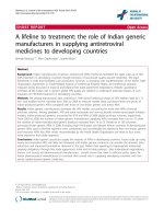
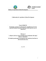
Fig. 1 A nomogram predicting the probability of metastatic lymph node involvement for patients with early gastric cancer. The probability of
metastatic lymph node involvement in early gastric cancer is calculated by drawing a line to the point on the axis for each of the following
variables: age, macroscopic type, size, histology, differentiation, ulcer, lymphovascular invasion and depth of invasion. The points for each variable
are summed and located on the total point line. Next, a vertical line is projected from the total point line to the predicted probability bottom
scale to obtain the individual probability of metastatic lymph node involvement
size, histology, differentiation, ulcer, lymphovascular invasion, and depth of invasion), and a total score was
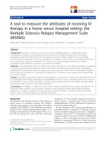
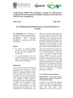
calculated from the nomogram. The total points corresponded to a predicted metastatic lymph node metastasis probability. Furthermore, we developed a ROC
curve to estimate the predictive accuracy of the model,
which had an AUC of 0.860 (95 % CI: 0.809–0.912), implying a good concordance (Fig. 2).
Discussion
Most surgeons consider D2 lymphadenectomy (dissection of all group I and group II lymph nodes) to be the
Fig. 2 A receiver operating characteristics (ROC) curve of the multivariate logistic regression model for predicting lymph node metastasis for patients
with early gastric cancer which had an AUC of 0.860 (95 % CI: 0.809–0.912), implying a good concordance
Zheng et al. BMC Cancer (2016) 16:92
standard and optimal surgical procedure for patients
with EGC within the past ten years [28, 29]. The lymph
node metastasis incidence of EGC is reported to be approximately 11 % to 18 %, and approximately 70 % to
80 % of patients will undergo overtreatment with D2
lymphadenectomy [29–32]. Recently, less invasive treatments have been performed for EGC, including endoscopic mucosal resection and endoscopic submucosal
dissection [33, 34]. Because of recent advances in surgical instrumentation and techniques, laparoscopic procedures have also been suggested as an alternative
minimally invasive treatment for EGC [35, 36]. For this
purpose, we retrospectively analyzed 597 EGC patients
based on clinical and routinely definitive pathological
characteristics to investigate the evidence used for making medical decisions. Many cancer clinicians are increasingly becoming attracted to simple tools such as
Nomograms to improve cancer treatment. In the present
study, we have developed a nomogram that could predict the incidence of lymph node metastasis in EGC
patients.
The lymph node metastasis incidence of all EGC patients was 9.7 % in the current study, 51.7 % in cancers
with lymphovascular invasion, 20.8 % in cancers with
an ulcer, 14.0 % in cancers with an undifferentiated
histology, 18.3 % in submucosal cancers, 13.1 % in larger sized tumors (≥2.0 cm), 17.9 % in macroscopic type
III/mixed, and 15.0 % in younger patients (<50 years
old), which are similar to or lower than previous results
[32, 38, 39]. In the multivariate analysis, the patients’
age at diagnosis, differentiation status, the presence of
an ulcer, lymphovascular invasion and depth of invasion
were independent factors for lymph node metastasis,
and the presence of lymphovascular invasion was considered the most important predictor. Previous surveys
have clarified the pathological characteristics of EGC
with or without nodal metastases. Histologic ulceration
of the tumor, larger size (≥20 mm) and submucosal
penetration were independent risk factors for regional
lymph node metastasis [39, 40]. Lymphovascular involvement and mixed histological type tumors have
previously been reported as risk factors for nodal metastases [41, 42]. In general, the depth of tumor invasion reflects the progression of a tumor originating
from the mucosal layer and is significantly associated
with the presence of regional lymph node metastasis in
EGC [18]. Our results were consistent with the above
studies. Based on the previous studies and our results,
the tumor size, macroscopic type and histology were
considered to have significant clinical meaning among
the risk factors of lymph node metastasis in EGC.
Therefore, we chose these characteristic features in our
nomogram for predicting the incidence of lymph node
metastasis in EGC. This figure could generate estimates
Page 6 of 8
of the likelihood of metastatic lymph node involvement.
Our nomogram appears to be simple and practical with
a relatively high area under the ROC curve of 0.860, thus
exhibiting a good performance related to a mean error
that never exceeded 5 %. These findings support our selection of variables for determining suitable treatment.
The gold standard in the curative treatment of gastric
cancer is a radical operation generally associated with
D2 lymphadenectomy which has a high success incidence in early cases [43]. However, some complications
and mortality are associated with this procedure that are
not always necessary [44]. Certain groups of patients
with EGC have a lower possibility of lymph node metastases, allowing less invasive treatment strategies to be
adopted for these situations [38]. Endoscopic resection
with curative intent is indicated only in tumors that fulfill the endoscopic resection criteria because these tumors rarely metastasize to lymph nodes [16]. These
treatments preserve bodily functions and maintain quality of life. Our investigation has provided a good and
helpful method to address this issue. For example, an
undifferentiated submucosal gastric cancer patient with
an ulcer and lymphovascular invasion, who is younger
than 50 years of age, has more than an 80 % possibility
of lymph node metastasis without considering other factors. This patient is suitable for a radical operation with
lymphadenectomy or laparoscopic lymph node dissection following endoscopic dissection. In contrast, a patient with opposite characteristics, such as differentiated
mucosal cancer, without an ulcer or lymphovascular invasion, and older than 50 years old, has almost no risk
of lymph node metastasis with respect to other patients
(less than 5 %), and should receive less invasive treatments. Therefore, we believe our nomogram will assist
surgeons in selecting the appropriate treatment for patients with EGC with regard to the probability of lymph
node metastasis.
To our knowledge, this is the first study providing a
nomogram to predict the incidence of lymph node metastasis for EGC. The potential limitations of this study
include the small cohort, and we should expand the
sample size to improve the nomogram. Additionally, this
is a single center retrospective study that needs further
external validation with different populations. In this
study, we did not use specific cutoff values of lymph
node metastasis for different treatments with EGC. Despite these limitations, this nomogram offers an effective
tool to predict the incidence of lymph node metastasis
for EGC patients, with which we could select the appropriate treatments for patients.
Conclusions
In conclusion, the present study constructed a nomogram to predict the probability of lymph node metastasis
Zheng et al. BMC Cancer (2016) 16:92
in EGC patients based on lymphovascular invasion,
depth of invasion, differentiation, age, macroscopic type,
size, and histology. This tool can assist clinicians and patients in quantifying the potential lymph node metastasis
incidence to make surgical decisions. Certain patients
are suitable for a radical operation or endoscopic dissection plus D2 lymphadenectomy, and some patients can
be selected for only endoscopic dissection (ESD, EMR).
For future studies, we should expand the sample size,
add additional centers to prove this nomogram, and determine the cutoff value of the lymph node metastasis
incidence for different treatments.
Page 7 of 8
6.
7.
8.
9.
10.
11.
Ethical statement
The study was approved by the institution Review Board
of Peking University Cancer Hospital. All patients provided written informed consent.
Abbreviations
EGC: early gastric cancer; EMR: endoscopic mucosal resection;
ESD: endoscopic submucosal dissection; ROC: operating characteristic curve;
AUC: area under the curve; CI: confidence intervals.
12.
13.
14.
Competing interest
The authors declare that they have no competing interests.
15.
Authors’ contributions
ZXZ helped collecting the data and drafting the manuscript. YNZ revised
both logic and grammar mistakes of the article and improved the statistical
analysis. LHZ, ZYL and XJW participated in the design of the study. YQL
contributed in managing and providing data. ZDB conceived of the study,
and participated in its design. JFJ, as the corresponding author, gave final
approval of the version to be published. All the authors have read and
approved the manuscript for publication. All authors read and approved the
final manuscript.
16.
17.
18.
Acknowledgments
The authors acknowledge the Department of Gastrointestinal Surgery and
Department of Pathology of the Peking University Cancer Hospital for the
data management.
19.
Author details
1
Department of Gastrointestinal Surgery, Key Laboratory of Carcinogenesis and
Translational Research (Ministry of Education), Peking University Cancer Hospital
& Institute, 52 Fu Cheng Road, Hai Dian District, 100142, Beijing, China.
2
Department of Pathology, Key Laboratory of Carcinogenesis and Translational
Research (Ministry of Education), Peking University Cancer Hospital & Institute,
Beijing, China.
20.
Received: 18 October 2015 Accepted: 7 February 2016
22.
21.
23.
References
1. Thrumurthy SG, Chaudry MA, Hochhauser D, Mughal M. The diagnosis and
management of gastric cancer. BMJ. 2013;347:f6367.
2. Chen W, Zheng R, Zhang S, Zhao P, Zeng H, Zou X, et al. Annual report on
status of cancer in China, 2010. Chin J Cancer Res. 2014;26:48–58.
3. Takahashi T, Saikawa Y, Kitagawa Y. Gastric cancer: current status of
diagnosis and treatment. Cancers (Basel). 2013;5:48–63.
4. Yamada T, Sugiyama H, Ochi D, Akutsu D, Suzuki H, Narasaka T, et al. Risk
factors for submucosal and lymphovascular invasion in gastric cancer
looking indicative for endoscopic submucosal dissection. Gastric Cancer.
2014;17:692–6.
5. Shen L, Shan Y-S, Hu H-M, Price TJ, Sirohi B, Yeh K-H, Yang Y-H, Sano T,
Yang H-K, Zhang X, Park SR, Fujii M, Kang Y-K, Chen L-T. Management of
24.
25.
26.
gastric cancer in Asia: resource-stratified guidelines. Lancet Oncol. 2013;
14(12):e535–47.
Bu Z, Ji J. Controversies in the diagnosis and management of early gastric
cancer. Chin J Cancer Res. 2013;25:263–6.
Saragoni L, Morgagni P, Gardini A, Marfisi C, Vittimberga G, Garcea D, et al.
Early gastric cancer: diagnosis, staging, and clinical impact. Evaluation of 530
patients. New elements for an updated definition and classification. Gastric
Cancer. 2013;16:549–54.
Lee HH, Yoo HM, Song KY, Jeon HM, Park CH. Risk of limited lymph node
dissection in patients with clinically early gastric cancer: indications of
extended lymph node dissection for early gastric cancer. Ann Surg Oncol.
2013;20:3534–40.
Baptista V, Singh A, Wassef W. Early gastric cancer: an update on endoscopic
management. Curr Opin Gastroenterol. 2012;28:629–35.
Gotoda T. Endoscopic resection of early gastric cancer. Gastric Cancer.
2007;10:1–11.
Ohnita K, Isomoto H, Shikuwa S, Yajima H, Minami H, Matsushima K,
et al. Early and long-term outcomes of endoscopic submucosal
dissection for early gastric cancer in a large patient series. Exp Ther
Med. 2014;7:594–8.
Ishikawa S, Togashi A, Inoue M, Honda S, Nozawa F, Toyama E, et al.
Indications for EMR/ESD in cases of early gastric cancer: relationship
between histological type, depth of wall invasion, and lymph node
metastasis. Gastric Cancer. 2007;10:35–8.
Yanai H, Matsumoto Y, Harada T, Nishiaki M, Tokiyama H, Shigemitsu T, et al.
Endoscopic ultrasonography and endoscopy for staging depth of invasion
in early gastric cancer: a pilot study. Gastrointest Endosc. 1997;46:212–6.
Chung I-K, Lee JH, Lee S-H, Kim S-J, Cho JY, Cho WY, et al. Therapeutic
outcomes in 1000 cases of endoscopic submucosal dissection for early
gastric neoplasms: Korean ESD Study Group multicenter study. Gastrointest
Endosc. 2009;69:1228–35.
Isomoto H, Shikuwa S, Yamaguchi N, Fukuda E, Ikeda K, Nishiyama H, et al.
Endoscopic submucosal dissection for early gastric cancer: a large-scale
feasibility study. Gut. 2009;58:331–6.
Gotoda T, Yanagisawa A, Sasako M, Ono H, Nakanishi Y, Shimoda T, et al.
Incidence of lymph node metastasis from early gastric cancer: estimation
with a large number of cases at two large centers. Gastric Cancer. 2000;3:
219–25.
Soetikno R, Kaltenbach T, Yeh R, Gotoda T. Endoscopic mucosal resection
for early cancers of the upper gastrointestinal tract. J Clin Oncol. 2005;23:
4490–8.
Abe N, Watanabe T, Suzuki K, Machida H, Toda H, Nakaya Y, et al. Risk
factors predictive of lymph node metastasis in depressed early gastric
cancer. Am J Surg. 2002;183:168–72.
Li C, Kim S, Lai JF, Oh SJ, Hyung WJ, Choi WH, et al. Risk factors for lymph
node metastasis in undifferentiated early gastric cancer. Ann Surg Oncol.
2008;15:764–9.
Klar M, Jochmann A, Foeldi M, Stumpf M, Gitsch G, Stickeler E, et al. The
MSKCC nomogram for prediction the likelihood of non-sentinel node
involvement in a German breast cancer population. Breast Cancer Res Treat.
2008;112:523–31.
Briganti A, Larcher A, Abdollah F, Capitanio U, Gallina A, Suardi N, et al.
Updated nomogram predicting lymph node invasion in patients with prostate
cancer undergoing extended pelvic lymph node dissection: the essential
importance of percentage of positive cores. Eur Urol. 2012;61:480–7.
Rahman R, Asombang AW, Ibdah JA. Characteristics of gastric cancer in Asia.
World J Gastroenterol. 2014;20:4483–90.
Santiago JMR, Sasako M, Osorio J. [TNM-7th edition 2009 (UICC/AJCC) and
Japanese Classification 2010 in Gastric Cancer. Towards simplicity and
standardisation in the management of gastric cancer]. Cir Esp. 2011;89:275–81.
Luomaranta A, Leminen A, Loukovaara M. Prediction of lymph node and
distant metastasis in patients with endometrial carcinoma: a new model
based on demographics, biochemical factors, and tumor histology. Gynecol
Oncol. 2013;129:28–32.
Wang SJ, Lemieux A, Kalpathy-Cramer J, Ord CB, Walker GV, Fuller CD, et al.
Nomogram for predicting the benefit of adjuvant chemoradiotherapy for
resected gallbladder cancer. J Clin Oncol. 2011;29:4627–32.
Van Zee KJ, Manasseh D-ME, Bevilacqua JLB, Boolbol SK, Fey JV, Tan LK,
et al. A nomogram for predicting the likelihood of additional nodal
metastases in breast cancer patients with a positive sentinel node biopsy.
Ann Surg Oncol. 2003;10:1140–51.
Zheng et al. BMC Cancer (2016) 16:92
Page 8 of 8
27. Ma H, Bandos AI, Gur D. On the use of partial area under the ROC curve for
comparison of two diagnostic tests. Biom J. 2015;57:304–20.
28. Borie F, Plaisant N, Millat B, Hay JM, Fagniez PL. French Associations for
Surgical Research: Appropriate gastric resection with lymph node dissection
for early gastric cancer. Ann Surg Oncol. 2004;11:512–7.
29. Nitti D, Marchet A, Mammano E, Ambrosi A, Belluco C, Mencarelli R, et al.
Extended lymphadenectomy (D2) in patients with early gastric cancer. Eur J
Surg Oncol. 2005;31:875–81.
30. Roviello F, Rossi S, Marrelli D, Pedrazzani C, Corso G, Vindigni C, et al.
Number of lymph node metastases and its prognostic significance in early
gastric cancer: a multicenter Italian study. J Surg Oncol. 2006;94:275–80.
discussion 274.
31. Pelz J, Merkel S, Horbach T, Papadopoulos T, Hohenberger W.
Determination of nodal status and treatment in early gastric cancer. Eur J
Surg Oncol. 2004;30:935–41.
32. Hyung WJ, Cheong JH, Kim J, Chen J, Choi SH, Noh SH. Application of
minimally invasive treatment for early gastric cancer. J Surg Oncol. 2004;85:
181–5. discussion 186.
33. Gotoda T, Yamamoto H, Soetikno RM. Endoscopic submucosal dissection of
early gastric cancer. J Gastroenterol. 2006;41:929–42.
34. Abe N, Watanabe T, Sugiyama M, Yanagida O, Masaki T, Mori T, Atomi Y.
Endoscopic treatment or surgery for undifferentiated early gastric cancer?
Am J Surg. 2004;188:181–4.
35. Noh SH, Hyung WJ, Cheong JH. Minimally invasive treatment for gastric
cancer: approaches and selection process. J Surg Oncol. 2005;90:188–93.
discussion 193–4.
36. Kitano S, Shiraishi N, Uyama I, Sugihara K, Tanigawa N. Japanese
Laparoscopic Surgery Study Group: A multicenter study on oncologic
outcome of laparoscopic gastrectomy for early cancer in Japan. Ann Surg.
2007;245:68–72.
37. Kitamura K, Yamaguchi T, Taniguchi H, Hagiwara A, Sawai K, Takahashi T.
Analysis of lymph node metastasis in early gastric cancer: rationale of
limited surgery. J Surg Oncol. 1997;64:42–7.
38. Sung C-M, Hsu C-M, Hsu J-T, Yeh T-S, Lin C-J, Chen T-C, et al. Predictive
factors for lymph node metastasis in early gastric cancer. World J
Gastroenterol. 2010;16:5252–6.
39. Yamao T, Shirao K, Ono H, Kondo H, Saito D, Yamaguchi H, et al. Risk factors
for lymph node metastasis from intramucosal gastric carcinoma. Cancer.
1996;77:602–6.
40. Hirasawa T, Gotoda T, Miyata S, Kato Y, Shimoda T, Taniguchi H, et al.
Incidence of lymph node metastasis and the feasibility of endoscopic
resection for undifferentiated-type early gastric cancer. Gastric Cancer.
2009;12:148–52.
41. Hanaoka N, Tanabe S, Mikami T, Okayasu I, Saigenji K. Mixed-histologic-type
submucosal invasive gastric cancer as a risk factor for lymph node
metastasis: feasibility of endoscopic submucosal dissection. Endoscopy.
2009;41:427–32.
42. Kwee RM, Kwee TC. Predicting lymph node status in early gastric cancer.
Gastric Cancer. 2008;11:134–48.
43. Sano T, Aiko T. New Japanese classifications and treatment guidelines for
gastric cancer: revision concepts and major revised points. Gastric Cancer.
2011;14:97–100.
44. Sasako M. Risk factors for surgical treatment in the Dutch Gastric Cancer
Trial. Br J Surg. 1997;84:1567–71.
Submit your next manuscript to BioMed Central
and we will help you at every step:
• We accept pre-submission inquiries
• Our selector tool helps you to find the most relevant journal
• We provide round the clock customer support
• Convenient online submission
• Thorough peer review
• Inclusion in PubMed and all major indexing services
• Maximum visibility for your research
Submit your manuscript at
www.biomedcentral.com/submit