The anti-apoptotic, antioxidant and antiinflammatory effects of curcumin on acrylamide-induced neurotoxicity in rats
Bạn đang xem bản rút gọn của tài liệu. Xem và tải ngay bản đầy đủ của tài liệu tại đây (12.15 MB, 10 trang )
Guo et al. BMC Pharmacology and Toxicology
/>
(2020) 21:62
RESEARCH ARTICLE
Open Access
The anti-apoptotic, antioxidant and antiinflammatory effects of curcumin on
acrylamide-induced neurotoxicity in rats
Jie Guo1,2, Xiaolu Cao1,2, Xianmin Hu1,2, Shulan Li1,2 and Jun Wang1,2*
Abstract
Background: Acrylamide (ACR) formed during heating of tobacco and carbohydrate-rich food as well as widely
applied in industries has been known as a well-established neurotoxic pollutant. Although the precise mechanism is
unclear, enhanced apoptosis, oxidative stress and inflammation have been demonstrated to contribute to the ACRinduced neurotoxicity. In this study, we assessed the possible anti-apoptotic, antioxidant and anti-inflammatory
effects of curcumin, the most active component in a popular spice known as turmeric, on the neurotoxicity caused
by ACR in rats.
Methods: Curcumin at the dose of 50 and 100 mg/kg was orally given to ACR- intoxicated Sprague-Dawley rats
exposed by ACR at 40 mg/kg for 4 weeks. All rats were subjected to behavioral analysis. The HE staining and
terminal deoxynucleotidyl transferase mediated dUTP nick end labelling (TUNEL) staining were used to detect
histopathological changes and apoptotic cells, respectively. The mRNA and protein expressions of apoptosis-related
molecule telomerase reverse transcriptase (TERT) were detected using real-time PCR and immunohistochemistry,
respectively. The contents of malondialdehyde (MDA) and glutathione (GSH) as well as the activities of superoxide
dismutase (SOD) and glutathione peroxidase (GSH-Px) were measured as the indicators for evaluating the level of
oxidative stress in brain. The levels of pro-inflammatory cytokinestumor necrosis factor-α (TNF-α) and interleukin-1β
(IL-1β) in the cerebral homogenates were detected using ELISA assay.
Results: ACR-induced weigh loss, deficits in motor function as well as pathological alterations in brains were
significantly improved in rats administrated with 50 and 100 mg/kg curcumin. TUNEL-positive apoptotic cells in
curcumin-treated ACR intoxicated brains were less than those in the ACR model group. Curcumin administration
especially at the dose of 100 mg/kg upregulated the TERT mRNA expression and enhanced the number of TERTpositive cells in ACR-intoxicated cortex tissues. Moreover, curcumin treatment reduced the concentrations of TNF-α,
IL-1β and MDA, while increased the GSH contents as well as the SOD and GSH-Px activities in the cerebral
homogenates, in comparison to ACR control group.
(Continued on next page)
* Correspondence:
1
Hubei Province Key Laboratory of Occupational Hazard Identification and
Control, Wuhan University of Science and Technology, Wuhan 430065, China
2
Department of Pharmacy, New Medicine Innovation and Development
Institute, College of Medicine, Wuhan University of Science and Technology,
Wuhan 430065, China
© The Author(s). 2020 Open Access This article is licensed under a Creative Commons Attribution 4.0 International License,
which permits use, sharing, adaptation, distribution and reproduction in any medium or format, as long as you give
appropriate credit to the original author(s) and the source, provide a link to the Creative Commons licence, and indicate if
changes were made. The images or other third party material in this article are included in the article's Creative Commons
licence, unless indicated otherwise in a credit line to the material. If material is not included in the article's Creative Commons
licence and your intended use is not permitted by statutory regulation or exceeds the permitted use, you will need to obtain
permission directly from the copyright holder. To view a copy of this licence, visit />The Creative Commons Public Domain Dedication waiver ( applies to the
data made available in this article, unless otherwise stated in a credit line to the data.
Guo et al. BMC Pharmacology and Toxicology
(2020) 21:62
Page 2 of 10
(Continued from previous page)
Conclusions: These data suggested the anti-apoptotic, antioxidant and anti-inflammatory effects of curcumin on
ACR-induced neurotoxicity in rats. Maintaining TERT-related anti-apoptotic function might be one mechanism
underlying the protective effect of curcumin on ACR-intoxicated brains.
Keywords: Acrylamide, Curcumin, Apoptosis, Antioxidant, Inflammation, Telomerase reverse transcriptase
Background
As a chemical formed during the high-temperature processing of tobacco and carbohydrate-rich foods, acrylamide (ACR) is well recognized as a human neurotoxin
which has posed significant public health concerns due
to its daily intake [1–3]. Moreover, ACR is widely
employed in various chemical and industrial processes
as a component to produce polymers used in gel chromatography, dye synthesis, production of paper, cosmetics and waste water management, etc [4, 5]. The
work-related ACR exposure has been demonstrated to
bring on neurotoxicity in occupationally exposed population, which is manifested as ataxia, skeletal muscle
weakness, gait abnormalities, skin abnormalities, as well
as numbness of hands and feet [4].
The exposure to monomeric form of ACR results in
multiple pathological changes in central and peripheral
nervous system. Among them, ACR-induced apoptosis
that subsequently leads to the death and loss of neurons
has been accepted as a fundamental and predominant
mechanism of neurotoxicity in ACR-exposed humans
and animals [6–8]. Telomerase reverse transcriptase
(TERT) is one of catalytic units of telomerase, importantly, acts as rate-limiting determinant and the most important regulator of telomerase activity [9, 10].
Telomerase is required to synthesize the telomeric DNA
strand thus maintain the length of telomeres, the latter
of which is a DNA-protein complex located at chromosome ends and has an ability of protecting against genome instability [9]. So far, the anti-apoptotic effect of
TERT has been revealed in neuronal cells influenced by
various risk factors such as oxidative stress, DNA damage and ischemia [9, 10]. In line with these findings, our
previous study [5] has demonstrated that TERT-related
anti-apoptotic function was significantly down-regulated
in rats with ACR-induced neurobehavioral deficits. The
mRNA and protein expression of TERT in the rat cerebral cortex was reduced by ACR treatment [5]. As the
critical events in chemical-induced neurodegeneration,
oxidative stress and enhanced lipid peroxidation are induced by exposure to ACR, which are also important
mechanisms underlying ACR-induced neurotoxicity [11,
12]. During ACR metabolism in the body, excessive
levels of reactive oxygen species (ROS) are certainly produced. Moreover, ACR may have deleterious effects on
antioxidant enzymes such as superoxide dismutase
(SOD) and glutathione peroxidase (GSH-Px) thus decrease the antioxidant defence in the brains [11, 12].
Furthermore, many evidences [12, 13] have shown the
production of inflammatory cytokines such as tumor necrosis factor-α (TNF-α) and interleukin-1β (IL-1β) was
enhanced after ACR intoxication.
Accordingly, in recent years, some agents with antiapoptosis, antioxidant and anti-inflammatory properties
have been expected to attenuate ACR-induced neurotoxicity [3, 8, 11–14]. As the most active constituent in turmeric, a common spice, with a strong safety record,
curcumin has been considered to be a potential natural
neuroprotective agent under limelight [15–18]. Based on
its known antioxidant, anti-inflammatory and antiapoptosis activities, curcumin has been shown to protect
the neurons against cerebral ischemia-reperfusion injury
[15, 16], dysfunction linked with Parkinson’s disease mediated by Bisphenol-A [19], sleep-deprivation induced
memory impairments [20], and depression [21], etc.
However, there is limited evidence in the possible ameliorative effect of curcumin against ACR-induced neurotoxicity. Prasad and Muralidhara [22] have demonstrated
the neuroprotective effect of curcumin in an ACR
model of neurotoxicity in an insect species, Drosophila
melanogaster. A recently published study [23] reported
that curcumin would exert a protective effect against
ACR-induced spatial memory impairment in rats. However, the anti-apoptotic, antioxidant and antiinflammatory activities of curcumin have not been well
evaluated in ACR-induced neurotoxicity. In the present
study, we identified whether curcumin could exert protective effects against neuron apoptosis, oxidative stress
and inflammatory response caused by ACR exposure in
rats.
Methods
Chemicals
ACR and curcumin were purchased from Amresco Co.
(Solon, OH, USA) and Sigma chemicals Co.(St. Louis,
MO, USA), respectively.
Experimental design
Male Sprague-Dawley rats, weighing 200–220 g, were
obtained from Hubei Experimental Animal Research
Center (Hubei, China). Rats were housed in standard
translucent cages (5 animals/cage) under controlled
Guo et al. BMC Pharmacology and Toxicology
(2020) 21:62
standard conditions (23 ± 2 °C, 55 ± 5% relative humidity,
12 h light/dark cycle) with restricted access to standard
rat chow and free access to tap water. After acclimation
for 1-week, healthy animals were randomly assigned into
4 groups (10 rats per group): normal control group;
ACR-intoxicated control group; low-dose (50 mg/kg)
curcumin treatment group and high-dose (100 mg/kg)
curcumin treatment group. A dose of 40 mg/kg ACR
(dissolved in normal saline) was intraperitoneally
injected every other day for 4 weeks in all animals except
the normal control group. The normal rats received saline as control. Meanwhile, rats in the curcumin treatment groups were daily administered with curcumin at
the corresponding oral administration dose for 4 weeks.
The doses of ACR and curcumin were chosen based on
the previous study [5] and preliminary experiments. The
normal and ACR-intoxicated control animals were orally
administered with the same volume of distilled water.
Body weight and behavioral alterations were monitored
once a week. At 24 h after the last administration, all animals were euthanized by CO2 asphyxiation, brain tissues were quickly collected.
Behavioral tests
All rats were subjected to behavioral analysis to assess
their motor functions.
In the hind limb splay examination [3, 5], the hind
paws of rats were inked, then the rats were placed in a
horizontal position of 30 cm high and dropped onto a
white paper. The distance between the center points of
right and left heels were recorded as the landing foot
spread distance.
In the movement initiation test [5, 24], the rat was
held by its hind limbs and its torso, one forelimb was
lifted above a table in order that the body weight was
supported by the other forelimb alone. Then, rat was
allowed to initiate stepping movements for one forelimb,
and then the other. The averaged time period to initiate
one step was recorded as the response latency for each
forelimb.
In the gait score test [3, 5], animals were placed on the
table and were observed for 3 min. Gait was scored as
follow: 1: normal gait; 2, slightly abnormal gait characterized by slight ataxia, weakness and foot splay; 3, moderately abnormal gait characterized by obvious ataxia
and foot splay with limb spread during ambulation; 4,
severely abnormal gait characterized by a combination
of all the above symptoms, dragging hind limbs and inability to support body weight.
Histopathological analysis
The collected brain tissues were fixed with 10% neutralbuffered formalin followed by dehydrating and paraffinembedding. Then, embedded brain sections (5-μm
Page 3 of 10
thickness) were stained with hematoxylin and eosin (HE)
for histopathological observation. The histopathological
changes in cerebral cortex, hippocampal CA1, CA3, and
dentate gyrus regions were analyzed.
TUNEL assay
The apoptotic neurons in the brain sections were detected using the terminal deoxynucleotidyl transferase
mediated dUTP nick end labelling (TUNEL) assay. After
deparaffinization and rehydration, the brain sections
were permeabilized with proteinase K solution, then exposed to the mixture of biotinylated nucleotide dUTP
and recombinant terminal deoxynucleotidyl transferase
(TdT) following the instruction manual of TUNEL
Apoptosis Assay Kit (Servicebio, Wuhan, China). Staining with 4,6-diamino-2-phenyl indole (DAPI) (Sigma, St.
Louis, USA) was performed to visualize nuclei. Images
were obtained under a fluorescent microscope (Olympus, Center Valley, USA).
Real-time PCR
Total RNA of brain cerebral cortex tissues was isolated
using TRIzol reagent (Invitrogen, Carlsbad, CA, USA).
The expression levels of TERT mRNA were measured
by real-time PCR using all-in-OneTM qPCR master mix
AOPR-1200 (GeneCopoeia, Rockville, MD, USA). The
sequences of primer sets for TERT were 5′-TGTTCC
TGTTCTGGCTAATGG- 3′(forward) and 5′-CCTCTT
GTGACAGTTCCCGT-3′ (reverse). β-actin gene was
applied as a reference.
Immunohistochemistry
Paraffin-embedded brain sections of 5-μm thickness were
incubated with a rabbit anti-TERT antibody (Servicebio,
Wuhan, China), then a biotinylated goat anti-rabbit secondary antibody (Servicebio, Wuhan, China). Immune
complexes were visualized by incubation with 3,3′-diaminobenzidine tetrachloride (DAB) and hematoxylin.
Measurement of parameters related to oxidative stress in
cerebral homogenates
The brain tissue were homogenized with 9 times the
volume of PBS on ice and then centrifuged to prepare
homogenates. The contents of malondialdehyde
(MDA) and glutathione (GSH) as well as the activities
of SOD and GSH-Px in the cerebral homogenates
were measured following the respective manufacturer’s protocols (Nanjing Jiancheng Bio-Engineering
Co., Ltd., Nanjing, China). Protein contents in the
cerebral homogenates were determined using the
bicinchoninic acid assay kit (Nanjing Jiancheng BioEngineering Co., Ltd., Nanjing, China).
Guo et al. BMC Pharmacology and Toxicology
(2020) 21:62
Page 4 of 10
Measurement of IL-1β and TNF-ɑ levels in cerebral
homogenates
Protective effect of curcumin on ACR-induced neuron
apoptosis
The concentrations of IL-1β and TNF-ɑ in cerebral homogenates were determined using using ELISA kits according to the manufacturer’s instructions (IL-1β:
PeproTech Inc., NJ, USA; TNF-ɑ: R&D Systems, Minneapolis, MN, USA).
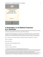
As showing in Fig. 3, immunofluorescent staining
showed that the number of TUNEL-positive apoptotic
nerve cells was significantly increased in the cortex and
hippocampus of ACR intoxicated rats. However, curcumin administration could effectively reduce the number
of apoptotic cells (P < 0.05; P < 0.01), suggesting its antiapoptotic activity in ACR-damaged neurons. TUNELpositive cells in curcumin-treated ACR intoxicated
brains had decreased to approximately 13.8–22.1% of
those in the ACR model group.
Statistical analysis
All experiments were conducted with two technical replicates. Data were expressed as the mean ± SD, and analyzed using one-way analysis of variance (ANOVA) with
post hoc Tukey test by SPSS 22.0 software. P < 0.05 or
P < 0.01 was considered statistically significant.
Results
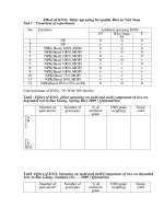
Effect of curcumin on ACR-induced body weight and
neurobehavioral changes
As shown in Fig. 1a, the animals in the ACR group
began to show slow growth compared to the normal
control group since 2 weeks of exposure (P < 0.05). At
the end of the 4-week exposure period, the average body
weight of ACR intoxicated rats was 73.4% of that of normal rats (P < 0.01). However, curcumin administration
protected the rats from ACR-induced weigh loss. Compared with the ACR model group, curcumin at the dose
of 50 mg/kg caused a significant weight gain at 4th week
(P < 0.05). And the body weight of rats administrated
with 100 mg/kg curcumin increased by 12.5 and 14.6%
at 3rd and 4th week, respectively (P < 0.01).
Landing foot spread distance was enlarged rapidly
from the first week of ACR exposure (Fig. 1b), and
significant differences were found between the ACR
intoxicated group and the normal control group
throughout the exposure period (P < 0.01). Similarly,
ACR intoxicated rats developed a progressive impairment of forelimb movement initiation (P < 0.01) (Fig.
1c) and significant gait abnormalities (Fig. 1d and e)
including obvious ataxia and foot splay, twisting of
hind limbs and inability to support body weight. Curcumin intervention in ACR intoxicated rats markedly
improved these neurobehavioral changes in a dosedependent manner (P < 0.05; P < 0.01).
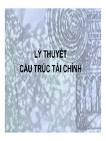
Effect of curcumin on ACR-induced histopathological
alterations in rat brains
The neuronal morphological characteristic in the cerebral cortex and hippocampus was identified using H&E
staining. As showing in Fig. 2, severe neuronal loss, condensed and fragmented nuclei were found in the cortex
and hippocampus of ACR intoxicated rats. Compared
with the ACR model group, there was more nerve cells
and less pathological alterations in the brain of rats administrated with curcumin.
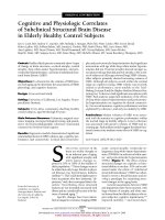
Effect of curcumin on ACR-inhibited TERT expression
Our previous study [5] suggested that TERT, an emerging anti-apoptotic molecule mainly expressed in cortical neurons, was down-regulated in the cerebral cortex
of ACR treated rats. In order to identify whether curcumin has regulative effect on ACR-inhibited TERT expression, the mRNA and protein expressions of TERT
were detected using real-time PCR and immunohistochemistry, respectively. As shown in Fig. 4, curcumin
treatment especially at the dose of 100 mg/kg increased TERT mRNA expression level (P < 0.01), and
enhanced the number of TERT-positive cells in ACRintoxicated cortex tissues, suggesting curcumin might
exert anti-apoptotic activity in ACR-induced neurotoxicity partly through maintaining TERT-related
anti-apoptotic function.
Effect of curcumin on oxidative stress caused by ACR
To explored the possible anti-oxidant effect of curcumin
on ACR-induced neurotoxicity in rats, the contents of
MDA, GSH and the activities of SOD, GSH-Px in the
cerebral homogenates were quantified as measures of
the level of oxidative stress in the brain. As shown in
Table 1, the content of MDA was markedly increased,
while the GSH level, the activities of SOD and GSHPX were markedly decreased in cerebral homogenates
of ACR-treated rats in comparison to the normal
control group (P < 0.01), suggesting ACR-induced oxidative stress in the brain. As expected, these alterations induced by ACR were significantly ameliorated
by curcumin treatment in a dose-dependent manner
(P < 0.05; P < 0.01), suggesting that the anti-oxidative
activity of curcumin might, at least partly, be responsible for its neuroprotective effect in ACR intoxicated
rats.
Effect of curcumin on cerebral contents of IL-1β and TNFɑ in ACR intoxicated rats
To explore the possible anti-inflammatory activity involved in curcumin mediated neuroprotection in ACR
intoxicated rats, the levels of pro-inflammatory
Guo et al. BMC Pharmacology and Toxicology
(2020) 21:62
Page 5 of 10
Fig. 1 Effect of curcumin on the body weights (a), landing foot spread distance (b), movement initiation test (c) and gait (d and e) in ACRtreated rats. Data are means±SD of 10 animals in each group. *P < 0.05, **P < 0.01 compared to the corresponding control rats. # P < 0.05, ## P <
0.01; compared with the corresponding ACR group
cytokines IL-1β and TNF-ɑ were detected in the cerebral homogenates. Our results show that, although
ACR exposure moderately stimulated the production
of pro-inflammatory cytokines in brain (P < 0.05),
curcumin at the dose of 100 mg/kg significantly decreased the levels of IL-1β and TNF-ɑ by 22.8 and
14.1%, respectively (P < 0.05) (Fig. 5), when compared
with the ACR group.
Discussion
Curcumin, with its neuroprotective effects and hardly
existing toxicity, have become an attractive alternative
treatment tool for various neurological disorders [15–20].
After systemic administration, curcumin can across the
blood–brain barrier, and exert its therapeutic efficacy in
the brain [25]. In the present study, we demonstrated the
anti-apoptotic, antioxidant and anti-inflammatory effects
Guo et al. BMC Pharmacology and Toxicology
(2020) 21:62
Page 6 of 10
Fig. 2 Effect of curcumin on the histopathological changes in cortex, CA1, CA3, dentate gyrus of ACR-treated rat brains. (H&E staining 200×)
of curcumin on ACR-induced neurotoxicity in rats, suggesting the use of curcumin to prevent or delay neurological damages induced by ACR exposure. In line with
the evidences from humans and animals [4, 5, 8, 11–14],
our study showed that the 4-week exposure of rats to
ACR at the dose of 40 mg/kg caused a significant body
weight loss, progressive deficits in motor function and adverse pathological outcome in the cortex and hippocampus of rats. Importantly, the present data revealed that
curcumin administration could efficiently rescue ACRinduced weight loss and neurobehavioral deficits, relieve
the neuropathological damages in brain.
As an important event of neuronal cell number control, apoptosis that is an inappropriate activation of the
neuronal cell-suicide program has been well-accepted as
a fundamental component in the development of various
brain diseases [26]. In particular, in view of the very limited regenerative capacity of the central nervous system
tissue, it is vitally important to prevent against neuronal
cell apoptosis, and then limit the brain damage caused
by neuronal death [26]. So far, apoptosis has become a
prime therapeutic target in the development of
neuroprotective agents. Treatment preventing the neuronal cell apoptosis can maintain the cell numbers, reduce the severity and progression of brain diseases. In
the present study, the anti-apoptotic potential of curcumin in ACR-intoxicated brains which was manifested by
the significant decreased TUNEL-positive apoptotic
nerve cells in the cortex and hippocampus might be an
important mechanism underlying its neuroprotective effect against exposure to ACR.
A variety of small molecules can act on crucial checkpoints of apoptosis [26]. In recent years, the role of TERT
in apoptosis has attracted considerable interest as an
emerging anti-apoptotic molecule involved in compensatory neuroprotective mechanism against neuronal cell
death [9, 10]. ACR intoxication significantly reduced the
expression of TERT in the brain, suggesting the TERTrelated anti-apoptotic function participated in the ACR
neurotoxicity [5]. Interestingly, some new evidences showing that curcumin up-regulates function of TERT have
emerged [27, 28]. Curcumin extracted with ethyl acetate
concentration-dependently up-regulated the TERT
mRNA expression in rat clone-9 hepatocytes [27].
Guo et al. BMC Pharmacology and Toxicology
(2020) 21:62
Page 7 of 10
Fig. 3 Effect of curcumin on the neuron apoptosis in ACR-treated rat brains. (TUNEL staining 400×). a Representative images. b Quantitative
assessment of neuronal density of TUNEL-positive cells (number of cells/mm2). Data are means±SD of 10 animals in each group. **P < 0.01
compared to the corresponding control rats. # P < 0.05, ## P < 0.01; compared with the corresponding ACR group
Pirmoradi et al. [28] reported that the TERT expression of
rat adipose tissue-derived stem cells was significantly increased in the presence of curcumin at concentrations of
1 and 5 μM. In line with these in vitro studies [27, 28], we
showed the curcumin-induced in vivo up-regulation of
TERT at the levels of gene and protein, which might be
one mechanism underlying the anti-apoptotic activity of
curcumin in ACR-intoxicated brains.
Guo et al. BMC Pharmacology and Toxicology
(2020) 21:62
Page 8 of 10
Fig. 4 Effect of curcumin on the expression of TERT in the cortex tissues of ACR-treated rats. a The mRNA expression was measured with Realtime PCR. b Immunohistochemical staining for the protein expression of TERT. Data are means ± SD of 10 animals in each group. **P < 0.01
compared to the corresponding control rats. ##P < 0.01; compared with the corresponding ACR group
Table 1 Effect of curcumin on the levels of MDA, GSH, SOD and GSH-Px in cerebral homogenates prepared from ACR intoxicated
rats (n = 10, mean ± SD)
Groups
MDA
(nmol/mg prot)
GSH
(mg/g prot)
SOD
(U/mg prot)
GSH-Px
(U/mg prot)
Normal
0.425 ± 0.141
4.41 ± 0.58
60.21 ± 5.38
14.81 ± 1.95
ACR
1.133 ± 0.352 **
2.30 ± 0.47**
52.72 ± 6.94 **
10.36 ± 1.84 **
ACR + curcumin 50 mg/kg
0.918 ± 0.322
2.77 ± 0.46 #
53.89 ± 8.02
12.58 ± 1.96 #
ACR + curcumin 100 mg/kg
0.854 ± 0.216
#
**P < 0.01 compared to the corresponding control rats. # P < 0.05,
2.92 ± 0.59
##
#
59.16 ± 6.46
P < 0.01; compared with the corresponding ACR group
#
13.15 ± 1.87 ##
Guo et al. BMC Pharmacology and Toxicology
(2020) 21:62
Page 9 of 10
Fig. 5 Effect of curcumin on cerebral contents of IL-1β and TNF-ɑ in ACR intoxicated rats. Data are means ± SD of 10 animals in each group.
*P < 0.05 compared to the corresponding control rats. # P < 0.05; compared with the corresponding ACR group
In addition, curcumin is well known for its classic and
strong anti-oxidative and anti-inflammatory activities
[29]. ACR exposure has been demonstrated to result in a
disturbance in the balance between the free radical formation and elimination, the latter of which is mediated
by antioxidant systems [11, 12]. The phenolic structure
in curcumin confers electron-capturing properties,
which destabilize ROS, explaining the well-accepted
antioxidant effects [30]. However, being similar to other
antioxidants including vitamin E, vitamin C, and carotenoids, curcumin has been found to show double-edged
roles in the level of intracellular ROS, which appeared to
be highly dependent on the cell type [30–32]. Curcumin
has been reported to elevate ROS levels in multiple cancer cells [30–32]. In this study, in line with the wellaccepted anti-oxidative activity of curcumin in normal
and non-malignant cells [29–32], 4-week exposure of
rats to 40 mg/kg ACR markedly enhanced the level of
MDA (an essential biomarker of oxidative stress and
lipid peroxidation), decreased the content of GSH (a biologically important intracellular thiol acting as a free
radical scavenger) and the activities of SOD and GSH-Px
(two important antioxidant enzymes) in the brain tissues. But curcumin alleviated the augmented production
of MDA and the reduction of antioxidant capacity induced by ACR, thus might play a role in the detoxification of reactive oxygen species generated by ACR.
Moreover, neuroinflammation has been demonstrated in
various pathologies of the brain including ACR-induced
neurotoxicity [33]. The 4-week exposure to ACR induced inflammatory responses in the brain tissues, evident by upregulated levels of IL-1β and TNF-ɑ, two
potent pro-inflammatory cytokines acting as master regulators of neuroinfammation in the central nerve system.
While curcumin could improve the ACR-induced neuroinflammation, which was in accord with its proven antiinflammatory property.
Conclusions
In summary, this study convinced the anti-apoptotic,
antioxidant and anti-inflammatory effects of curcumin
on ACR-induced neurotoxicity in rats. And maintaining
TERT-related anti-apoptotic function might be one
mechanism underlying the protective effect of curcumin
on ACR-intoxicated brains.
Abbreviations
ACR: Acrylamide; GSH: Glutathione; GSH-Px: Glutathione peroxidase;
HE: Hematoxylin and eosin; IL-1β: Interleukin-1β; MDA: Malondialdehyde;
ROS: Reactive oxygen species; SOD: Superoxide dismutase; TERT: Telomerase
reverse transcriptase; TNF-α: Tumor necrosis factor-α; TUNEL: Terminal
deoxynucleotidyl transferase mediated dUTP nick end labelling
Acknowledgements
Not applicable.
Authors’ contributions
JW and XC contributed to the design of the research. JG,XH and SL
performed the research. JG, CX and JW analyzed the data. JW prepared the
article. All authors read and approved the final manuscript.
Funding
This study was financially supported by the National Natural Science Funding
of China (Nos. 71974153, Nos. 81602108). The study funder had no further
role in the study design, data collection, analyses, interpretation of results,
writing of the article, or the decision to submit it for publication.
Availability of data and materials
The datasets supporting the conclusions of this article are included within
the article. The raw data can be requested from the corresponding author.
Ethics approval and consent to participate
Animal experiments were approved by the Animals Care and Use
Committee of Medicine College, Wuhan University of Science and
Guo et al. BMC Pharmacology and Toxicology
(2020) 21:62
Page 10 of 10
Technology (resolution number 2019078), and accomplished in line with the
guidelines of the National Health and Medical Research Council of China.
20.
Consent for publication
Not applicable.
Competing interests
The authors declare that they have no competing interests.
Received: 21 October 2019 Accepted: 11 August 2020
References
1. Erkekoglu P, Baydar T. Acrylamide neurotoxicity. Nutr Neurosci. 2014;17(2):
49–57.
2. Koszucka A, Nowak A, Nowak I, Motyl I. Acrylamide in human diet, its
metabolism, toxicity, inactivation and the associated European Union legal
regulations in food industry. Crit Rev Food Sci Nutr. 2019;25:1–16.
3. Su B, Guan Q, Wang M, Liu N, Wei X, Wang S, Yang X, Jiang W, Xu M, Yu S.
Calpeptin is neuroprotective against acrylamide-induced neuropathy in rats.
Toxicology. 2018;400–401:1–8.
4. Pennisi M, Malaguarnera G, Puglisi V, Vinciguerra L, Vacante M,
Malaguarnera M. Neurotoxicity of acrylamide in exposed workers. Int J
Environ Res Public Health. 2013;10(9):3843–54.
5. Wang J, Zhang MY, Xu SQ, Cheng J, Yu ZJ, Hu XM. Down-regulation of
telomerase reverse transcriptase-related anti-apoptotic function in a rat
model of acrylamide induced neurobehavioral deficits. Biotech Histochem.
2018;93(7):512–8.
6. Sahinturk V, Kacar S, Vejselova D, Kutlu HM. Acrylamide exerts its
cytotoxicity in NIH/3T3 fibroblast cells by apoptosis. Toxicol Ind Health.
2018;34(7):481–9.
7. Lee JG, Wang YS, Chou CC. Acrylamide-induced apoptosis in rat primary
astrocytes and human astrocytoma cell lines. Toxicol in Vitro. 2014;28(4):
562–70.
8. Sun G, Wang X, Li T, Qu S, Sun J. Taurine attenuates acrylamide- induced
apoptosis via a PI3K/AKT-dependent manner. Hum Exp Toxicol. 2018;37(12):
960327118765335.
9. Liu MY, Nemes A, Zhou QG. The emerging roles for telomerase in the
central nervous system. Front Mol Neurosci. 2018;11:160.
10. Li J, Tang B, Qu Y, Mu D. Telomerase reverse transcriptase: a novel
neuroprotective mechanism involved in neonatal hypoxic-ischemic brain
injury. Int J Dev Neurosci. 2011;29(8):867–72.
11. Goudarzi M, Mombeini MA, Fatemi I, Aminzadeh A, Kalantari H, Nesari A,
Najafzadehvarzi H, Mehrzadi S. Neuroprotective effects of Ellagic acid
against acrylamide-induced neurotoxicity in rats. Neurol Res. 2019;41(5):419–
28.
12. Santhanasabapathy R, Vasudevan S, Anupriya K, Pabitha R, Sudhandiran G.
Farnesol quells oxidative stress, reactive gliosis and inflammation during
acrylamide-induced neurotoxicity: behavioral and biochemical evidence.
Neuroscience. 2015;308:212–27.
13. Acaroz U, Ince S, Arslan-Acaroz D, Gurler Z, Kucukkurt I, Demirel HH, Arslan
HO, Varol N, Zhu K. The ameliorative effects of boron against acrylamideinduced oxidative stress, inflammatory response, and metabolic changes in
rats. Food Chem Toxicol. 2018;118:745–52.
14. Adewale OO, Brimson JM, Odunola OA, Gbadegesin MA, Owumi SE, Isidoro
C, Tencomnao T. The potential for plant derivatives against acrylamide
neurotoxicity. Phytother Res. 2015;29(7):978–85.
15. Mukherjee A, Sarkar S, Jana S, Swarnakar S, Das N. Neuro-protective role of
nanocapsulated curcumin against cerebral ischemia-reperfusion induced
oxidative injury. Brain Res. 1704;2019:164–73.
16. Xu L, Ding L, Su Y, Shao R, Liu J, Huang Y. Neuroprotective effects of
curcumin against rats with focal cerebral ischemia-reperfusion injury. Int J
Mol Med. 2019;43(4):1879–87.
17. Baj T, Seth R. Role of Curcumin in regulation of TNF-α mediated brain
inflammatory responses. Recent Patents Inflamm Allergy Drug Discov. 2018;
12(1):69–77.
18. Zhao XC, Zhang L, Yu HX, Sun Z, Lin XF, Tan C, Lu RR. Curcumin protects
mouse neuroblastoma Neuro-2A cells against hydrogen-peroxide-induced
oxidative stress. Food Chem. 2011;129(2):387–94.
19. Akintunde JK, Farouk AA, Mogbojuri O. Metabolic treatment of syndrome
linked with Parkinson's disease and hypothalamus pituitary gonadal
21.
22.
23.
24.
25.
26.
27.
28.
29.
30.
31.
32.
33.
hormones by turmeric curcumin in Bisphenol-a induced neuro-testicular
dysfunction of wistar rat. Biochem Biophys Rep. 2018;17:97–107.
Noorafshan A, Karimi F, Kamali AM, Karbalay-Doust S, Nami M. Restorative
effects of curcumin on sleep-deprivation induced memory impairments
and structural changes of the hippocampus in a rat model. Life Sci. 2017;
189:63–70.
Fan C, Song Q, Wang P, Li Y, Yang M, Yu SY. Neuroprotective effects of
Curcumin on IL-1β-induced neuronal apoptosis and depression-like
behaviors caused by chronic stress in rats. Front Cell Neurosci. 2019;12:516.
Prasad SN. Muralidhara. Neuroprotective effect of geraniol and curcumin in
an acrylamide model of neurotoxicity in Drosophila melanogaster:
relevance to neuropathy. J Insect Physiol. 2014;60:7–16.
Yan D, Yao J, Liu Y, Zhang X, Wang Y, Chen X, Liu L, Shi N, Yan H. Tau
hyperphosphorylation and P-CREB reduction are involved in acrylamideinduced spatial memory impairment: suppression by curcumin. Brain Behav
Immun. 2018;71:66–80.
Pang Y, Lin S, Wright C, Shen J, Carter K, Bhatt A, Fan LW. Intranasal insulin
protects against substantia nigra dopaminergic neuronal loss and
alleviatesmotor deficits induced by 6-OHDA in rats. Neuroscience. 2016;318:
157–65.
Goozee KG, Shah TM, Sohrabi HR, Rainey-Smith SR, Brown B, Verdile G,
Martins RN. Examining the potential clinical value of curcumin in the
prevention and diagnosis of Alzheimer's disease. Br J Nutr. 2016;115(3):449–
65.
Cavallucci V, D'Amelio M. Matter of life and death: the pharmacological
approaches targeting apoptosis in brain diseases. Curr Pharm Des. 2011;
17(3):215–29.
Pan MH, Wu JC, Ho CT, Badmaev V. Effects of water extract of Curcuma
longa (L.) roots on immunity and telomerase function. J Complement Integr
Med. 2017;14(3):20150107.
Pirmoradi S, Fathi E, Farahzadi R, Pilehvar-Soltanahmadi Y, Zarghami N.
Curcumin affects adipose tissue-derived Mesenchymal stem cell aging
through TERT gene expression. Drug Res (Stuttg). 2018;68(4):213–21.
He Y, Yue Y, Zheng X, Zhang K, Chen S, Du Z. Curcumin, inflammation, and
chronic diseases: how are they linked? Molecules. 2015;20(5):9183–213.
Willenbacher E, Khan SZ, Mujica SCA, Trapani D, Hussain S, Wolf D,
Willenbacher W, Spizzo G, Seeber A. Curcumin: new insights into an ancient
ingredient against Cancer. Int J Mol Sci. 2019;20(8):1808.
Tong R, Wu X, Liu Y, Liu Y, Zhou J, Jiang X, Zhang L, He X, Ma L. Curcumininduced DNA Demethylation in human gastric Cancer cells is mediated by
the DNA-damage response pathway. Oxidative Med Cell Longev. 2020;2020:
2543504.
Liczbiński P, Michałowicz J, Bukowska B. Molecular mechanism of curcumin
action in signaling pathways: Review of the latest research. Phytother Res.
2020. />Zong C, Hasegawa R, Urushitani M, Zhang L, Nagashima D, Sakurai T,
Ichihara S, Ohsako S, Ichihara G. Role of microglial activation and
neuroinflammation in neurotoxicity of acrylamide in vivo and in vitro.
Arch Toxicol. 2019;93(7):2007–19.
Publisher’s Note
Springer Nature remains neutral with regard to jurisdictional claims in
published maps and institutional affiliations.