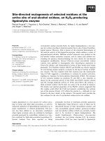
Diacerein retards cell growth of chondrosarcoma cells at the G2/M cell cycle checkpoint via cyclin B1/CDK1 and CDK2 downregulation
Bạn đang xem bản rút gọn của tài liệu. Xem và tải ngay bản đầy đủ của tài liệu tại đây (2.38 MB, 11 trang )
Lohberger et al. BMC Cancer (2015) 15:891
DOI 10.1186/s12885-015-1915-4
RESEARCH ARTICLE
Open Access
Diacerein retards cell growth of
chondrosarcoma cells at the G2/M cell
cycle checkpoint via cyclin B1/CDK1 and
CDK2 downregulation
Birgit Lohberger1*, Andreas Leithner1, Nicole Stuendl1, Heike Kaltenegger1, Werner Kullich2
and Bibiane Steinecker-Frohnwieser2
Abstract
Background: Chondrosarcoma is characterized for its lack of response to conventional cytotoxic chemotherapy,
propensity for developing lung metastases, and low rates of survival. Research within the field of development and
expansion of new treatment options for unresectable or metastatic diseases is of particular priority. Diacerein, a
symptomatic slow acting drug in osteoarthritis (SYSADOA), implicates a therapeutic benefit for the treatment of
chondrosarcoma by an antitumor activity.
Methods: After treatment with diacerein the growth behaviour of the cells was analyzed with the xCELLigence
system and MTS assay. Cell cycle was examined using flow cytometric analysis, RT-PCR, and western blot analysis of
specific checkpoint regulators. The status for phosophorylation of mitogen-activated protein kinases (MAPKs) was
analyzed with a proteome profiler assay. In addition, the possible impact of diacerein on apoptosis was investigated
using cleaved caspase 3 and Annexin V/PI flow cytometric analysis.
Results: Diacerein decreased the cell viability and the cell proliferation in two different chondrosarcoma cell lines in
a dose dependent manner. Flow cytometric analysis showed a classical G2/M arrest. mRNA and protein analysis
revealed that diacerein induced a down-regulation of the cyclin B1-CDK1 complex and a reduction in CDK2 expression.
Furthermore, diacerein treatment increased the phosphorylation of p38α and p38β MAPKs, and Akt1, Akt2, and Akt 3 in
SW-1353, whereas in Cal-78 the opposite effect has been demonstrated. These observations accordingly to our cell cycle
flow cytometric analysis and protein expression data may explain the G2/M phase arrest. In addition, no apoptotic
induction after diacerein treatment, neither in the Cal-78 nor in the SW-1353 cell line was observed.
Conclusions: Our results demonstrate for the first time that the SYSADOA diacerein decreased the viability of
human chondrosarcoma cells and induces G2/M cell cycle arrest by CDK1/cyclin B1 down-regulation.
Keywords: Chondrosarcoma, Diacerein, Cell cycle arrest, Cyclin B1, CDK
* Correspondence:
1
Department of Orthopedic Surgery, Medical University Graz,
Auenbruggerplatz 5, A-8036 Graz, Austria
Full list of author information is available at the end of the article
© 2015 Lohberger et al. Open Access This article is distributed under the terms of the Creative Commons Attribution 4.0
International License ( which permits unrestricted use, distribution, and
reproduction in any medium, provided you give appropriate credit to the original author(s) and the source, provide a link to
the Creative Commons license, and indicate if changes were made. The Creative Commons Public Domain Dedication waiver
( applies to the data made available in this article, unless otherwise stated.
Lohberger et al. BMC Cancer (2015) 15:891
Background
Diacerein represents a symptomatic slow acting drug in
osteoarthritis (SYSADOA) from the anthraquinone chemical class and as the general term implicates, efficacy
against the symptoms of osteoarthritis (OA) has been
demonstrated [1]. As an anti-rheumatic drug, the potential of diacerein lies in the in vitro inhibition of the synthesis of interleukin-1 and its activity within the synthesis of
proteoglycans, glycosaminoclycans, and hyaluronuic acid,
principle components of cartilage extracellular matrix [2].
By using an experimental canine model of OA, an effective reduction in chondrocyte DNA fragmentation and cell
death, due to a diacerein induced reduction of caspase-3
activity has been observed [3]. Within the early lesions of
experimental OA the activation of the caspase cascade has
been connected to chondrocyte death, whereas caspase as
well as MEK1/2 and p38MAPK inhibitors reveal a marked
deterioration of the programmed cell death and attenuate
the severity of cartilage lesions [4, 5]. Studying the cell
proliferation and cell viability characteristics of C28/I2
chondrocytes, strikingly increased concentrations of diacerein significantly decreases cell growth and viability [6].
These observed growth-inhibiting qualities of diacerein,
when applied at higher concentrations, might implicate
a therapeutic benefit for the treatment of chondrosarcoma [7]. While diacerein has proved to be effective in
the treatment of OA, Qin et al described that a diacerein
α-aminophosphonate conjugate has anti-proliferative activities on tumor cells [8].
Chondrosarcomas constitute a heterogeneous group
of neoplasms, tumor cells with the common characteristics in terms of the production of components of the
extracellular matrix within the cartilage [9]. With an
incidence of 1:50,000, chondrosarcoma typically occurs
in adults in their 3rd to 6th decade of life and represent
the second most common primary malignant bone
tumor in large epidemiologic series [10]. Wide surgical
excision remains the best available treatment for intermediate- to high-grade tumors since they are relatively
chemo- and radiotherapy resistant because of their
extracellular matrix, low percentage of dividing cells,
and poor vascularity, [11–14]. However, for high-grade
chondrosarcoma, the prognosis is poor even after adequate surgery [15]. From the clinical point of view it is
a huge challenge within the field of cancer treatment,
to prevent recurrence and to find better treatment options for unresectable or metastatic diseases, such as
chondrosarcoma.
The aim of this study was to show if diacerein is able
to generate a reduction in cell growth and if this decline
is generated by cell cycle arrest or apoptosis. Therefore,
the effect of diacerein on cell proliferation, cell cycle distribution, and apoptosis of two human chondrosarcoma
cell lines was investigated.
Page 2 of 11
Methods
Cell culture
Human chondrosarcoma cell lines SW-1353 (CLS,
Eppelheim, Germany) and Cal-78 (DSMZ, Braunschweig,
Germany) were cultured in Dulbecco’s-modified Eagle’s
medium (DMEM-F12; GIBCO®, Invitrogen, Darmstadt,
Germany), containing 5 % fetal bovine serum (FBS), 1 % Lglutamine, 100 units/ml Penicillin, 100 μg/ml Streptomycin,
and 0.25 μg Amphotericin B (all GIBCO®, Invitrogen). Both
cell lines were verified by short tandem repeat analysis
using PowerPlex 16 System Kit (Promega, Mannheim,
Germany). Cells were kept at 37 °C in a humidified atmosphere of 5 % CO2 and were passaged by trypsinization after
reaching 80–90 % confluence.
Sample preparation
Pure Diacerein (TRB Chemedica International, Geneva,
Switzerland) was dissolved in DMSO and diluted with
culture medium. The final DMSO concentration was
max. 0.5 %, which did not affect the behavior of the cells
as observed by benchmark experiments.
Cell proliferation
MTS assay (Brand, Voerde-Friedrichsfeld, Germany) was
used to measure the metabolic activity of cells: 5 × 10³
cells per well were seeded into 96 well plates and treated
with 0–500 μM diacerein. The cells were treated for
24 h and 48 h, thereafter a CellTiter 96 AQueous Assay
(Promega, Mannheim, Germany) was performed following the manufacturers’ instructions; untreated cells were
used for control (ctrl).
The xCELLigence DP device from Roche Diagnostics
(Mannheim, Germany) was used to monitor cell proliferation in realtime after cells were seeded on electronic
microtiter plates (E-Plate; Roche Diagnostic) [16]. Cells
were treated with 0, 30, 100, and 300 μM diacerein and
the proliferation rate was measured for 24 h. Cell index
(CI) measurements were performed in triplicates with a
signal detection set for every 20 min. The cell index (CI)
is a measure for the cell density of cells and was normalized to the time point when diacerein was added. Subsequent to the continuous xCELLigence cell monitoring,
the slope (1/h) representing the rate of change of the cell
index was calculated from time 7–24 h. Acquisition and
analysis was performed with the RTCA software (Version
1.2, Roche Diagnostics).
Flow cytometry for cell cycle analysis
SW1353 and Cal-78 cells were treated with 30, 100, and
300 μM diacerein. After a treatment time of 24 and
48 h, respectively, the cells were harvested by trypsinization and fixed with 70 % ice-cold ethanol for 10 min at
4 °C. Next to washing, the cell pellet was re-suspended
in PI-staining buffer (50 μl/ml PI, RNAse A, Beckman
Lohberger et al. BMC Cancer (2015) 15:891
Coulter, Brea, CA) and was incubated for 15 min at 37 °C.
Cell cycle distribution was analyzed by FACS Calibur (BD
Biosciences, San Diego, CA) using ModFit software.
Reverse transcription polymerase chain reaction (RT-PCR)
Total ribonucleic acid (RNA) was isolated from treated and
untreated cells with the RNeasy Mini Kit and DNase-I
treatment according to the manufacturer’s manual (Qiagen,
Hilden, Germany). One microgram RNA was reverse transcribed using a RevertAid cDNA Synthesis Kit (Fermentas).
Amplification was achieved with the Platinum SYBR Green
Super Mix with ROX (Invitrogen) on AB7900HT (Applied
Biosystems, Invitrogen). Each qPCR run consisted of a
standard 3-step PCR temperature protocol (annealing
temperature of 60 °C) followed by a melting curve
protocol to confirm a single gene-specific peak and to
detect primer dimerization. Relative quantification of
expression levels were obtained by the ΔΔCt method
based on the geometric mean of the internal controls
glyceraldehyde 3-phosphate dehydrogenase (GAPDH),
β-actin (ACTB), and hypoxanthine phosphoribosyltransferase (HPRT-1), respectively. The following primers
were used for real time RT-PCR: QuantiTect primer
assays (Qiagen) for cyclin B1 (ID QT00006615), CDK1
(ID QT00042672), and CDK2 (ID QT00005586). The expression level (CT) of the target gene was normalized to
the reference genes (ΔCt), the ΔCt of the test sample was
normalized to the ΔCt of the control (ΔΔCt). Finally, the
expression ratio was calculated with the 2-ΔΔCt method
(*p < 0.05).
Page 3 of 11
MAPK array
For the Proteome Profiler Human Phospho-MAPK Array
Kit (ARY002B; R&D Systems, Minneapolis; MN), cells
were treated with 30 μM diacerein for 24 h and whole cell
protein extracts were prepared with lysis buffer. Capture
and control antibodies of the major families of mitogenactivated protein kinases (MAPKs), the extracellular signalregulated kinases (ERK1/2), c-Jun N-terminal kinases
(JNK1-3), and different p38 isoforms (α/β/δ/γ), have been
spotted in duplicate on nitrocellulose membranes. To analyzing the phosphorylation status the array was performed
following the manufacturers’ instructions; untreated cells
were used for control.
Caspase-3 apoptosis assay
After incubation with with 30, 100, and 300 μM diacerein for 48 h, cells were harvested by trypsinization, fixed
with formaldehyde for 10 min at 37 °C (2 × 10⁶ cells/ml),
permeabilized with methanol, and finally re-suspended
in incubation-buffer (FBS:PBS 1:200). Caspase-3, a marker
for cells that undergo apoptosis, is activated by the proteolytic processing of its inactive zymogen into the activated
p17 and p12 fragments, respectively. The FITC-conjugated
monoclonal cleaved caspase-3 (Asp175) antibody (#9661;
Cell Signaling Technology) detects endogenous levels of
the large fragment (17/19 kDa), released after the activation of caspase-3. The antibody does not recognize full
length caspase-3 or other cleaved caspases. Cells were analyzed by flow cytometry (FACS Calibur, BD Biosciences)
performed with FACSDiva software. Histograms were created using FCS3 express software (De Novo software, Los
Angeles, CA). Untreated cells were used as negative
control.
Western blot analysis
For immunoblotting, whole cell protein extracts were
prepared with lysis buffer (50 mM Tris–HCl pH 7.4,
150 mM NaCl, 50 mM NaF, 1 mM EDTA, 10 % NP-40,
1 % Triton-X, and protease inhibitors), subjected to
SDS-PAGE (10 or 12 %) and blotted onto PVDF membrane (Roth, Karlsruhe, Germany). Primary antibodies
against cyclin B1 (sc-245), CDK1 (sc-54), CDK2 (sc6248), and p53 (sc-126) were purchased from Santa Cruz
(Santa Cruz Biotechnology, Santa Cruz, CA), phosphoHistone H2A.X (#9718), Akt (#9272), phospho-Akt
(Ser473) (#9271), p38 MAPK (#9212), and phospho-p38
MAPK (Thr180/Tyr182) (#4511) antibodies from Cell
Signaling Technology (Cell Signaling Technology,
Danvers, MA) and β-actin (A4700) from Sigma-Aldrich
(Vienna, Austria). Blots were developed using horseradish
peroxidase-conjugated secondary antibodies (Dako, Jena,
Germany) at room temperature for 1 h and the SuperSignal® West Pico Chemoluminescent Substrate (Thermo
Scientific, Rockford, IL), in accordance with the manufacturers’ protocol.
Annexin V/PI apoptosis assay
The APC Annexin V Apoptosis Detection Kit (BioLegends,
San Diego, CA) was performed following the manufacturers’ instructions. Apoptotic cells were identified by the
incubation of 1 × 10⁵ cells in 100 μl Annexin V binding
buffer containing 5 μl Annexin V-APC and 5 μl PI for
15 min at room temperature. Flow cytometry analysis was
performed with FACS Calibur (BD Biosciences); 10,000
events were collected. Cells were identified in the side scatter and forward scatter with linear scale. Fluorescence signals were shown with logarithmic scale. Compensation
was performed by single Annexin and PI measurements
and analyzed by FCS3 express software (De Novo software). Untreated cells were used as negative control.
Statistical analysis
All values are expressed as mean values ± SD. Student’s
unpaired t-test was used to evaluate differences between
treated groups and their respective controls. The significance of dose or time responses was assessed by repeated
Lohberger et al. BMC Cancer (2015) 15:891
measures analysis. Graphic data were prepared and calculated with SigmaPlot® (Systat Software Inc., San Jose, CA).
Results
Diacerein reduced cell proliferation and viability of
chondrosarcoma cells
Chondrosarcoma cells were treated with 30, 100, and
300 μM diacerein for 48 h. During this period, cell
growth curves were automatically recorded in real time
by the xCELLigence System (Fig. 1a). Diacerein inhibited
cell growth in a concentration dependent manner in
both cell lines. While an effect on Cal-78 cells could
only be observed at a concentration of 300 μM diacerein, causing a complete reduction of cell proliferation,
SW-1353 cells demonstrated a higher sensitivity for
Page 4 of 11
diacerein illustrated by a considerably reduced cell index
at a concentration of 30 and 100 μM. A block of cell
growth or rather an induced cell death may contribute
to this decrease in cell index. In Cal-78 cells slope values
changed under the influence of diacerein from 0.022 ±
0.007 (ctrl) to 0.025 ± 0.006 (30 μM), -0.007 ± 0.016
(100 μM; p = 0.001), and −0.035 ± 0.007 (300 μM; p =
8.77E-11). In SW-1353 cells a change from 0.144 ± 0.026
(ctrl) to 0.166 ± 0.082 (30 μM), 0.028 ± 0.055 (100 μM;
p = 0.012), and −0.028 ± 0.013 (300 μM; p = 1.01E-10)
could be observed.
To investigate the influence on cell viability, chondrosarcoma cell lines were exposed to 0, 3, 10, 30, 100, and
300 μM of diacerein for 24 and 48 h. After the equivalent treatment, cells were measured by the MTS assay
Fig. 1 Influence of diacerein on cell viability and cell proliferation of chondrosarcoma cells. a Dynamic proliferation curves for Cal-78 and SW-1353 cells
in the presence of 0 (black circles), 30 μM (grey circles), 100 μM (light grey triangles), and 300 μM (bicolour squares) diacerein. Data shown are representatives
from three independent experiments (n = 3, measured in duplicates). b After incubation over 24 and 48 h diacerein inhibited cell growth in a
concentration dependent manner. Untreated cells were measured as controls (n = 12, mean ± S.D.)
Lohberger et al. BMC Cancer (2015) 15:891
(n = 12). Figure 1b shows the time- and dose-dependent
inhibition of cell viability.
Diacerine caused a cell cycle G2/M arrest
To investigate the effects of diacerein on cell cycle,
chondrosarcoma cells were exposed to 30, 100, and
300 μM diacerein. Untreated cells were measured as
controls. The cell cycle distribution for both cell lines
(Cal-78 and SW-1353) is summarized in form of the
stacked bar chart given in Fig. 2a. In SW-1353 cells, diacerein caused after 48 h exposure a pronounced decrease in the number of cells in the G1 (grey bars)
phase, accompanied by a significant increase of the
number of S (black bars) and G2/M phase (striated bars)
cells, indicating a G2/M arrest. In the Cal-78 cell line
only a moderate tendency towards G2/M phase could be
Page 5 of 11
observed. Representative measurements of untreated
(control) and diacerein treated SW-1353 cells are depicted
to highlight the differences (Fig. 2b). All values of three
individual experiments (% of gated cells) are listed in
Table 1.
Diacerein decreased cyclin B1, CDK1, and CDK2 levels
Relative mRNA expression levels of cyclin B1, CDK1, and
CDK2 were analyzed by real time RT-PCR after treatment
of 30 and 100 μM diacerein for 48 h (Fig. 3a). Untreated
control cells served as reference value (ratio = 1). In the
case of Cal-78 cells treatment with diacerein (100 μM)
induced a small, but not significant change, for the expression of CDK1, whereas the cyclin B1 and CDK2 levels
were significantly down-regulated (cyclin B1: 0.612 ± 0.22
(p = 0.039); CDK2: 0.673 ± 0.16 (p = 0.027)) within the
Fig. 2 Graphical presentation of the cell cycle distribution. a Treatment of SW-1353 cells with 100 and 300 μM diacerein caused a significant
decrease in the number of cells in G1 phase (grey) after 48 h which was accompanied by a pronounced increase of cells in S phase (black) and
G2/M phase (dashed lines), while only small changes were observed in Cal-78 cells. Values are expressed as percentage of the cell population in
the G1, S, and G2/M phase of cell cycle. b Representative measurements of untreated control and 300 μM diacerein treated SW-1353 cells
Lohberger et al. BMC Cancer (2015) 15:891
Page 6 of 11
Table 1 Cell cycle distribution of chondrosarcoma cell lines after 24 and 48 h exposure to different concentrations of diacereina
Cell line
Treatment
h
G0/G1 (%)
S (%)
G2/M (%)
Cal-78
control
24 h
57.48 ± 8.19
23.79 ± 5.84
18.73 ± 2.51
SW-1353
diacerein 30 μM
24 h
57.57 ± 8.30
25.23 ± 4.69
17.22 ± 3.68
diacerein 100 μM
24 h
62.04 ± 5.54
22.01 ± 2.64
15.94 ± 2.85
diacerein 300 μM
24 h
54.15 ± 7.33
26.76 ± 8.21
19.08 ± 3.67
control
48 h
61.08 ± 3.03
21.23 ± 2.31
17.68 ± 0.89
diacerein 30 μM
48 h
61.31 ± 1.16
23.66 ± 2.55
15.03 ± 1.80
diacerein 100 μM
48 h
69.42 ± 2.80*
17.79 ± 2.38
12.78 ± 3.92
diacerein 300 μM
48 h
56.95 ± 12.37
24.51 ± 9.02
18.53 ± 4.09
control
24 h
51.14 ± 7.05
22.73 ± 6.01
26.12 ± 1.35
diacerein 30 μM
24 h
49.05 ± 5.46
27.15 ± 6.08
23.79 ± 1.23
diacerein 100 μM
24 h
55.95 ± 4.11
19.53 ± 6.81
24.52 ± 6.83
diacerein 300 μM
24 h
45.32 ± 12.41
25.35 ± 8.41
29.33 ± 7.06
control
48 h
64.77 ± 7.67
15.65 ± 6.65
19.56 ± 1.03
diacerein 30 μM
48 h
65.54 ± 5.75
15.37 ± 5.80
19.08 ± 0.31
diacerein 100 μM
48 h
54.36 ± 5.92
22.16 ± 10.54
23.47 ± 4.86
diacerein 300 μM
48 h
44.38 ± 10.52*
25.73 ± 7.23
29.89 ± 5.33*
Asterisks represent significant differences between control and treated cells (p < 0.0.5; n = 3; mean ± S.D.)
a
observation period. In SW-1353 cells a highly significant
down-regulation by 100 μM diacerein in the expression of
cyclin B1 to 0.095 ± 0.03 (p = 1.28E-05), CDK1 levels to
0.154 ± 0.02 (p = 9.82E-06), and the CDK2 levels to 0.443 ±
0.12 (p = 0.002) could be demonstrated.
In order to substantiate our observations, western blot
analysis for the specific regulatory proteins responsible
for the G2/M transition under the exposure of 30, 100,
and 300 μM diacerein for 48 h were performed (Fig. 3b).
Corresponding to the real time RT-PCR data, the expression of cyclin B1 and its corresponding cyclin-dependent
kinases CDK1 and CDK2 were clearly declined in diacerein treated SW-1353 cells, whereas diacerein affected the
expression of these G2/M checkpoint regulator proteins
in Cal-78 cells only to a small extent. Figure 3c represents
the quantitative evaluation of the western blot analysis of
three independent experiments. All values were normalized to their corresponding β-actin.
Phosphorylation of MAPKs under diacerein treatment
Treatment with 30 μM diacerein for 24 h increased the
phosphorylation of p38α (17.07 % change) and p38β
(34.01 %) MAPKs in SW-1353 cells, whereas in Cal-78
the observed phosphorylation changed to the contrary
(p38α: −22.78 % and p38β: −27.41 %). Likewise, the
MKK3 and MKK6 phosphorylation increased considerably
(MKK3: 28.19 % and MKK6: 22.59 %) in SW-1353 cells,
and decreased in Cal-78 cells (MKK3: −26.75 % and
MKK6: −18.87 %). The same picture is given for members
of the Akt family: a down-regulation for the Cal-78 cells
(Akt1: −23.34 %, Akt2: −16.70 %, and Akt3: −20.13 %) and
an augmentation when tested in SW1353 cells (Akt1:
12.85 %, Akt2: 19.29 %, and Akt3: 9.23 %) (Fig. 4a).
Western blot analysis confirmed the results from the
proteome profiler phospho-MAPK array (Fig. 4b). Phosphorylation of Akt (pAkt) and p38 (pp38) were upregulated after the treatment with different concentrations of
diacerein in SW-1353 cells, whereas, only minor changes
can be observed in the Cal-78 cells. The used phosphoAkt antibody detects endogenous levels of Akt1, Akt2,
and Akt3 only when phosphorylated at Ser473. The used
p38 MAPK antibody detects endogenous levels of total
p38α, -β and -γ MAPK protein. In addition, the expression of p53 was diminished in Cal-78 cells. The crosslinking of interesting genes of cell cycle regulation and
MAPK pathways is given in Fig. 4c using IPA (Ingenuity
Pathway Analysis) software.
Diacerein did not induce apoptosis
Apoptosis induction was investigated by flow cytometric
analysis of caspase-3 cleavage (Fig. 5a) and Annexin V/PI
staining (Fig. 5b). Figure 4a shows the cleaved caspase-3
measurements after a 48 h exposure of the cells to 30,
100, and 300 μM diacerein. The flow cytometric analysis
histograms represent untreated cells (black filled) versus
30 μM diacerein (striated lines), 100 μM diacerein (blue
lanes), and 300 μM diacerein (margenta lanes) treated
cells. Green lanes represent the Staurosporin positive control. In both cell lines only minimal caspase-3 cleavage
could be detected, worth mentioning is the minor dose
response detectable for the curve shift to the right within
Cal-78 cells. Although, induction of apoptosis was further
Lohberger et al. BMC Cancer (2015) 15:891
Page 7 of 11
Fig. 3 Regulation of the cell cycle checkpoints. a Relative mRNA expression of the cell cycle regulators cyclin B1, CDK1, and CDK2 after 48 h
incubation with 30 and 100 μM diacerein. Asterisks represent significant differences between control and treated cells (* p < 0.05; ** p < 0.01; ***
p < 0.001). b Total protein analysis after 48 h of treatment revealed a significant down-regulation of the G2/M arrest regulator proteins cyclin B1,
CDK1, and CDK2 in 100 and 300 μM diacerein treated chondrosarcoma cells. β-actin was used as loading control. c Quantitative evaluation of the
western blot analysis of cell cycle regulator proteins
verified by Annexin V/PI staining, in accordance with the
caspase-3 data, chondrosarcoma cells did not elicit an
increase in Annexin positive cells. Studying the phosphoHistone H2A.X DNA damage marker, a slight concentration
dependent increase in expression was detected for Cal-78
cells under the treatment with diacerein, while the SW-1353
cells did not show a response at the H2A.X expression level
(Fig. 5c). These sets of data imply that diacerein do not
significantly influence the induction of apoptosis.
Discussion
Chondrosarcoma is characterized for its lack of response
to conventional cytotoxic chemotherapy, propensity for
developing lung metastases, and poor survival. Therefore
research within the field of development and expansion of
new treatment options is of particular priority.
The applied concentration of 100 μM diacerein reduced
the cell viability of human chondrocytes by 20 % and accordingly induced a decrease in cell growth, while these
Lohberger et al. BMC Cancer (2015) 15:891
Fig. 4 (See legend on next page.)
Page 8 of 11
Lohberger et al. BMC Cancer (2015) 15:891
Page 9 of 11
(See figure on previous page.)
Fig. 4 Phosphorylation of MAPKs under diacerein treatment. a MAPKs phosphorylation of Cal-78 and SW-1353 chondrosarcoma cell lines in percent
change after 30 μM diacerein for 24 h. b Western blot analysis confirmed the results of the proteome profiler phospho-MAPK array and showed a
significant increase of phospho-Akt and phospho-p38 after the treatment with different concentrations of diacerein in SW-1353 cells, whereas, no
significant changes can be observed in the Cal-78 cells. The expression of p53 was diminished in Cal-78 cells. c Relationship of interesting genes using
IPA (Ingenuity Pathway Analysis)
two observations point to the fact that diacerein modulates
cellular physiology [6]. Rhein, the metabolite of diacerein,
has been demonstrated to induce anti-catabolic and antiproliferative effects on chondrocytes stimulated by IL-1β at
similar concentrations. Both effects were interpreted by
alterations in cell cycle regulation and not by action of
apoptosis [17]. However, the effect on chondrosarcoma has
not been examined yet.
In initial experiments we showed, that in two different
chondrosarcoma cell lines diacerein decreased the cell
viability and the cell proliferation in a dose dependent
manner. To identify the mechanism behind, within this
study we reviewed the effect of diacerein on cell cycle
distribution, the expression of cell cycle checkpoint proteins, the mitogen-activated protein kinases (MAPKs)
pathways, and the induction of apoptosis in SW-1353
and Cal-78 cell lines.
In eukaryotes, the cell cycle is regulated by cyclins and
cyclin-dependent kinases (CDKs). Cell cycle checkpoints
enable cellular repair or may result in the activation of
apoptosis signalling, if the cellular damages are significantly intense to be repaired properly [18, 19]. In particular, cyclin B and CDK1 proteins participate in the
regulation of the progression of G2/M phase [20]. It is
Fig. 5 Cleaved caspase-3 and Annexin V/PI apoptosis assays. a Cal-78 and SW-1353 cells were treated with 300 μM diacerein. Cleavage of caspase-3
was detected after 48 h by flow cytometry. The y-axis denotes cell counts and the x-axis represents fluorescence intensity of APC antibody. Black filled
histogram represents untreated control cells, striated histogram represents 30 μM diacerein, blue lanes showed 100 μM diacerein, and margenta lanes
showed 300 μM diacerein treated cells. Green lanes represented the Staurosporin positive control. b Likewise, AnnexinV/PI staining under the influence
of 300 μM diacerein for 48 h confirmed the lack of apoptosis induction. c In Cal-78 cells, the DNA damage marker H2AX was marginally increased.
β-actin was used as loading control
Lohberger et al. BMC Cancer (2015) 15:891
widely known that cells are blocked in the G2/M phase
during DNA damage, and cells are more susceptible to
the cytotoxic effects of radiotherapy in the G2/M phase
[21]. Increasing induced G2/M phase arrest allows cell
death which may be a useful strategy in cancer therapeutics [22].
The results of FACS analysis of SW-1353 cells treated
with diverse concentrations of diacerein showed a decrease in the number of cells in G1 and S phase which
was accompanied by a significant increase of the number
of G2/M phase cells, strongly indicating a classical G2/
M arrest. Interestingly, in the Cal-78 cell line only small
changes in cell cycle distribution could be detected, a
fact possibly ascribed to the differences in the origin of
the two cell lines. SW-1353 cells were obtained from a
primary grade II chondrosarcoma of the right humerus,
whereas the Cal-78 cells were established from the
recurrence of a dedifferentiated chondrosarcoma (grade
III) of the muscle.
We further explored the effect of diacerein on the key
regulators in the cell cycle checkpoints including CDK1,
CDK2, and cyclin B1 in Cal-78 and SW-1353 cells. The
CDK1-cyclin B1 complex is pivotal in regulating the G2/
M phase transition and mitosis. We observed a significant decrease in the mRNA and protein expression
levels of cyclin B1 and CDK1 in SW-1353, whereas in
Cal-78 cells only the protein expression of cyclin B1 was
significantly affected. The diacerein induced downregulation of the cyclin B1-CDK1 complex might explain
the observed reduction in cell growth in chondrosarcoma cells. Cyclin B1 is responsible for the transition of
the cell from the G2 to the M phase but changes to a
disruption in cancer cells where overexpression of cyclin
B1 can lead to uncontrolled cell growth [19]. In addition
both cell lines feature a significant reduction of CDK2
expression verified at the RNA and protein level, respectively. Altogether we are able to postulate, that
diacerein influences cell cycle of chondrosarcoma cells,
whereas the extent of the expression level of regulatory
proteins involved appear to depend on the source of the
cells. In our study Cal-78 cells are obvious more insensitive to diacerein regarding the cell cycle controls than
the SW-1353 cells.
MAPKs are signaling components that are important
in converting extracellular stimuli into a wide range of
cellular responses. Signaling network is increasingly
important for our comprehension of cell proliferation.
The p38 MAPK as an important stress kinase is involved
in the regulation of inflammation, cell growth and differentiation, cell cycle, and cell death [23]. Four isoforms of
p38, known as p38α, p38β, p38γ, and p38δ have been
identified, which can all be phosphorylated by the
MAPK kinase MKK6 (SKK3) and MKK3, respectively
[24]. p38 can negatively regulate cell cycle progression
Page 10 of 11
both at the G1/S and the G2/M transitions by several
mechanisms, including the down-regulation of cyclins,
up-regulation of CDK inhibitors and modulation of the
tumor suppressor p53 [25]. Moreover, we also provided
evidence about the existence of a crosstalk between the
p38 MAPK and Akt/mTOR signaling pathways in SW1353 cells exposed to diacerein. In the present study, we
have observed that diacerein treatment significantly
increases the phosphorylation of p38α and p38β MAPKs
in SW-1353, whereas Cal-78 shows a decrease of these
p38-MAPKs. These observations are in accordance with
our cell cycle FACS and protein expression data. The
serine-threonine Akt kinase family is well-known as crucial regulators of cell survival, proliferation, metabolism,
and migration [26]. Deregulation of Akt kinases is frequently associated with human diseases such as cancer
[27]. It has been reported previously that Akt activity is
high in the G2/M phase of the cell cycle in epithelial
cells [28]. Akt activity protects cells from apoptosis during the G2/M transition and is necessary for efficient
changeover to mitosis during unperturbed cell cycles
[29]. In addition to its role in cell cycle progression, Aktmediated phosphorylation and cytoplasmic translocation
of CDK2 is also important for apoptosis induced by
stresses such as methotrexate and docetaxel [30]. Therefore, an increased phosphorylation of Akt1, Akt2, and
Akt 3 in the SW-1353 cells after diacerein treatment
explained the G2/M phase arrest.
These observed growth-inhibiting characteristics of higher
concentrations of diacerein might implicate a therapeutic
benefit for the treatment of chondrosarcoma. To elucidate
the apoptotic potential of diacerein on chondrosarcoma
cells, we have performed two different apoptosis assays.
Diacerein treatment showed no apoptotic induction, neither
in the Cal-78 nor in the SW-1353 cell line.
Conclusion
Our results demonstrate for the first time that diacerein
decreased the viability of human chondrosarcoma cells
and induces G2/M cell cycle arrest by CDK1/cyclin B1
down-regulation. In summary, our findings strongly support diacerein as an interesting target for further investigation and development of novel therapeutics in sarcoma
research.
Abbreviations
ACTB: β-actin; CDK: Cyclin-dependent kinase; CI: Cell index; ERK1/2: Extracellular
signal-regulated kinases 1/2; GAPDH: Glyceraldehyde 3-phosphate
dehydrogenase; HPRT-1: Hypoxanthine phosphoribosyl-transferase; JNK1-3:
c-Jun N-terminal kinases 1-3; MAPKs: Mitogen-activated protein kinases;
OA: Osteoarthritis; RNA: Ribonucleic acid; RT-PCR: Reverse transcription
polymerase chain reaction; SYSADOA: Symptomatic slow acting drug in
osteoarthritis.
Competing interests
The authors declare that they have no competing interests.
Lohberger et al. BMC Cancer (2015) 15:891
Authors’ contributions
BL and BSF conceived and supervised the study. NS, HK, and BSF performed
the experiments. BL and BSF analyzed and interpreted the data. BL, BSF, and
AL drafted and revised the manuscript. WK and AL provided technical support.
All authors read and approved the final manuscript.
Acknowledgement
Financial support from the Medical University of Graz is gratefully acknowledged.
Author details
1
Department of Orthopedic Surgery, Medical University Graz,
Auenbruggerplatz 5, A-8036 Graz, Austria. 2Ludwig Boltzmann Institute for
Rehabilitation of Internal Diseases, Ludwig Boltzmann Cluster for
Rheumatology, Balneology and Rehabilitation, Saalfelden, Austria.
Received: 30 July 2015 Accepted: 6 November 2015
References
1. Bjornsson J, McLeod RA, Unni KK, Ilstrup DM, Pritchard DJ. Primary
chondrosarcoma of long bones and limb girdles. Cancer.
1998;83(10):2105–19.
2. Cho HJ, Oh YJ, Han SH, Chung HJ, Kim CH, Lee NS, et al. Cdk1 proteinmediated phosphorylation of receptor-associated protein 80 (RAP80) serine
677 modulates DNA damage-induced G2/M checkpoint and cell survival.
J Biol Chem. 2013;288(6):3768–76.
3. D’Adamo DR. Appraising the current role of chemotherapy for the
treatment of sarcoma. Semin Oncol. 2011;38 Suppl 3:S19–29.
4. D’Lima D, Hermida J, Hashimoto S, Colwell C, Lotz M. Caspase inhibitors
reduce severity of cartilage lesions in experimental osteoarthritis. Arthritis
Rheum. 2006;54(6):1814–21.
5. Damron TA, Ward WG, Stewart A. Osteosarcoma, chondrosarcoma, and
Ewing’s sarcoma: National Cancer Data Base Report. Clin Orthop Relat Res.
2007;459:40–7.
6. Fletcher CDM BJ, Hogendoorn PCW, Mertens F, editors. World Health
Organization: World Health Organization classification of tumours of soft
tissue and bone. 4th ed. Lyon: IARC Press; 2013. p. 264–74.
7. Gao Y, Moten A, Lin HK. Akt: a new activation mechanism. Cell Res.
2014;24(7):785–6.
8. Giuffrida AY, Burgueno JE, Koniaris LG, Gutierrez JC, Duncan R, Scully SP.
Chondrosarcoma in the United States (1973 to 2003): an analysis of 2890
cases from the SEER database. J Bone Joint Surg Am. 2009;91(5):1063–72.
9. Ichijo H. From receptors to stress-activated MAP kinases. Oncogene.
1999;18(45):6087–93.
10. Jian-Mei Q, Jian-Fei L, Man-Yi Y, Ri-Zheng H, Qing X, Ying-Ming P, Heng-Shan
W, Gui-Yang Y. Synthesis and antitumor activities of novel diacerein αaminophosphonates conjugates. Indian Journal of Chemistry 2014;
53B(12):1584-95.
11. Kandel ES, Skeen J, Majewski N, Di Cristofano A, Pandolfi PP, Feliciano CS,
et al. Activation of Akt/protein kinase B overcomes a G(2)/m cell cycle
checkpoint induced by DNA damage. Mol Cell Biol. 2002;22(22):7831–41.
12. Leeb BF. Clinical efficacy and safety of diacerein in osteoarthritis. european
musculoskeletal review. 2010.
13. Legendre F, Heuze A, Boukerrouche K, Leclercq S, Boumediene K, Galera P,
et al. Rhein, the metabolite of diacerhein, reduces the proliferation of
osteoarthritic chondrocytes and synoviocytes without inducing apoptosis.
Scand J Rheumatol. 2009;38(2):104–11.
14. Maddika S, Ande SR, Wiechec E, Hansen LL, Wesselborg S, Los M. Aktmediated phosphorylation of CDK2 regulates its dual role in cell cycle
progression and apoptosis. J Cell Sci. 2008;121(Pt 7):979–88.
15. Martel-Pelletier J, Mineau F, Jolicoeur FC, Cloutier JM, Pelletier JP. In vitro
effects of diacerhein and rhein on interleukin 1 and tumor necrosis factoralpha systems in human osteoarthritic synovium and chondrocytes.
J Rheumatol. 1998;25(4):753–62.
16. Montenegro MF, Sanchez-del-Campo L, Fernandez-Perez MP, Saez-Ayala M,
Cabezas-Herrera J, Rodriguez-Lopez JN. Targeting the epigenetic machinery
of cancer cells. Oncogene. 2015;34(2):135–43.
17. Nishida K, Furumatsu T, Takada I, Kawai A, Yoshida A, Kunisada T, et al.
Inhibition of human chondrosarcoma cell growth via apoptosis by
peroxisome proliferator-activated receptor-gamma. Br J Cancer.
2002;86(8):1303–9.
Page 11 of 11
18. Pawlik TM, Keyomarsi K. Role of cell cycle in mediating sensitivity to radiotherapy.
Int J Radiat Oncol Biol Phys. 2004;59(4):928–42.
19. Pelletier JP, Fernandes JC, Jovanovic DV, Reboul P, Martel-Pelletier J.
Chondrocyte death in experimental osteoarthritis is mediated by MEK 1/2
and p38 pathways: role of cyclooxygenase-2 and inducible nitric oxide
synthase. J Rheumatol. 2001;28(11):2509–19.
20. Pelletier JP, Mineau F, Boileau C, Martel-Pelletier J. Diacerein reduces the level
of cartilage chondrocyte DNA fragmentation and death in experimental dog
osteoarthritic cartilage at the same time that it inhibits caspase-3 and inducible
nitric oxide synthase. Clin Exp Rheumatol. 2003;21(2):171–7.
21. Sheth DS, Yasko AW, Johnson ME, Ayala AG, Murray JA, Romsdahl MM.
Chondrosarcoma of the pelvis. Prognostic factors for 67 patients treated
with definitive surgery. Cancer. 1996;78(4):745–50.
22. Shtivelman E, Sussman J, Stokoe D. A role for PI 3-kinase and PKB activity in
the G2/M phase of the cell cycle. Curr Biol. 2002;12(11):919–24.
23. Soderstrom M, Ekfors TO, Bohling TO, Teppo LH, Vuorio EI, Aro HT. No
improvement in the overall survival of 194 patients with chondrosarcoma in
Finland in 1971–1990. Acta Orthop Scand. 2003;74(3):344–50.
24. Steinecker-Frohnwieser B, Weigl L, Kullich W, Lohberger B. The disease modifying
osteoarthritis drug diacerein is able to antagonize pro inflammatory state
of chondrocytes under mild mechanical stimuli. Osteoarthritis Cartilage.
2014;22(7):1044–52.
25. Vermeulen K, Berneman ZN, Van Bockstaele DR. Cell cycle and apoptosis.
Cell Prolif. 2003;36(3):165–75.
26. Vermeulen K, Van Bockstaele DR, Berneman ZN. The cell cycle: a review of
regulation, deregulation and therapeutic targets in cancer. Cell Prolif.
2003;36(3):131–49.
27. Wagner EF, Nebreda AR. Signal integration by JNK and p38 MAPK pathways
in cancer development. Nat Rev Cancer. 2009;9(8):537–49.
28. Xing JZ, Zhu L, Jackson JA, Gabos S, Sun XJ, Wang XB, et al. Dynamic
monitoring of cytotoxicity on microelectronic sensors. Chem Res Toxicol.
2005;18(2):154–61.
29. Xu N, Lao Y, Zhang Y, Gillespie DA. Akt: a double-edged sword in cell
proliferation and genome stability. J Oncol. 2012;2012:951724.
30. Zhang W, Liu HT. MAPK signal pathways in the regulation of cell
proliferation in mammalian cells. Cell Res. 2002;12(1):9–18.
Submit your next manuscript to BioMed Central
and take full advantage of:
• Convenient online submission
• Thorough peer review
• No space constraints or color figure charges
• Immediate publication on acceptance
• Inclusion in PubMed, CAS, Scopus and Google Scholar
• Research which is freely available for redistribution
Submit your manuscript at
www.biomedcentral.com/submit