Investigation of the feasibility of elective irradiation to neck level Ib using intensitymodulated radiotherapy for patients with nasopharyngeal carcinoma: A retrospective analysis
Bạn đang xem bản rút gọn của tài liệu. Xem và tải ngay bản đầy đủ của tài liệu tại đây (674.94 KB, 10 trang )
Zhang et al. BMC Cancer (2015) 15:709
DOI 10.1186/s12885-015-1669-z
RESEARCH ARTICLE
Open Access
Investigation of the feasibility of elective
irradiation to neck level Ib using intensitymodulated radiotherapy for patients with
nasopharyngeal carcinoma: a retrospective
analysis
Fan Zhang1†, Yi-Kan Cheng2†, Wen-Fei Li1, Rui Guo1, Lei Chen1, Ying Sun1, Yan-Ping Mao1, Guan-Qun Zhou1,
Xu Liu1, Li-Zhi Liu3, Ai-Hua Lin4, Ling-Long Tang1* and Jun Ma1*
Abstract
Background: To assess the feasibility of elective neck irradiation to level Ib in nasopharyngeal carcinoma (NPC)
using intensity-modulated radiation therapy (IMRT).
Methods: We retrospectively analyzed 1438 patients with newly-diagnosed, non-metastatic and biopsy-proven NPC
treated with IMRT.
Results: Greatest dimension of level IIa LNs (DLN-IIa) ≥ 20 mm and/or level IIa LNs with extracapsular spread (ES),
oropharynx involvement and positive bilateral cervical lymph nodes (CLNs) were independently significantly
associated with metastasis to level Ib LN at diagnosis. No recurrence at level Ib was observed in the 904 patients
without these characteristics (median follow-up, 38.7 months; range, 1.3–57.8 months), these patients were classified
as low risk. Level Ib irradiation was not an independent risk factor for locoregional failure-free survival, distant
failure-free survival, failure-free survival or overall survival in low risk patients. The frequency of grade ≥ 2 subjective
xerostomia at 12 months after radiotherapy was not significantly different between low risk patients who received
level Ib-sparing, unilateral level Ib-covering or bilateral level Ib-covering IMRT.
Conclusion: Level Ib-sparing IMRT should be safe and feasible for patients without a DLN-IIa ≥ 20 mm and/or level
IIa LNs with ES, positive bilateral CLNs or oropharynx involvement at diagnosis. Further investigations based on
specific criteria for dose constraints for the submandibular glands are warranted to confirm the benefit of elective
level Ib irradiation.
Keywords: Nasopharyngeal neoplasms, Intensity-modulated radiotherapy, Elective neck irradiation, Level Ib
Background
Nasopharyngeal carcinoma (NPC) is one of the most
common head and neck malignancies in Southeast Asia.
Radiotherapy is the mainstay treatment modality for
NPC. Intensity-modulated radiation therapy (IMRT) has
* Correspondence: ;
†
Equal contributors
1
Department of Radiation Oncology, State Key Laboratory of Oncology in
South China, Collaborative Innovation Center for Cancer Medicine, Sun
Yat-sen University Cancer Center, No. 651 Dongfeng Road East, Guangzhou
510060, People’s Republic of China
Full list of author information is available at the end of the article
gradually replaced two-dimensional radiation therapy
(2D-RT) as it offers improved target conformity, arousing a need for evidence of how to feasibly reduce specific
radiation fields and provide better protection of adjacent
organs at risk (OARs) without jeopardizing disease control [1, 2]. Xerostomia is the most common side effect of
radiotherapy in NPC. Most stimulated saliva is secreted
by the parotid glands (PGs), while the submandibular
glands (SMGs) produce most of the unstimulated saliva
and mucins, which may influence the degree of a dry
mouth sensation [3]. Preliminary data demonstrated that
© 2015 Zhang et al. Open Access This article is distributed under the terms of the Creative Commons Attribution 4.0
International License ( which permits unrestricted use, distribution, and
reproduction in any medium, provided you give appropriate credit to the original author(s) and the source, provide a link to
the Creative Commons license, and indicate if changes were made. The Creative Commons Public Domain Dedication waiver
( applies to the data made available in this article, unless otherwise stated.
Zhang et al. BMC Cancer (2015) 15:709
IMRT can spare the PGs to aid recovery of secretion [4, 5]
and confirmed protection of the SMGs can speed up the
recovery of salivary flow and reduce xerostomia [6–10].
Therefore preservation of SMG function during IMRT is
crucial to reduce xerostomia.
The SMGs are located in neck node level Ib. Previous
studies revealed that level Ib is not a regular region of
direct drainage [11, 12] and skip metastasis in the cervical nodes is extremely infrequent in NPC [11, 13, 14].
The incidence of level Ib lymph node (LN) involvement
is low in NPC (range 2–4 %) [11, 13–15]. Therefore, it
may be safe to selectively omit level Ib irradiation in certain groups of patients with NPC treated using IMRT.
However, there is no consensus on this issue. Some
studies routinely irradiate level Ib [1, 16–18], which exposes the SMGs to radiation; whereas others selectively
spare level Ib with different criteria [11, 19–21]. Data on
elective neck irradiation to level Ib in patients with NPC
treated with IMRT is scarce. Chen and colleagues [22]
reported that regional LN recurrence alone is rare in patients with negative level Ib LNs after level Ib-sparing
IMRT; however, suitable criteria for elective irradiation
of neck level Ib need to be re-evaluated due to the small
sample size investigated.
To provide the optimal balance between preservation
of the SMGs and regional control, it necessary to investigate which cohorts of patients can be spared level Ib irradiation. Therefore, we conducted a retrospective study
to assess the feasibility of elective level Ib irradiation in a
large cohort of patients with NPC treated with IMRT.
Methods
Patients
Approval for retrospective analysis of the patient data
was obtained from the ethics committee of Sun Yatsen University Cancer Center. Informed consent was
obtained from each patient for their consent to have their
information used in research without affecting their treatment option or violating their privacy. Selection criteria
were: (1) patients with newly-diagnosed, histologicallyconfirmed NPC; (2) with no evidence of distant metastasis
(M0); (3) who completed the planned course of radical
IMRT; (4) and for whom full treatment plan data was
available, including the isodose distribution and dosevolume histogram (DVH). Exclusion criteria included: (1)
prior or other current malignancy; (2) prior RT, chemotherapy or surgery (except for diagnostic procedures) to
the primary tumor or nodes. Between November 2009
and December 2012, 1811 consecutive patients with
newly-diagnosed, non-metastatic, biopsy-proven NPC
were treated with IMRT at our center. All patients underwent a pretreatment evaluation, including complete history, physical and neurologic examinations, hematology
and biochemistry profiles, MRI scans of the nasopharynx
Page 2 of 10
and neck, chest radiography, abdominal sonography and
single photon emission computed tomography (SPECT).
Furthermore, 29.2 % (528/1811) underwent positron
emission tomography (PET)-CT. Medical records and
imaging studies were analyzed retrospectively. All patients were restaged according to the 7th edition of
the American Joint Committee on Cancer (AJCC) staging system for NPC. Of these, 373 (20.5 %) patients
were eliminated from the study, as their treatment
plans were incomplete due to data loss (damage to
hard disk) and unavailable for further analyses. The
resulting 1438 patients were included in this study.
Image assessment
All MRI materials and clinical records were retrospectively reviewed to minimize heterogeneity in restaging.
All scans were separately evaluated by two radiologists
specializing in head-and-neck cancer (Ying Sun and LiZhi Liu,); all disagreements were resolved by consensus.
Nodal size data (for example, the maximal axial diameter
and minimal axial diameter), necrosis and extracapsular
spread (ES) for positive LNs were documented. The
diagnostic criteria for retropharyngeal lymph node
(RLN) and cervical lymph node (CLN) metastases included (1) any visible LN in the median RLNs, a shortest
axial dimension ≥ 5 mm in the lateral RLNs, ≥ 11 mm
for the jugulodigastric region and ≥ 10 mm in other cervical regions, or a group of three LNs that were borderline in size; or (2) LNs of any size in the presence of
necrosis or ES [23, 24]. The criteria for the diagnosis of
central necrosis on MRI were a focal area of high signal
intensity on T2-weighted images or a focal area of low
signal intensity on T1-weighted images with or without
a surrounding rim of enhancement; the criteria for
extracapsular spread were the presence of indistinct LN
margins, irregular LN capsular enhancement, or infiltration into the adjacent fat or muscle [24]. Lymph node
locations were based on the International Consensus
Guidelines for neck level delineation [12].
Radiotherapy
All patients received IMRT. All patients were immobilized in the supine position with a thermoplastic mask.
After administration of intravenous contrast material, 3
mm CT slices were acquired from the head to the level
2 cm below the sternoclavicular joint. Target volumes
were defined in accordance with International Commission on Radiation Units and Measurements reports 50
and 62. All target volumes were delineated slice-by-slice
on the treatment planning computed tomography scan
as follows:
(i) GTV (Gross Tumor Volume): determined from
MRI, clinical information, and endoscopic findings.
Zhang et al. BMC Cancer (2015) 15:709
Gross disease at the primary site together with
enlarged RLNs was designated as the GTVnx and
clinically-involved gross LNs were designated as the
GTVnd.
(ii) CTV (clinical target volumes): were individually
delineated on the basis of the tumor invasion
pattern [14]. The first clinical tumor volume (CTV1) was defined as the GTVnx plus a 5–10-mm
margin for the high-risk regions of microscopic extension encompassing the entire nasopharynx. The
second CTV (CTV-2) was defined by adding a 5–10
mm margin to CTV-1 for low-risk regions of microscopic extension (this margin could be reduced
where CTV-2 was in close proximity to critical
structures) and included the entire nasopharynx, anterior half to two-thirds of the clivus (or entire clivus, if involved), skull base (bilateral foramen ovale
and rotundum), pterygoid fossae, parapharyngeal
space, inferior sphenoid sinus (in T3-T4 disease, the
entire sphenoid sinus), posterior quarter to third of
the nasal cavity, maxillary sinuses (to ensure pterygopalatine fossae coverage), the levels of the LNs located, and the elective neck area. Neck levels were
contoured according to the International Consensus
Guidelines for the CT-based delineation of neck
levels published in 2003 [12]. The elective neck area
included either partial neck irradiation of levels II,
III and VA or whole neck irradiation of level II-V.
This decision was made by the individual doctors for
each case. In respect of neck irradiation of neck
node level Ib for the 1398 patients without metastasis to the level Ib LNs at diagnosis, 31.7 % (443/
1398) patients received irradiation of level Ib (level
Ib-covering IMRT, including unilateral level Ib in
Page 3 of 10
16.5 % [231/1398] and bilateral level Ib in 15.2 %
[212/1398]); the remainder (68.3 %, 955/1398) received level Ib-sparing IMRT.
(iii)The OARs: included the brainstem, spinal cord,
temporal lobe, optic nerves, optic chiasm, lens, eyes,
parotid glands, mandible, temporomandibular joints,
middle-ears and larynx.
The prescribed radiation doses were: a median total
dose of 68 Gy (range, 66–72 Gy) in 30–33 fractions to the
planning target volume (PTV) of GTV-P, 64 Gy (range,
64–70 Gy) to the PTV of the nodal gross tumor volume
(GTV-N), 60 Gy (range, 60–63 Gy) to the PTV of CTV-1,
and 54 Gy (range 54–56 Gy) to the PTV of CTV-2 (lowrisk regions and neck nodal regions). The constraints for
the OARs were as per the Radiation Therapy Oncology
Group (RTOG) guidelines as reported in a previous study
(Brain stem: Dmax ≤ 54 Gy, Brain stem PRV: D1% ≤ 60
Gy; Spinal cord: Dmax ≤ 45 Gy, Spinal cord PRV: D1% ≤
50 Gy; Optic nerves, Chiasm: Dmax ≤ 54 Gy; Parotid
glands: Dmean ≤ 26 Gy, V30 ≤ 50 %) [25]. However, as delineation of the SMGs was described in the protocol of
our centre, there was no dose constraint for the SMGs.
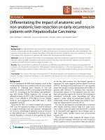
Fig. 1 shows the ≥ 40 Gy isodose distributions for the posterior and anterior regions of the SMGs. All patients were
treated with one fraction daily 5 days per week. Intracavitary after-loading treatment with iridium-192 was used to
address local persistence at 3–4 weeks after external RT at
15 to 20 Gy in three to five fractions every 2 days.
Chemotherapy
During the study period, institutional guidelines recommended no chemotherapy in stage I–IIA, concurrent chemoradiotherapy in stage IIB, and concurrent
Fig. 1 Isodose distributions for the submandibular glands. The 40 Gy and higher isodose distributions for the posterior part of the SMGs and anterior
part of the SMGs in patients with NPC who received level Ib-sparing IMRT (a), unilateral level Ib-covering IMRT (b), and bilateral level Ib-covering IMRT
(c). CTV-2, blue shadow; GTV-LN, red shadow; 66 Gy isodose, brown line; 60 Gy isodose, orange line; 54 Gy isodose, yellow line; 45 Gy isodose, green line; 40
Gy isodose, blue line
Zhang et al. BMC Cancer (2015) 15:709
Page 4 of 10
chemoradiotherapy with or without induction/adjuvant chemotherapy for stage III–IVA-B, as defined by
the 7th edition of the UICC/AJCC Staging System. Overall, 203/1438 patients (14.1 %) were treated with RT only,
and 1235/1438 patients (85.9 %) received induction,
concurrent, or adjuvant chemotherapy (concurrent
alone, 35.5 % [511/1235]; induction-concurrent, 37.4 %
[538/1235]; concurrent-adjuvant, 1.1 % [14/1235]; 0.9 %
induction-concurrent-adjuvant [13/1235]; 10.9 % induction alone, [156/1235]). In total, 93.0 % (996/1071) of
patients with stage III–IV disease received chemotherapy. Deviations from institutional guidelines were due
to organ dysfunction (suggesting intolerance to
chemotherapy) or patient’s refusal.
and test independent significance by backward elimination of insignificant explanatory variables.
To investigate whether irradiation of level Ib was associated with xerostomia, regional and subsequent distant
control, the Chi-square test (or Fisher’s exact test, if indicated) was used to evaluate the baseline clinical characteristics and the degree of xerostomia. Actuarial
survival rates were estimated by the Kaplan-Meier
method and compared using the log-rank test. Multivariable analyses using the Cox proportional hazards model
were used to estimate hazard ratios (HR) and test independent significance by backward elimination of insignificant explanatory variables. Statistical significance was
defined as P <0.05 based on two-sided tests.
Follow-up and xerostomia assessment
Results
Follow-up was measured from first day of treatment to
day of last examination or death. During the first two
years, patients were evaluated every three months, and
every six months thereafter for 3 year or until death.
Generally, follow-up included physical and neurologic
examinations, chest radiography, abdominal sonography,
single photon emission CT whole body bone scan, and
head and neck MRI. All local recurrences were diagnosed by soft-tissue swelling in fiberoptic endoscopy or
MRI of the nasopharynx and confirmed by biopsy, except for recurrence at the skull base which was confirmed by progressive bone erosion on MRI. Regional
recurrences were diagnosed by clinical examination or
neck MRI and confirmed by biopsy. Distant metastases
were diagnosed by clinical symptoms, physical examinations, and imaging methods including chest radiography,
bone scan, MRI, CT and abdominal sonography. Xerostomia related to radiation therapy was graded at approximately 12 months after radiotherapy according to
the Radiation Morbidity Scoring Criteria of the RTOG.
Predictors for metastasis to the level Ib lymph nodes at
diagnosis
Statistical analysis
All analyses were conducted using Statistical Package for
the Social Sciences 19.0 (SPSS; Chicago, IL, USA). All
events were measured from the first day of treatment. The
following endpoints (interval to the first defining event)
were evaluated: locoregional failure-free survival (LR-FFS),
distant failure-free survival (D-FFS), failure-free survival
(FFS) and overall survival (OS). LR-FFS was calculated
from the first date of treatment to first locoregional
failure; D-FFS, to first remote failure; FFS, to the date
of tumor relapse or death from any cause, whichever
occurred first; and OS, to last examination or death.
To investigate predictors for neck level Ib metastasis
at diagnosis, the Chi-square test (or Fisher’s exact test, if
indicated) was employed for univariable analyses to
examine associations and a logistic regression model, for
multivariable analyses to estimate hazard ratios (HR)
Univariable analysis of 1438 patients revealed that more
advanced N disease (for example, greatest dimension of
the level IIa LNs [DLN-IIa] ≥ 20 mm or level IIa LNs
with ES [P <.001]) and orpharynx involvement (P =
.001) were significantly associated with metastasis to the
level Ib LNs at diagnosis (Table 1).
Multivariable analysis to adjust for various risk factors demonstrated a DLN-IIa ≥ 20 mm or level IIa
LNs with ES (HR 2.21; 95 % confidence interval [CI]
1.10–4.46; P = .026) and oropharynx involvement (HR
2.59; 95 % CI 1.18–5.69; P = .018) were independently significantly associated with metastasis to the
level Ib LNs at diagnosis, while positive bilateral
CLNs (HR 1.95; 95 % CI 0.97–3.92; P = .061) had a
borderline significant association with metastasis to
the level Ib LNs at diagnosis (Table 2). In the 1193
patients with positive LNs in this series, univariable and
multivariable analyses confirmed that a DLN-IIa ≥ 20 mm
and/or level IIa LNs with ES (HR 2.41; 95 % CI 1.22–4.76;
P = .011), oropharynx involvement (HR 2.50; 95 % CI
1.13–5.56; P = .024) and positive bilateral CLNs (HR 2.11;
95 % CI 1.06–4.20; P = .034) were independently significantly associated with metastasis to the level Ib LNs at
diagnosis.
The percentage of positive level Ib LNs at diagnosis
in patients with and without a DLN-IIa ≥ 20 mm or
level IIa LNs with ES were 6.9 % vs. 1.7 % (P <.001);
with and without oropharynx involvement, 7.8 % vs.
2.3 % (P = .001); and with and without positive bilateral CLNs, 6.7 % vs. 1.8 % (P <.001), respectively.
Regional control at level Ib
Three patients experienced recurrence at level Ib, including two in-field recurrences (inside CTV2) and one
out-of-field recurrence (outside CTV2). Table 3 shows
the features of the three patients who suffered regional
Zhang et al. BMC Cancer (2015) 15:709
Page 5 of 10
Table 1 Univariable analyses of factors related to level IB LNs metastases at diagnosis in 1438 patients
Variable
Metastasis to level Ib LNs at diagnosis, N (%)
(−), n = 1398
*P
(+), n = 40
Sex
Male
1052 (75.3)
33 (82.5)
346 (24.7)
7 (17.5)
<50 years
950 (68.0)
23 (57.5)
≥50 years
448 (32.0)
17 (42.5)
Female
.294
Age
.163
Histologic type
Keratinizing squamous cell carcinoma
Nonkeratinizing carcinoma
5 (0.4)
0
1393 (99.6)
40 (100.0)
T1
247 (17.7)
5 (12.5)
T2
207 (14.8)
6 (15.0)
T3
679 (48.6)
18 (45.0)
T4
265 (19.0)
11 (27.5)
1133 (81.0)
29 (72.5)
265 (19.0)
11 (27.5)
(−)
1291 (92.3)
31 (77.5)
(+)
107 (7.7)
9 (22.5)
(−)
918 (65.7)
22 (55.0)
(+)
480 (34.3)
18 (45.0)
1.000
’
T stage
.537
T classification
T1-3
T4
.176
Oropharynx involvement
.001
Nasal cavity involvement
.162
N classification
N0
235 (16.8)
0
N1
823 (58.9)
19 (47.5)
N2
216 (15.5)
13 (32.5)
N3
124 (8.9)
8 (20.1)
<.001
Positive RLNs
(−)
387 (27.7)
3 (7.5)
(+)
1011 (72.3)
37 (92.5)
(−)
570 (40.8)
4 (10.0)
(+)
828 (59.2)
36 (90.0)
(−)
1054 (75.4)
22 (55.0)
(+)
344 (24.6)
18 (45.0)
(−)
1051 (75.2)
26 (65.0)
(+)
347 (24.8)
14 (35.0)
(−)
1247 (89.2)
34 (85.0)
(+)
151 (10.8)
6 (15.0)
.005
Positive CLNs
<.001
LN necrosis
<.001
LNs with ES
.143
DLN-IIa ≥30 mm or level IIa LNs with ES
.435
Zhang et al. BMC Cancer (2015) 15:709
Page 6 of 10
Table 1 Univariable analyses of factors related to level IB LNs metastases at diagnosis in 1438 patients (Continued)
DLN-IIa ≥20 mm or level IIa LNs with ES
(−)
1113 (79.6)
19 (47.5)
(+)
285 (20.4)
21 (52.5)
(−)
1196 (85.6)
26 (65.0)
(+)
202 (14.4)
14 (35.0)
(−)
1121 (80.2)
20 (50.0)
(+)
277 (19.8)
20 (50.0)
(−)
1318 (94.3)
21 (80.0)
(+)
80 (5.7)
8 (20.0)
<.001
MAD of LNs ≥30 mm
<.001
Positive bilateral CLNs
<.001
Positive CLNs at supraclavicular fossa
<.001
Abbreviations: LNs, lymph nodes; WHO, World Health Organization; RLNs, retropharyngeal lymph nodes; CLNs, cervical lymph nodes; LNs, lymph nodes; DLN-IIa,
greatest dimension of level IIa lymph nodes; MAD, maximal axial diameter; ES, extra-capsular spread
*P-values were calculated using an unadjusted chi-square test (or Fisher’s exact test, if indicated)
recurrence at level Ib; all three patients had a DLN-IIa ≥
20 mm and/or level IIa LNs with ES, oropharynx involvement and/or positive bilateral CLNs at diagnosis.
Therefore, the 904 patients without a DLN-IIa ≥ 20 mm
level IIa LNs with ES, oropharynx involvement or positive bilateral CLNs at diagnosis were classified as patients at a low risk of metastasis to the level Ib LNs (low
risk patients).
Clinical characteristics of low risk patients
Table 3 shows the clinical characteristics of the 904 patients at low risk: 79.7 % (722/904) received level Ibsparing IMRT and 20.1 % (182/904) received level Ibcovering IMRT. Significantly higher numbers of younger
patients and patients with advanced N disease received
level Ib-covering IMRT, and a significantly higher number of patients treated with level Ib-covering IMRT received chemotherapy (Table 4).
Patterns of failure for low risk patients
Median follow-up time for the low risk patients was 38.7
months (range, 1.3–57.8 months); 63.6 % (631/904) were
followed up for ≥ 3 years. In total, 11.4 % (113/904) of
the low risk patients developed treatment failure: distant
metastasis was the most common pattern of failure (65/
904 patients; 7.2 %); 3.3 % (30/904) experienced local
failure; 2.1 % (19/904) experienced regional recurrence,
including 1/23 (5.3 %) at level Ia, 0/23 at level Ib (0 %),
11/19 at level II (57.9 %), 4/19 at level III (21.0 %), 2/19
at level IV (10.5 %), 1/19 at level V (10.5 %). Twelve of
Table 2 Multivariable analysis of predictors for level IB LNs metastases at diagnosis in 1438 patients
Variable
HR
95 % CI
P*
Age, ≧50 years vs. <50 years
1.51
0.78–2.94
.219
T classification, T4 vs. T1-3
1.16
0.53–2.52
.708
Nasal cavity involvement, (+) vs. (−)
1.31
0.65–2.64
.446
Oropharynx involvement, (+) vs. (−)
2.59
1.18–5.69
.018
Positive RLNs, (+) vs. (−)
2.85
0.86–9.50
.088
Positive CLNs, (+) vs. (−)
2.53
0.80–8.01
.113
LN necrosis, (+) vs. (−)
1.22
0.59–2.52
.594
LNs with ES, (+) vs. (−)
0.57
0.27–1.19
.131
DLN-IIa ≥ 20 mm or level IIa LNs with ES, (+) vs. (−)
2.21
1.10–4.46
.026
MAD of LNs ≥30 mm, (+) vs.(−)
1.51
0.70–3.25
.293
Positive bilateral CLNs, (+) vs.(−)
1.95
0.97–3.92
.061
Positive CLNs at supraclavicular fossa, (+) vs. (−)
2.04
0.87–4.82
.103
Abbreviations: LNs, lymph nodes; HR, hazard ratio; 95 % CI, 95 % confidence interval; RLNs, retropharyngeal lymph nodes; CLNs, cervical lymph nodes; DLN-IIa,
greatest dimension of level IIa lymph nodes; MAD, maximal axial diameter; ES, extra-capsular spread
*P-values were calculated using a binary logistic regression model
Zhang et al. BMC Cancer (2015) 15:709
Page 7 of 10
Table 3 Features of the three patients with recurrence at the level Ib LNs after intensity-modulated radiotherapy
Case 1
Case 2
Case 3
Staging
T4N3a
T3N2
T4N3b
Positive bilateral CLNs
Yes
Yes
Yes
DLN-IIa ≥20 mm or level IIa lymph nodes with
ES
None
Right
Right
Oropharynx involvement
Left
None
None
Bilateral
Right
Right
Tumor involvement
Irradiation of neck level Ib
Recurrence at neck level Ib
Laterality
Left
Right
Left
Other regional recurrence
IA + IIb + IV + Vb
IIa + IIb + III
Ib
Concomitant failure
Axillary LNs
-
Paranasophrynx+skull base
Time to recurrence
12 months
12 months
23 months
Salvage treatment
Chemo
Chemo + surgery
Chemo + RT
Treatment response
PD
PD
PR
Sequential failure
Death due to multiple
metastasis
Axillary and mediastinal
LNs
Death due to intractable
epistaxis
Abbreviations: LNs, lymph nodes. DLN-IIa, greatest dimension of level IIa lymph nodes; ES, extra-capsular spread; chemo, chemotherapy; RT, radiotherapy; PD, progressive disease; PR, partial response
the 904 low risk patients (1.3 %) developed both distant
failure and locoregional recurrence. At last follow-up, 39
deaths had been recorded in the 904 low risk patients
(4.3 %), with the majority (31/39, 88.6 %) attributed to
NPC.
Survival outcomes of low risk patients
The estimated 3-year LR-FFS, D-FFS, FFS, and OS rates
for low risk patients were 95.5 %, 92.8 %, 89.2 %, and
96.4 %, respectively. Significant differences were observed in the estimated 3-year survival rates between
low risk patients who received level Ib-sparing IMRT
and level Ib-covering IMRT (LR-FFS: 96.2 % vs. 92.0 %
[HR 1.92; 95 % CI 1.04–3.56; P = .013]; D-FFS: 93.9 %
vs. 88.2 % [HR 1.92; 95 % CI 1.14–3.23; P = .012]; FFS:
90.6 % vs. 84.1 % [HR 1.64; 95 % CI 1.08–2.51; P = .022];
OS: 96.5 % vs. 96.1 % [HR 1.18; 95 % CI 0.56–2.49; P =
.662], respectively, Table 5). However, in multivariable
analyses, irradiation of level Ib was not an independent
risk factor for LR-FFS, D-FFS, FFS or OS (Table 5).
Xerostomia in low risk patients
In total, 50.7 % (463/913) of the low risk patients experienced subjective xerostomia at 12 months after
radiotherapy, which was predominately mild (grade III, 98.7 %). No significant difference was observed in
the frequency of grade ≥ 2 subjective xerostomia at
12 months after radiotherapy among low risk patients
who received level Ib-sparing, unilateral level Ibcovering or bilateral level Ib-covering IMRT (10.1 %
vs. 14.0 % vs. 18.0 %, P = .056).
Discussion
This is the largest-sample observational cohort study to
assess clinical predictors of metastasis to the level Ib
LNs in patients with NPC at diagnosis and furthermore,
first to compare disease control and xerostomia after
level Ib-sparing IMRT and level Ib-covering IMRT. We
found that a DLN-IIa ≥ 20 mm and/or level IIa LNs with
ES, oropharynx involvement and positive bilateral CLNs
were independently significantly associated with metastasis to the level Ib LNs at diagnosis. These pretreatment
factors effectively identify patients at low risk of recurrence at the level Ib LNs. For low risk patients, irradiation of level Ib was not an independent risk factor for
LR-FFS, D-FFS, FFS or OS.
The incidence of level Ib LN metastasis in this study
was only 2.8 %, which is similar to previous studies [11,
13–15]. Based on previous research [26–28], we hypothesized primary tumor invasion and nodal disease may be
related to metastasis to the level Ib LNs. In our analyses,
a DLN-IIa ≥ 20 mm and/or level IIa LNs with ES, oropharynx involvement and positive bilateral CLNs were
independently significantly associated with level Ib LN
involvement at diagnosis, in accordance with previous
studies [26–28]. The level Ib LNs receive efferent lymphatic drainage from the submental LNs, medial canthus,
lower nasal cavity, hard and soft palates, maxillary and
mandibular alveolar ridges, cheek, upper and lower lips,
and most of the anterior tongue [12, 29]. The level Ib
LNs are at risk of developing metastases from cancers of
the oral cavity, anterior nasal cavity, soft tissue structures of the middle face, and SMGs. Therefore, we
Zhang et al. BMC Cancer (2015) 15:709
Page 8 of 10
Table 4 Clinical features at diagnosis for low risk patients who
received level Ib-sparing and -covering IMRT
Variable
Irradiation of level Ib, N (%)
(−), n = 722
(+), n = 182
Male
536 (74.2)
122 (67.0)
Female
186 (25.8)
60 (33.0)
<50 years
447 (66.1)
139 (76.4)
≧50 years
245 (33.9)
43 (23.6)
T1
157 (21.7)
36 (19.8)
T2
108 (15.0)
38 (20.9)
T3
332 (46.0)
83 (45.6)
T4
125 (17.3)
25 (13.7)
N0
206 (28.5)
21 (11.5)
N1
493 (68.7)
137 (75.3)
N3
20 (2.8)
24 (13.2)
(−)
256 (35.5)
45 (24.7)
(+)
466 (64.5)
137 (75.3)
P*
Sex
.051
Age
.008
T classification
.208
N classification
<.001
Positive RLNs
.006
Positive CLNs
(−)
479 (66.3)
60 (33.0)
(+)
243 (33.7)
122 (67.0)
<.001
Positive CLNs at supraclavicular fossa
(−)
710 (98.3)
168 (92.3)
(+)
12 (1.7)
14 (7.7)
<.001
Chemotherapy
(−)
147 (20.4)
15 (8.2)
(+)
575 (79.6)
167 (20.1)
<.001
Abbreviations: IMRT, intensity-modulated radiotherapy; RLNs, retropharyngeal
lymph nodes; CLNs, cervical lymph nodes; ES, extra-capsular spread
* P-values were calculated using unadjusted chi-square test (or Fisher’s exact
test, if indicated)
concluded that level Ib is not a regular region of direct
drainage for the primary tumor in NPC. We speculate
level Ib involvement may result from retrograde tumor
spread after blockage of the normal routes of lymphatic
drainage (for example, massive level IIa LNs or bilateral
positive CLNs), or metastasis from tumors involving
anatomical sites that drain to level Ib (for example, the
oropharynx, which is adjacent to the soft palate). However, similarly to previous studies [26–28], nasal cavity
involvement did not correlate with metastasis to level Ib
in this study. This may be explained by the fact that the
above-mentioned studies did not include involvement of
the anterior nasal cavity as a variable for analysis. Nasal
cavity involvement did not exceed the posterior third in
axial plane on MRI scans in most cases in this study,
and only the anterior third of the nasal cavity drains to
level Ib [12].
Though various protocols of level Ib delineation and
dose definitions for IMRT have been reported at different treatment centers over the years [1, 11, 16–21, 30],
there is little evidence to address the association between elective irradiation and disease control at level Ib.
Chen and colleagues [22] investigated 120 patients with
NPC and negative level Ib LNs at diagnosis who received
level Ib-sparing IMRT and observed no regional recurrence at level Ib, and regional LN recurrence alone was
rare. They concluded that level Ib-sparing IMRT is feasible in patients with negative level Ib LNs [22]. Yi et al.
[27] developed a risk score model for metastasis to the
level Ib LNs and found that level Ib-sparing irradiation
was an independent risk factor for locoregional recurrence in 190 high risk patients (involvement of level II/
III/IV LNs, carotid sheath involvement and the maximal
axial diameter [MAD] of the CLNs ≥ 20 mm). However,
level Ib-sparing irradiation did not affect locoregional recurrence in the 137 low risk patients in the same study.
However, it should be noted that all of the 327 patients
received three-dimensional conventional radiation therapy (3D-CRT), which is inferior to IMRT in terms of
OAR protection [31, 32], and data on xerostomia was
not available to confirm the advantage of level Ibsparing irradiation [27].
Interestingly, all the three cases of level Ib LN recurrences in this study occurred in patients with a
DLN-IIa ≥ 20 mm, level IIa LNs with ES, oropharynx
involvement and/or positive bilateral CLNs at diagnosis. According to our previous analysis, though 79 %
of low risk patients were treated with level Ib-sparing
IMRT, none of these patients experienced recurrence
at level Ib. Our multivariable analyses also showed
that irradiation of level Ib was not an independent
risk factor for LR-FFS, D-FFS, FFS or OS. Omitting
irradiation of level Ib did not significantly jeopardize
disease control at level Ib nor compromise locoregional control, distant control or OS in low risk patients
in this study. Therefore, we conclude that level Ibsparing IMRT should be safe in patients without a
DLN-IIa ≥ 20 mm, level IIa LNs with ES, oropharynx
involvement or positive bilateral CLNs. Our results
are in accordance with previous studies [22, 27] and
provide further meaningful evidence for elective sparing of level Ib in the IMRT era.
Previous studies have reported level Ib-sparing IMRT
reduces xerostomia in patients with head and neck cancer [6–8, 10]. However, this study did not observe a significant difference in the frequency of grade ≥ 2
subjective xerostomia at 12 months after IMRT between
patients who received level Ib-sparing, unilateral level
Zhang et al. BMC Cancer (2015) 15:709
Page 9 of 10
Table 5 Multivariate analyses of prognostic factors in low risk patients (n = 904)
Variable
LR-FFS
D-FFS
FFS
OS
HR (95 % CI)
P*
HR (95 % CI)
P*
HR (95 % CI)
P*
HR (95 % CI)
P*
Sex, female vs. male
0.68 (0.34–1.38)
.290
0.82 (0.46–1.42)
.459
0.82 (0.52–1.29)
.384
0.77 (0.37–1.63)
.499
Age, ≥50 vs. <50 years
1.27 (0.69–2.32)
.445
1.44 (0.87–2.37)
.155
1.60 (1.08–2.37)
.020
2.44 (1.29–4.60)
.006
T classification
1.51 (1.11–2.07)
.009
1.32 (1.03–1.70)
.029
1.33 (1.08–1.64)
.007
1.60 (1.12–2.28)
.009
Positive RLNs, (+) vs. (−)
1.70 (0.77–3.73)
.185
1.43 (0.76–2.70)
.266
1.55 (0.94–2.58)
.089
1.17 (0.53–2.57)
.694
Positive CLNs, (+) vs. (−)
2.16 (1.20–3.89)
.010
2.35 (1.40–3.96)
.001
2.01 (1.34–3.04)
.001
2.76 (1.44–5.32)
.002
Positive CLNs at SCF, (+) vs. (−)
1.16 (0.27–5.04)
.846
3.00 (1.24–7.18)
.014
2.12 (0.96–4.71)
.064
2.69 (0.79–9.12)
.113
Chemotherapy, (+) vs. (−)
1.14 (0.38–3.41)
.816
1.18 (0.48–2.91)
.719
0.89 (0.46–1.71)
.717
0.52 (0.20–1.34)
.174
Irradiation of level Ib, (+) vs. (−)
1.68 (0.88–3.19)
.114
1.43 (0.82–2.49)
.207
1.31 (0.83–2.05)
.247
0.88 (0.39–1.95)
.744
Abbreviations: LR-FFS, locoregional failure-free survival; D-FFS, distant failure-free survival; FFS, failure-free survival; OS, overall survival; HR, hazard ratio; 95 % CI, 95
% confidence interval; RLNs, retropharyngeal lymph nodes; CLNs, cervical lymph nodes; DLN-IIa, greatest dimension of level IIa lymph nodes; LNs, lymph nodes; ES,
extra-capsular spread
*
P-values were calculated using an adjusted Cox proportional-hazards model
Ib-covering or bilateral level Ib-covering IMRT. The
main reason for this result is that the dose constrains for
the SMGs were not included in the treatment planning
protocol of our centre. Even when the SMGs were excluded from the CTV2, the 40 Gy isodose line still
exceeded the anterior two-thirds of the SMGs in this
series, while previous studies reported that the SMG
salivary flow rate depends on the mean dose to the
SMGs up to a threshold of 39 Gy, with recovery over
time [8]. Investigations of SMG-sparing IMRT also
found it feasible to substantially reduce the dose to
the SMG to below a threshold of 39 Gy without target underdosing [8]. Therefore, we believe that proper
dose constrains for the SMGs should be studied in
the future for level Ib-sparing IMRT in certain cohorts of patients with NPC.
This is the largest sample size study to investigate the
feasibility of elective level Ib-sparing IMRT. However,
this study inevitably bears the inherent limitations of its
retrospective nature. Firstly, the identification of low risk
patients who may not need irradiation to level Ib was
not based on pathologic evidence but assessment of pretreatment MRI scans. For example, ES was diagnosed on
the basis of radiographic findings, which is a common
and difficult problem for NPC research due to the lack
of pathologic confirmation of LN metastases in patients
with NPC. Secondly, irradiation of level Ib was not randomly assigned but decided by the individual physicians
for each patient, based on their recognition of the delineation protocols from reports of different centers. Bias
towards more patients with advanced N disease receiving level Ib-covering IMRT was inevitable. Thirdly, delineation of the SMGs was not described in the
treatment planning protocol of our centre; therefore,
further analyses of the relationship between the degree
of xerostomia and dose to the SMGs was not possible
for this cohort. Further investigations based on more
specific criteria for dose constraints for the SMGs are
warranted to confirm the benefit of elective level Ib
irradiation.
Conclusion
Level Ib-sparing IMRT should be safe and feasible for
patients without a DLN-IIa ≥ 20 mm and/or level IIa
LNs with ES, positive bilateral CLNs or oropharynx involvement at diagnosis. Further investigations based on
specific criteria for dose constraints for the SMGs are
warranted to confirm the benefit of elective level Ib
irradiation.
Abbreviations
NPC: Nasopharyngeal carcinoma; IMRT: Intensity-modulated radiation
therapy; LN: Lymph node; CLN: Cervical lymph nodes; RLN: Retropharyngeal
lymph node; DLN-IIa: Greatest dimension of level IIa LNs; ES: Extracapsular
spread; PGs: Parotid glands; SMGs: Submandibular glands; GTV: Gross tumor
volume; CTV: Clinical target volumes; PTV: Planning target volume.
Competing interests
We declare that we have no conflict of interests.
Authors’ contributions
The authors contributions are as follows: Fan Zhang (MD) and Yi-Kan Cheng
(MD) contributed to the literature research, study design, data collection, data
analysis, interpretation of findings and writing of the manuscript. Wen-Fei Li
(MD), Rui Guo (MD), Lei Chen (MD), Ying Sun (PhD, professor), Guan-Qun Zhou
(MD), Yan-Ping Mao (MD), Xu Liu (MD) and Li-Zhi Liu (MD) contributed to data
collection. Ai-Hua Lin (PhD, professor) contributed data analyses. Ling-Long
Tang (MD) and Jun Ma (PhD, professor) contributed to data collection, study
design, critical review of data analyses, interpretation of findings and critical
editing of the manuscript. All authors read and approved the final manuscript.
Acknowledgments
This work was supported by grants from the Health & Medical
Collaborative Innovation Project of Guangzhou City, China (No.
201400000001), the National Science & Technology Pillar Program during
the Twelfth Five-year Plan Period (No. 2014BAI09B10), the Planned
Science and Technology Project of Guangdong Province (No.
2013B021800175), and the Key Laboratory Construction Project of
Guangzhou City, China, (No.121800085), Sun Yat-Sen University Clinical
Research 5010 Program (No. 2012011).
Zhang et al. BMC Cancer (2015) 15:709
Author details
1
Department of Radiation Oncology, State Key Laboratory of Oncology in
South China, Collaborative Innovation Center for Cancer Medicine, Sun
Yat-sen University Cancer Center, No. 651 Dongfeng Road East, Guangzhou
510060, People’s Republic of China. 2Department of Radiation Oncology, The
Sixth Affiliated Hospital of Sun Yat-sen University, Guangzhou 510655,
People’s Republic of China. 3State Key Laboratory of Oncology in South
China, Collaborative Innovation Center for Cancer Medicine, Imaging
Diagnosis and Interventional Center, Sun Yat-sen University Cancer Center,
Guangzhou 510060, People’s Republic of China. 4Department of Medical
Statistics and Epidemiology, School of Public Health, Sun Yat-sen University,
Guangzhou 510080, People’s Republic of China.
Page 10 of 10
16.
17.
18.
Received: 1 January 2015 Accepted: 1 October 2015
19.
References
1. Lee N, Xia P, Quivey JM, Sultanem K, Poon I, Akazawa C, et al. Intensitymodulated radiotherapy in the treatment of nasopharyngeal carcinoma: an
update of the UCSF experience. Int J Radiat Oncol Biol Phys. 2002;53(1):12–22.
2. Kwong DL, Sham JS, Leung LH, Cheng AC, Ng WM, Kwong PW, et al.
Preliminary results of radiation dose escalation for locally advanced
nasopharyngeal carcinoma. Int J Radiat Oncol Biol Phys. 2006;64(2):374–81.
3. Milne RW, Dawes C. The relative contributions of different salivary glands to
the blood group activity of whole saliva in humans. Vox Sang.
1973;25(4):298–307.
4. Hsiung C-Y, Ting H-M, Huang H-Y, Lee C-H, Huang E-Y, Hsu H-C. Parotidsparing intensity-modulated radiotherapy (IMRT) for nasopharyngeal
carcinoma: Preserved parotid function after IMRT on quantitative salivary
scintigraphy, and comparison with historical data after conventional
radiotherapy. International Journal of Radiation Oncology*Biology*Physics.
2006;66(2):454–61.
5. Nutting CM, Morden JP, Harrington KJ, Urbano TG, Bhide SA, Clark C, et al.
Parotid-sparing intensity modulated versus conventional radiotherapy in
head and neck cancer (PARSPORT): a phase 3 multicentre randomised
controlled trial. Lancet Oncol. 2011;12(2):127–36.
6. Saarilahti K, Kouri M, Collan J, Kangasmäki A, Atula T, Joensuu H, et al. Sparing
of the submandibular glands by intensity modulated radiotherapy in the
treatment of head and neck cancer. Radiother Oncol. 2006;78(3):270–5.
7. Houweling AC, Dijkema T, Roesink JM, Terhaard CHJ, Raaijmakers CPJ.
Sparing the contralateral submandibular gland in oropharyngeal cancer
patients: A planning study. Radiother Oncol. 2008;89(1):64–70.
8. Murdoch-Kinch C-A, Kim HM, Vineberg KA, Ship JA, Eisbruch A. Dose-Effect
Relationships for the Submandibular Salivary Glands and Implications for
Their Sparing by Intensity Modulated Radiotherapy. Int J Radiat Oncol Biol
Phys. 2008;72(2):373–82.
9. Doornaert P, Verbakel WF, Rietveld DH, Slotman BJ, Senan S. Sparing the
contralateral submandibular gland without compromising PTV coverage by
using volumetric modulated arc therapy. Radiat Oncol. 2011;6:74.
10. Wang ZH, Yan C, Zhang ZY, Zhang CP, Hu HS, Tu WY, et al. Impact of
salivary gland dosimetry on post-IMRT recovery of saliva output and
xerostomia grade for head-and-neck cancer patients treated with or
without contralateral submandibular gland sparing: a longitudinal study. Int
J Radiat Oncol Biol Phys. 2011;81(5):1479–87.
11. Tang L, Mao Y, Liu L, Liang S, Chen Y, Sun Y, et al. The volume to be
irradiated during selective neck irradiation in nasopharyngeal carcinoma.
Cancer. 2009;115(3):680–8.
12. Gregoire V, Levendag P, Ang KK, Bernier J, Braaksma M, Budach V, et al.
CT-based delineation of lymph node levels and related CTVs in the nodenegative neck: DAHANCA, EORTC, GORTEC, NCIC, RTOG consensus
guidelines. Radiother Oncol. 2003;69(3):227–36.
13. Ho FCH, Tham IWK, Earnest A, Lee K, Lu JJ. Patterns of regional lymph node
metastasis of nasopharyngeal carcinoma: A meta-analysis of clinical
evidence. BMC Cancer. 2012;12(1):98.
14. Li WF, Sun Y, Chen M, Tang LL, Liu LZ, Mao YP, et al. Locoregional
extension patterns of nasopharyngeal carcinoma and suggestions for
clinical target volume delineation. Chin J Cancer. 2012;31(12):579–87.
15. Li W-F, Sun Y, Mao Y-P, Chen L, Chen Y-Y, Chen M, et al. Proposed Lymph
Node Staging System Using the International Consensus Guidelines for
Lymph Node Levels Is Predictive for Nasopharyngeal Carcinoma Patients
20.
21.
22.
23.
24.
25.
26.
27.
28.
29.
30.
31.
32.
From Endemic Areas Treated With Intensity Modulated Radiation Therapy.
Int J Radiat Oncol Biol Phys. 2013;86(2):249–56.
Kam MK, Teo PM, Chau RM, Cheung KY, Choi PH, Kwan WH, et al.
Treatment of nasopharyngeal carcinoma with intensity-modulated
radiotherapy: the Hong Kong experience. Int J Radiat Oncol Biol Phys.
2004;60(5):1440–50.
Wolden SL, Chen WC, Pfister DG, Kraus DH, Berry SL, Zelefsky MJ. Intensitymodulated radiation therapy (IMRT) for nasopharynx cancer: Update of the
Memorial Sloan-Kettering experience. Int J Radiat Oncol Biol Phys.
2006;64(1):57–62.
Lee N, Harris J, Garden AS, Straube W, Glisson B, Xia P, et al. IntensityModulated Radiation Therapy With or Without Chemotherapy for
Nasopharyngeal Carcinoma: Radiation Therapy Oncology Group Phase II
Trial 0225. J Clin Oncol. 2009;27(22):3684–90.
Tham IW, Hee SW, Yeo RM, Salleh PB, Lee J, Tan TW, et al. Treatment of
nasopharyngeal carcinoma using intensity-modulated radiotherapy-the
national cancer centre singapore experience. Int J Radiat Oncol Biol Phys.
2009;75(5):1481–6.
Commitee. CNCSW. Intensity-modulated radiotherapy target delineation
and dose delivery for nasopharyngeal carcinoma- 2010 expert consensus
guidelines. Chin J Radiat Oncol. 2011;20:267–79.
Lee NY, Zhang Q, Pfister DG, Kim J, Garden AS, Mechalakos J, et al. Addition
of bevacizumab to standard chemoradiation for locoregionally advanced
nasopharyngeal carcinoma (RTOG 0615): a phase 2 multi-institutional trial.
Lancet Oncol. 2012;13(2):172–80.
Chen J, Ou D, He X, Hu C. Sparing level Ib lymph nodes by intensitymodulated radiotherapy in the treatment of nasopharyngeal carcinoma. Int
J Clin Oncol. 2013.
van den Brekel MW, Stel HV, Castelijns JA, Nauta JJ, van der Waal I, Valk J, et
al. Cervical lymph node metastasis: assessment of radiologic criteria.
Radiology. 1990;177(2):379–84.
Tang L, Li L, Mao Y, Liu L, Liang S, Chen Y, et al. Retropharyngeal lymph
node metastasis in nasopharyngeal carcinoma detected by magnetic
resonance imaging. Cancer. 2008;113(2):347–54.
Sun Y, Guo R, Yin WJ, Tang LL, Yu XL, Chen M, et al. Which T category of
nasopharyngeal carcinoma may benefit most from volumetric modulated
arc therapy compared with step and shoot intensity modulated radiation
therapy. PLoS One. 2013;8(9), e75304.
Yi W, Liu XM, Xia YF, Liu Q, Li JT. Influence of level-Ib lymphadenopathy on the
prognosis of nasopharyngeal carcinoma. Chin J Cancer. 2010;29(1):87–93.
Yi W, Li X, Liu Z, Jiang C, Niu D, Xia Y. A risk score model for the metastasis of
level Ib lymph node based on the clinicopathological features of
nasopharyngeal carcinoma in a large sample. Mol Clin Oncol. 2014;2(5):789–97.
Yuan G, Zheng X, Zhu X, Wang Z, Song W, Zhang H, et al. Risk factors of
level Ib lymphadenopathy in nasopharyngeal carcinoma. Nan Fang Yi Ke Da
Xue Xue Bao. 2014;34(7):983–7.
Grégoire V, Ang K, Budach W, Grau C, Hamoir M, Langendijk JA, et al.
Delineation of the neck node levels for head and neck tumors: A 2013
update. DAHANCA, EORTC, HKNPCSG, NCIC CTG, NCRI, RTOG, TROG
consensus guidelines. Radiother Oncol. 2014;110(1):172–81.
Lin S, Pan J, Han L, Zhang X, Liao X, Lu JJ. Nasopharyngeal Carcinoma
Treated With Reduced-Volume Intensity-Modulated Radiation Therapy:
Report on the 3-Year Outcome of a Prospective Series. Int J Radiat Oncol
Biol Phys. 2009;75(4):1071–8.
Cozzi L, Fogliata A, Bolsi A, Nicolini G, Bernier J. Three-dimensional
conformal vs. intensity-modulated radiotherapy in head-and-neck cancer
patients: comparative analysis of dosimetric and technical parameters. Int J
Radiat Oncol Biol Phys. 2004;58(2):617–24.
Longobardi B, De Martin E, Fiorino C, Dell’oca I, Broggi S, Cattaneo GM, et
al. Comparing 3DCRT and inversely optimized IMRT planning for head and
neck cancer: Equivalence between step-and-shoot and sliding window
techniques. Radiother Oncol. 2005;77(2):148–56.