Endometriosis as a risk factor for ovarian or endometrial cancer - results of a hospitalbased case - control study
Bạn đang xem bản rút gọn của tài liệu. Xem và tải ngay bản đầy đủ của tài liệu tại đây (510.98 KB, 8 trang )
Burghaus et al. BMC Cancer (2015) 15:751
DOI 10.1186/s12885-015-1821-9
RESEARCH ARTICLE
Open Access
Endometriosis as a risk factor for ovarian or
endometrial cancer — results of a hospitalbased case–control study
Stefanie Burghaus1†, Lothar Häberle1,2†, Michael G. Schrauder1, Katharina Heusinger1, Falk C. Thiel1,3,
Alexander Hein1, David Wachter4, Johanna Strehl4, Arndt Hartmann4, Arif B. Ekici5, Stefan P. Renner1,
Matthias W. Beckmann1 and Peter A. Fasching1,6*
Abstract
Background: No screening programs are available for ovarian or endometrial cancer. One reason for this is the low
incidence of the conditions, resulting in low positive predictive values for tests, which are not very specific. One
way of addressing this problem might be to use risk factors to define subpopulations with a higher incidence. The
aim of this study was to investigate the extent to which a medical history of endometriosis can serve as a risk
factor for ovarian or endometrial cancer.
Methods: In a hospital-based case–control analysis, the cases represented patients with endometrial or ovarian
cancer who were participating in studies aimed at assessing the risk for these diseases. The controls were women
between the age of 40 and 85 who were invited to take part via a newspaper advertisement. A total of 289 cases
and 1016 controls were included. Using logistic regression models, it was tested whether self-reported endometriosis is
a predictor of case–control status in addition to age, body mass index (BMI), number of pregnancies and previous oral
contraceptive (OC) use.
Results: Endometriosis was reported in 2.1 % of the controls (n = 21) and 4.8 % of the cases (n = 14). Endometriosis
was a relevant predictor for case–control status in addition to other predictive factors (OR 2.63; 95 % CI, 1.28 to 5.41).
Conclusion: This case–control study found that self-reported endometriosis may be a risk factor for endometrial
or ovarian cancer in women between 40 and 85 years. There have been very few studies addressing this issue,
and incorporating it into a clinical prediction model would require a more precise characterization of the risk
factor of endometriosis.
Keywords: Endometriosis, Ovarian cancer, Endometrial cancer, Risk factor
Background
Ovarian cancer is associated with a high mortality rate
in comparison with other cancers. In the United States,
the incidence of ovarian cancer is estimated to be
around 22,200 annually. About 14,000 of these women
* Correspondence:
†
Equal contributors
1
Department of Gynecology and Obstetrics, Erlangen University Hospital,
Friedrich Alexander University of Erlangen–Nuremberg, Comprehensive
Cancer Center Erlangen-EMN, Erlangen, Germany
6
Division of Hematology and Oncology, Department of Medicine, David
Geffen School of Medicine, University of California at Los Angeles, Los
Angeles, CA, USA
Full list of author information is available at the end of the article
are expected to die of the disease [1]. In Germany the
corresponding figures are 7400 and 5500 [2]. This high
mortality rate is mainly the consequence of ineffective
early detection or screening programs. Most of the cancers are diagnosed at advanced stages. Uterine endometrial cancer is the most frequent type of gynecological
cancer. In Germany, there are approximately 11,600 new
diagnoses every year and 2400 disease-related deaths [2].
Although the mortality due to endometrial cancer is
fairly low, there are no established early detection
methods or screening programs for this disease. Earlier
detection would result in much less invasive surgery and
© 2015 Burghaus et al. Open Access This article is distributed under the terms of the Creative Commons Attribution 4.0
International License ( which permits unrestricted use, distribution, and
reproduction in any medium, provided you give appropriate credit to the original author(s) and the source, provide a link to
the Creative Commons license, and indicate if changes were made. The Creative Commons Public Domain Dedication waiver
( applies to the data made available in this article, unless otherwise stated.
Burghaus et al. BMC Cancer (2015) 15:751
less use of radiotherapy and chemotherapy, leading to
substantial benefits for the patients.
With regard to ovarian cancer, effective risk-reducing strategies have been described. Bilateral salpingo-oophorectomy
has been shown to reduce the risk among BRCA mutation carriers by 71–96 % [3–5]. Numbers of live births,
oral contraceptive use, and tubal ligation are also associated with a significant reduction in the lifetime risk of
ovarian cancer.
There are no established screening programs for endometrial cancer, but risk-modifying strategies are known
that allow the risk of endometrial cancer to be controlled — such as weight control, physical activity, and
no exogenous unopposed estrogen [6–9].
Risk factors are therefore of special interest for both
diseases, since accurate risk prediction might make
individualized early detection or screening programs
possible. Risk factors for ovarian cancer include reproductive behavior and use of hormonal therapies. Pregnancies and the use of oral contraceptives can reduce
the incidence of ovarian cancer [10]. Mutations in the
BRCA1 and BRCA2 genes are reported to lead to a lifetime risk of about 20–40 and 15–25 %, respectively
[11]. Large-scale genotyping efforts have recently identified and confirmed a total of 11 low-penetrance risk
loci that are common in the population [12–20].
Endometrial cancer risk factors include hormonal
and metabolic factors such as obesity, tamoxifen use,
diabetes, hypertension, and high dietary fat consumption [21]. With regard to genetic risk factors, endometrial cancer is the most common malignancy in women,
with mutations associated with Lynch syndrome [22].
Genome-wide association studies have identified some
low-penetrance loci, but large-scale confirmation studies are still pending [23–25].
In this study endometriosis is evaluated as a risk factor
for ovarian- or endometrial cancer. Endometriosis is a
chronic disease that affects 4–30 % of all women during
the reproductive age [26–28]. Furthermore it is one of
the most frequent gynecological diseases. However it
can reasonably be assumed, that the prevalence is about
10 % [28]. The pathogenesis of endometriosis is considered to be complex. Historically a metaplastic transformation of peritoneal cells or the still favourably retrograde
menstruation of cells through the tubes into the peritoneal cavity are discussed [29]. On a molecular level different pathways such as the estrogen and progesterone
pathway, vasculogenesis, sphingolipids, prostaglandins,
and cytokines appear to be involved.
Pelvic pain during menstruation is the main symptom in patients with endometriosis. Other symptoms
can be chronic lower abdominal pain, dysuria, dyschezia and/ or dyspareunia. The disease is characterized
by endometrial cells outside the uterus and is located
Page 2 of 8
mainly in the retrouterine pouch. The diagnosis
occurs in gynecological examination and especially
during laparoscopic surgeries with histological verification [30]. Therapy options comprise mainly medication and surgical therapy. The surgical removal of the
lesion is often the first line therapy [31].
An association between endometriosis and both diseases has been suggested, and in the case of ovarian
cancer the connection is clearly established [32–35].
Patients with endometriosis tend to be younger and to
be diagnosed at earlier stages and with lower-grade
ovarian cancer lesions [36, 37]. With regard to endometrial cancer, the evidence is less clear. A reduced risk
of endometrial cancer was even found in a nested case–
control study including 39 patients with endometrial
cancer and 211 controls (OR 0.58; 95 % CI, 0.42 to
0.81) [37]. In a different nested case–control study, patients were found to have a relative risk (RR) of 1.23
(95 % CI, 0.63 to 2.38) [38]. However, most of the relevant studies only include a small number of events, so
that definitive conclusions about associations cannot as
yet be drawn [39–42].
The aim of the present case–control study was to investigate the extent to which a medical history of endometriosis represents a risk factor for ovarian or endometrial
cancer in addition to age, body mass index (BMI), number
of pregnancies, and previous oral contraceptive (OC) use.
Methods
A series of case–control and cohort studies have been
conducted in the Department of Gynecology and Obstetrics at Erlangen University Hospital in an effort to
identify risk factors for breast cancer and gynecological
cancer, as well as prognostic factors. These are: 1. The
Bavarian Ovarian Cancer Study (BAV) which was conducted from 2002 to 2011 and was affiliated to largescale research consortia working on identifying genetic
and epidemiologic risk factors [13–17, 19, 20], as well as
prognostic factors [43–45]. 2. The Bavarian Endometrial
Cancer Study (BECS) conducted from 2002 to 2013 also
affiliated to larger research consortia [23–25]. 3. The
Bavarian Breast Cancer Cases and Controls Study
(BBCC) [17, 46–51] conducted from 2002 to 2013. The
corresponding controls were recruited using local newspaper advertisements inviting women over the age of 40
without breast, ovarian or endometrial cancer anamnesis, respectively.
Cases of this study were patients with histologically
confirmed current or former endometrial or invasive
epithelial ovarian cancer disease who were treated at
Erlangen University Hospital. The controls originate
from the three studies mentioned above. Women who
had any other types of cancer were not eligible for inclusion in the study. All subjects had to complete the
Burghaus et al. BMC Cancer (2015) 15:751
same self-reported medical history form and the same
study questionnaire. The age criteria of cases and controls were to be over 40 and less than 85 years. The
ethics committee of the medical faculty at Friedrich
Alexander University, Erlangen, approved the study and
all of the patients and healthy participants provided
written informed consent.
Data acquisition
A standardized questionnaire including modules on
pregnancy history, previous use of hormonal contraceptives and hormone replacement therapy, medical history,
family history, and lifestyle was filled out by the patients
and healthy control individuals, and was completed in a
structured interview with trained medical personnel if
any questions had not been fully answered. The question
about a history of endometriosis was expressed in a
“yes/no/don’t know” form, and was answered by cases
and controls in the same way when completing the
questionnaire. Additional information for patients was
obtained from the patient charts, such as information
about medical procedures, histology of the tumor, and
concomitant medication.
Statistical considerations
The primary objective was to investigate whether information about endometriosis can be used to assess the
risk for ovarian or endometrial cancer, in addition to
other well-known risk factors. For this purpose, a multiple logistic regression model was fitted with cancer
case–control status as a binary outcome (yes vs. no) and
the following predictors: endometriosis status (categorical; yes vs. no), age (continuous), BMI (continuous),
number of pregnancies (integer), and oral contraceptive
use (categorical; yes vs. no). The Wald test was performed for endometriosis status. A significant P value
would indicate that endometriosis information is an additional risk factor for ovarian or endometrial cancer. The
regression model was also used to estimate adjusted
odds ratios (ORs), particularly for endometriosis status.
Patients for whom outcome data were lacking and patients with missing information on age or endometriosis
were excluded. Missing predictor values were imputed
using single “best guesses” (median value of continuous
or integer predictors, the most common value of categorical or ordinal predictors) based on nonmissing data
across all subjects. Continuous predictors were used as
natural cubic spline functions to describe nonlinear
effects [52]. The number of degrees of freedom (1 or 2)
of each predictor was determined as done recently
in [53].
The performance of the logistic regression model in
terms of discrimination and calibration (“goodness of
fit”) was assessed using the area under the receiver
Page 3 of 8
operating characteristic curve (AUC) and the Hosmer–
Lemeshow statistic applied to the case–control design
[54]. The AUC ranges from 0.5 (no discrimination between cases and controls) to 1 (perfect discrimination).
It can be interpreted as representing the probability
that the model will give a person who has disease a
higher probability of being diseased than it gives to a
randomly chosen healthy person. In accordance with
Hosmer and Lemeshow, patients were ranked with respect to the predicted conditional probability of ovarian
or endometrial cancer and categorized into equal-sized
groups based on percentiles. Frequencies of predicted
events in each group were compared with frequencies
of observed events in each group using a scatter plot
and the Hosmer–Lemeshow χ2 test. A large P value indicates satisfactory calibration.
Model building was evaluated by 10-fold crossvalidation with 20 repetitions to address overfitting. For
this purpose, the model-building process (i.e., determination of cubic spline functions and estimation of regression coefficients) was carried out on each training
set, resulting in several logistic regression models (one
model per set), which were then used to calculate the
AUCs on the corresponding validation data sets. The
average of all these AUCs was taken as an evaluation
measure. This cross-validated AUC may be regarded as
an estimation of the expected probability of two randomly chosen future ill or healthy subjects being correctly classified as ill or healthy, respectively, using the
main regression model described above.
As sensitivity analysis, a simple logistic regression model
was fitted to get an unadjusted OR for endometrioses
status.
All of the tests were two-sided, and a P value of < 0.05
was regarded as statistically significant. Calculations were
carried out using the R system for statistical computing
(version 3.0.1; R Development Core Team, Vienna, Austria,
2013).
Results
Descriptive statistics
A total of 1305 participants were included in the analyses, of whom 165 were patients with ovarian cancer,
131 were patients with endometrial cancer, and 1016
were control individuals. Complete information with all
variables was available for 90 % of the participants. The
proportions of missing predictor values were between
5.5 and 6.5 %. The missing values were imputed, as described above. Descriptive statistics are shown in Table 1.
Endometriosis was noted by 2.1 % of the controls (n =
21) and by 4.8 % of the cases (n = 14). The mean age of
subjects with endometriosis was 53.2 years for cases and
57.7 years for controls. Endometriosis was present in 4.2 %
of the ovarian cancer patients (seven of 165 patients) and
Burghaus et al. BMC Cancer (2015) 15:751
Page 4 of 8
Table 1 Characteristics of the study participants, showing mean and standard deviation (SD) for continuous characteristics and
frequency and percentage for categorical characteristics
Characteristic
Controls (n = 1016)
Cases (n = 289)
Ovarian cancer cases (n = 165)
Endometrial cancer cases (n = 131)a
Mean or n
SD or %
Mean or n
SD or %
Mean or n
SD or %
Mean or n
SD or %
Age [years]
60.9
9.3
62.1
11.1
59.5
11.1
65.6
10.5
Body mass index [kg/m2]
25.5
4.3
27
5.8
26
4.7
28.3
6.9
Self-reported endometriosis
No
995
97.9
275
95.2
158
95.8
124
94.7
Yes
21
2.1
14
4.8
7
4.2
7
5.3
No
275
27.1
141
48.8
72
43.6
71
54.2
Yes
741
72.9
148
51.2
93
56.4
60
45.8
0
121
11.9
41
14.2
14
8.5
27
20.6
1
165
16.2
62
21.5
31
18.8
32
24.4
2
373
36.7
100
34.6
66
40.0
37
28.2
3
219
21.6
52
18.0
35
21.2
19
14.5
4+
138
13.6
34
11.8
19
11.5
16
12.2
Oral contraceptive use
Pregnancies (n)
a
Summed up numbers of ovarian and endometrial cancer cases is larger than 289, because there were cases with both ovarian and endometrial cancer
in 5.3 % of the endometrial cancer patients (seven of 131
patients).
Prediction of ovarian or endometrial cancer
The preliminary logistic regression analyses showed
that the continuous predictors of age and BMI fitted
best as cubic spline functions both with two degrees
of freedom. The main logistic regression analyses indicated that endometriosis status is a risk factor for
ovarian or endometrial cancer (P < 0.01, Wald test), in
addition to well-known risk factors. Women with a
history of endometriosis had an increased risk of developing ovarian or endometrial cancer when all other
predictors were also considered (Table 2).
Oral contraceptive use was protective, but the number
of pregnancies did not appear to influence the risk of
cancer in this study. Both younger women and older
women had a higher risk than medium-aged women.
There were no relevant differences between older and
younger women. Women with a high BMI had a higher
risk than women with a medium or low BMI. There
were no relevant differences between women with a low
and medium BMI (Table 2).
The logistic regression model appeared to be wellcalibrated (P = 0.44, Hosmer–Lemeshow χ2 test). The
AUC on the whole data set was 0.685; the crossvalidated AUC was slightly smaller (0.675), indicating
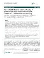
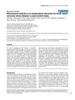
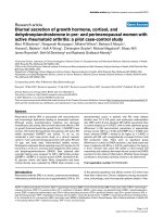
slight overfitting. Figure 1 shows that there was a good
correlation between the observed frequencies of ovarian
or endometrial cancer cases and the frequencies predicted by the regression model.
The sensitivity analysis yielded a similar result. The
unadjusted OR for endometriosis status was 2.41 (95 %
CI, 1.21 to 4.81) indicating that the predictors of the multiple regression model behaved unsuspiciously.
Discussion
In this case–control study, self-reported endometriosis
was confirmed as a risk factor for a combined group of
ovarian or endometrial cancer patients between the age
Table 2 Logistic regression analyses, showing adjusteda odds
ratios (ORs), with the corresponding 95 % confidence intervals
(CIs) in brackets
Predictor
Ageb
BMIc
Oral contraceptive use
OR (95 % CI)
Younger vs. medium
1.36 (1.11, 1.66)
Older vs. medium
1.24 (1.07, 1.43)
Older vs. younger
0.91 (0.70, 1.18)
Low vs. medium
0.99 (0.83, 1.17)
High vs. medium
1.26 (1.09, 1.46)
High vs. low
1.28 (0.95, 1.72)
Yes vs. no
0.43 (0.32, 0.58)
No. of pregnancies
Per-pregnancy increase
0.93 (0.84, 1.02)
Self-reported endometriosis
yes vs. no
2.63 (1.28, 5.41)
BMI body mass index
a
ORs were estimated using a multiple logistic regression model, with the
predictors listed in the first column of the table
b
Age was used as a nonlinear continuous predictor. It was evaluated at the
first sextile (“young” — i.e., 51 years), median (“medium” — i.e., 62 years), and
fifth sextile (“older” — i.e., 70 years)
c
BMI was used as a nonlinear continuous predictor. It was evaluated at the
first sextile (“low” — i.e., 21.7 kg/m2), median (“medium” — i.e., 25.0 kg/m2),
and fifth sextile (“high” — i.e., 30.1 kg/m2)
Burghaus et al. BMC Cancer (2015) 15:751
Page 5 of 8
Fig. 1 Observed and predicted frequencies of ovarian or endometrial cancer cases. The patients were ranked according to the predicted
conditional probability of being a case by the logistic regression model, and grouped into 10 categories based on deciles. Numbers of observed
cancer cases in each category (“observed events”) are plotted against the summed-up predicted probabilities of being a case in each category
(“predicted events”). Points below the gray line indicate when the regression model overestimates the cancer risk, and points above it indicate
underestimation. A perfect prediction model would have all points on the gray line
of 40 and 85. In addition, other already well-known risk
factors for ovarian and endometrial cancer such as age
and BMI were confirmed.
Endometriosis has been identified as a risk factor for subtypes of ovarian cancer [55, 56]. In a large, multicenter
study including more than 1500 patients with endometriosis, 7900 patients with ovarian carcinoma and 13,200 control patients, endometriosis was identified as a risk factor
for clear cell, endometrioid, and low-grade serous ovarian
carcinoma [57]. Clear increases in risk were found in the
group of endometriosis patients for clear cell ovarian carcinoma (OR 3.75; 95 % CI, 3.04 to 4.58), for endometrioid
ovarian carcinoma (OR 2.32; 95 % CI, 1.94 to 2.78),
and for low-grade serous ovarian carcinoma (OR 2.02;
95 % CI, 1.38 to 2.97). These findings did not apply
to high-grade serous ovarian carcinoma (OR 1.11; 95 %
CI, 0.96 to 1.29). In a national in-patient registry in
Sweden from 1969 to 1983 a cohort of 64,492 patients with a hospital diagnosis of endometriosis was
also found to have a significantly elevated risk for ovarian
cancer [58].
Until now, endometriosis has not been defined as a
risk factor for endometrial cancer. There are currently no data from population-based studies suggesting
an association between endometriosis and endometrial
cancer. A retrospective case–control study including 1399
patients did not show any association between endometrial cancer and endometriosis [59]. Previously reported data on endometriosis as a risk factor for
endometrial cancer are inconclusive [37–42]. The studies mentioned have limited case numbers in comparison
with the present study, which confirmed an increased
risk.
As mentioned above, an increased risk of epithelial ovarian cancer in patients with endometriosis has been shown
in numerous epidemiologic studies, but the pathogenesis
is poorly understood [35]. Current molecular studies have
sought to link the two conditions via pathways related to
oxidative stress, inflammation, and hyperestrogenism. As
a result of repetitive hemorrhage, with an accumulation of
heme and free iron in endometriotic lesions, reactive oxygen species are produced and play a role in the development of ovarian carcinoma [60]. Similarly, cytokines and
mediators are responsible for the microenvironment of
endometriosis and endometriosis-associated ovarian
carcinoma.
Although endometriosis is not yet established as a risk
factor for endometrial cancer, recent studies have discussed an influence of the epithelial-to-mesenchymal
transition and stem cells in endometrial cancer [61].
Burghaus et al. BMC Cancer (2015) 15:751
Endometrial stem cells are frequent in endometrial tissue during menstruation. It may therefore be speculated
that endometrial stem cells may play an important role
in the development of endometriotic implants [62] and
thus in endometriosis and endometrial cancer.
A molecular pathway cable of confirming the hypothesis is not currently known. An epigenetic analysis has
identified HNF1B as a subtype-specific susceptibility
gene for ovarian cancer [16]. Different variants in
HNF1B are associated with the risk of serous or clear
cell epithelial cancer. HNF1B is also overexpressed in
endometriosis [16], supporting the hypothesis that the
gene may have an oncogenic role in initiating specific
subtypes of ovarian cancer in patients with endometriosis. HNF1B might also be the link to endometrial cancer. A genome-wide association study has linked minor
alleles of certain single nucleotide polymorphisms in
HNF1B with a decreased risk of endometrial cancer
[23]. Further research is needed in order to define a
molecular pathway.
It has been hypothesized that endometriosis develops
from stem/progenitor cells. It would be of great interest
to associate the technique for identifying stem/progenitor cells in endometriotic tissues with an analysis of genetic/epigenetic changes in these cells that may possibly
affect their molecular signature and activity [63]. This
might make it possible to identify a molecular pathway
for the development of ovarian or endometrial cancer in
patients with endometriosis.
This study is the first case–control study to confirm
the influence of endometriosis on ovarian and endometrial cancer in a population in Germany. One advantage
of this case–control study was the validated epidemiological questionnaire that was used. A limitation is the
small number of cases, due to the low incidences of
ovarian and endometrial cancer, at 18.6 patients per
100,000 population and 26.9 patients per 100,000 population, respectively. Similarly in this study there are
fewer patients with reported endometriosis than expected by a prevalence of 10 % in reproductive age. This
effect can be caused by a notoriously underdiagnosed
disease and the prespecified age range from 40 to 85 in
our cases and controls with a consecutively decrease in
symptoms [64], which leads probably to a reduced description in the medical history form and the study
questionnaire. The number of 2.1 % endometriosis in
controls respectively 4.8 % in cases is congruent with the
data of the Iowa Women’s Health Study with a cohort of
more than 40,000 postmenopausal women, which publicated a number of 3.8 % of self-reported history of endometriosis [40]. Self-reported endometriosis is an inexact
and inaccurate method of assessment and may force up
the case numbers for endometriosis. Also the higher
number of self-reported endometriosis in patients with
Page 6 of 8
ovarian- or endometrial-cancer could originate in a better knowledge and remembering of their previous
gynecological diseases. Further limitations are the retrospective analysis of the data and the combined analysis
of ovarian and endometrial cancer cases. The reason for
the combined analysis was the low rate of seven patients
with endometriosis in each group of patients with ovarian (n = 158) or endometrial cancer (n = 124). Statistical
analyses were performed for each group, and there was a
significantly higher risk in the group of patients with
endometrial cancer and no significance in the patients
with ovarian cancer. However, these data are not shown,
due to the small number of cases of endometriosis in
each group. Our results do not necessarily hold for subjects younger than 40, because women of this age were
excluded from this study.
Conclusions
There have been few studies addressing the question of
whether endometriosis is a risk factor for ovarian or
endometrial cancer, and incorporating this into a clinical
prediction model would require precise characterization
of endometriosis as a risk factor. Larger studies are
needed in order to confirm the data for subgroups - especially for a younger population than the described one -,
to examine molecular pathways, and to understand the
pathogenesis.
Abbreviations
AUC: Area under the receiver operating characteristic curve; BAV: Bavarian
Ovarian Cancer Study; BBCC: Bavarian Breast Cancer Cases and Controls
Study; BECS: Bavarian Endometrial Cancer Study; BMI: Body mass index;
OC: Oral contraceptive; RR: Relative risk; OR: Odds ratio.
Competing interests
The authors declare that they have no competing interests.
Authors’ contributions
SB and LH contributed equally to this work. SB designed the study,
interpreted the data, helped to draft the manuscript and edited the paper.
LH designed the study, performed the statistical analysis, interpreted the
data and helped to draft the manuscript. MGS, KH, FCT and AH acquired
clinical data. DW, JS, AH and ABE acquired histological data. SPR and MWB
participated in the design the study. PAF designed the study, interpreted the
data and supervised research. All authors read and approved the final
manuscript.
Acknowledgments
We acknowledge support by Deutsche Forschungsgemeinschaft and
Friedrich-Alexander-Universität Erlangen-Nürnberg (FAU) within the funding
programme Open Access Publishing.
Author details
1
Department of Gynecology and Obstetrics, Erlangen University Hospital,
Friedrich Alexander University of Erlangen–Nuremberg, Comprehensive
Cancer Center Erlangen-EMN, Erlangen, Germany. 2Biostatistics Unit,
Department of Gynecology and Obstetrics, Erlangen University Hospital,
Friedrich Alexander University of Erlangen-Nuremberg, Erlangen, Germany.
3
Current address: ALB FILS KLINKEN GmbH, Goeppingen, Germany. 4Institute
of Pathology, Erlangen University Hospital, Friedrich Alexander University of
Erlangen–Nuremberg, Comprehensive Cancer Center Erlangen-EMN,
Erlangen, Germany. 5Institute of Human Genetics, Erlangen University
Hospital, Friedrich Alexander University of Erlangen–Nuremberg,
Burghaus et al. BMC Cancer (2015) 15:751
Comprehensive Cancer Center Erlangen-EMN, Erlangen, Germany. 6Division
of Hematology and Oncology, Department of Medicine, David Geffen School
of Medicine, University of California at Los Angeles, Los Angeles, CA, USA.
Page 7 of 8
22.
Received: 29 December 2014 Accepted: 16 October 2015
23.
References
1. American Cancer Society. Cancer facts & figures 2013. Atlanta: American
Cancer Society; 2013.
2. Waldmann A, Eisemann N, Katalinic A. Epidemiology of malignant cervical,
corpus uteri and ovarian tumours - current data and epidemiological
trends. Geburtsh Frauenheilk. 2013;73:123–9.
3. Rebbeck TR, Lynch HT, Neuhausen SL, Narod SA, Van’t Veer L, Garber JE,
et al. Prophylactic oophorectomy in carriers of BRCA1 or BRCA2 mutations.
N Engl J Med. 2002;346:1616–22.
4. Kauff ND, Satagopan JM, Robson ME, Scheuer L, Hensley M, Hudis CA, et al.
Risk-reducing salpingo-oophorectomy in women with a BRCA1 or BRCA2
mutation. N Engl J Med. 2002;346:1609–15.
5. Rebbeck TR, Kauff ND, Domchek SM. Meta-analysis of risk reduction
estimates associated with risk-reducing salpingo-oophorectomy in BRCA1 or
BRCA2 mutation carriers. J Natl Cancer Inst. 2009;101:80–7.
6. Renehan AG, Tyson M, Egger M, Heller RF, Zwahlen M. Body-mass index
and incidence of cancer: a systematic review and meta-analysis of
prospective observational studies. Lancet. 2008;371:569–78.
7. Fader AN, Arriba LN, Frasure HE, von Gruenigen VE. Endometrial cancer and
obesity: epidemiology, biomarkers, prevention and survivorship. Gynecol
Oncol. 2009;114:121–7.
8. Friedenreich CM, Neilson HK, Lynch BM. State of the epidemiological
evidence on physical activity and cancer prevention. Eur J Cancer.
2010;46:2593–604.
9. McTiernan A, Irwin M, Vongruenigen V. Weight, physical activity, diet, and
prognosis in breast and gynecologic cancers. J Clin Oncol. 2010;28:4074–80.
10. Tung KH, Goodman MT, Wu AH, McDuffie K, Wilkens LR, Kolonel LN, et al.
Reproductive factors and epithelial ovarian cancer risk by histologic type: a
multiethnic case–control study. Am J Epidemiol. 2003;158:629–38.
11. Antoniou A, Pharoah PD, Narod S, Risch HA, Eyfjord JE, Hopper JL, et al.
Average risks of breast and ovarian cancer associated with BRCA1 or BRCA2
mutations detected in case Series unselected for family history: a combined
analysis of 22 studies. Am J Hum Genet. 2003;72:1117–30.
12. Fasching PA, Gayther S, Pearce L, Schildkraut JM, Goode E, Thiel F, et al.
Role of genetic polymorphisms and ovarian cancer susceptibility. Mol
Oncol. 2009;3:171–81.
13. Song H, Ramus SJ, Tyrer J, Bolton KL, Gentry-Maharaj A, Wozniak E, et al.
A genome-wide association study identifies a new ovarian cancer
susceptibility locus on 9p22.2. Nat Genet. 2009;41:996–1000.
14. Bolton KL, Tyrer J, Song H, Ramus SJ, Notaridou M, Jones C, et al. Common
variants at 19p13 are associated with susceptibility to ovarian cancer. Nat
Genet. 2010;42:880–4.
15. Goode EL, Chenevix-Trench G, Song H, Ramus SJ, Notaridou M, Lawrenson
K, et al. A genome-wide association study identifies susceptibility loci for
ovarian cancer at 2q31 and 8q24. Nat Genet. 2010;42:874–9.
16. Shen H, Fridley BL, Song H, Lawrenson K, Cunningham JM, Ramus SJ, et al.
Epigenetic analysis leads to identification of HNF1B as a subtype-specific
susceptibility gene for ovarian cancer. Nat Commun. 2013;4:1628.
17. Bojesen SE, Pooley KA, Johnatty SE, Beesley J, Michailidou K, Tyrer JP, et al.
Multiple independent variants at the TERT locus are associated with
telomere length and risks of breast and ovarian cancer. Nat Genet.
2013;45:371–84.
18. Fasching PA, Ekici AB, Wachter DL, Hein A, Bayer CM, Haberle L, et al. Breast
cancer risk – from genetics to molecular understanding of pathogenesis.
Geburtsh Frauenheilk. 2013;73:1228–35.
19. Pharoah PD, Tsai YY, Ramus SJ, Phelan CM, Goode EL, Lawrenson K, et al.
GWAS meta-analysis and replication identifies three new susceptibility loci
for ovarian cancer. Nat Genet. 2013;45:362–70.
20. Permuth-Wey J, Lawrenson K, Shen HC, Velkova A, Tyrer JP, Chen Z, et al.
Identification and molecular characterization of a new ovarian cancer
susceptibility locus at 17q21.31. Nat Commun. 2013;4:1627.
21. Smith RA, von Eschenbach AC, Wender R, Levin B, Byers T, Rothenberger D,
et al. American Cancer Society guidelines for the early detection of cancer:
update of early detection guidelines for prostate, colorectal, and
24.
25.
26.
27.
28.
29.
30.
31.
32.
33.
34.
35.
36.
37.
38.
39.
40.
41.
42.
43.
44.
45.
endometrial cancers. Also: update 2001–testing for early lung cancer
detection. CA Cancer J Clin. 2001;51:38–75. quiz 77–80.
Aarnio M, Sankila R, Pukkala E, Salovaara R, Aaltonen LA, de la Chapelle A, et al.
Cancer risk in mutation carriers of DNA-mismatch-repair genes. Int J Cancer.
1999;81:214–8.
Spurdle AB, Thompson DJ, Ahmed S, Ferguson K, Healey CS, O’Mara T, et al.
Genome-wide association study identifies a common variant associated
with risk of endometrial cancer. Nat Genet. 2011;43:451–4.
Long J, Zheng W, Xiang YB, Lose F, Thompson D, Tomlinson I, et al.
Genome-wide association study identifies a possible susceptibility locus for
endometrial cancer. Cancer Epidemiol Biomarkers Prev. 2012;21:980–7.
Delahanty RJ, Xiang YB, Spurdle A, Beeghly-Fadiel A, Long J, Thompson D,
et al. Polymorphisms in inflammation pathway genes and endometrial
cancer risk. Cancer Epidemiol Biomarkers Prev. 2013;22:216–23.
Bulun SE. Endometriosis. N Engl J Med. 2009;360:268–79.
Signorello LB, Harlow BL, Cramer DW, Spiegelman D, Hill JA. Epidemiologic
determinants of endometriosis: a hospital-based case–control study. Ann
Epidemiol. 1997;7:267–741.
Vigano P, Parazzini F, Somigliana E, Vercellini P. Endometriosis:
epidemiology and aetiological factors. Best Pract Res Clin Obstet Gynaecol.
2004;18:177–200.
Sampson JA. Metastatic or Embolic Endometriosis, due to the Menstrual
Dissemination of Endometrial Tissue into the Venous Circulation. Am J
Pathol. 1927;3:93–110. 43.
Walter AJ, Hentz JG, Magtibay PM, Cornella JL, Magrina JF. Endometriosis:
correlation between histologic and visual findings at laparoscopy. Am J
Obstet Gynecol. 2001;184:1407–11. discussion 1411–1403.
Abbott J, Hawe J, Hunter D, Holmes M, Finn P, Garry R. Laparoscopic
excision of endometriosis: a randomized, placebo-controlled trial. Fertil
Steril. 2004;82:878–84.
Sayasneh A, Tsivos D, Crawford R. Endometriosis and ovarian cancer: a
systematic review. ISRN Obstet Gynecol. 2011;2011:140310.
Munksgaard PS, Blaakaer J. The association between endometriosis and
gynecological cancers and breast cancer: a review of epidemiological data.
Gynecol Oncol. 2011;123:157–63.
Pearce CL, Templeman C, Rossing MA, Lee A, Near AM, Webb PM, et al.
Association between endometriosis and risk of histological subtypes of ovarian
cancer: a pooled analysis of case–control studies. Lancet Oncol. 2012;13:385–94.
Worley MJ, Welch WR, Berkowitz RS, Ng SW. Endometriosis-associated
ovarian cancer: a review of pathogenesis. Int J Mol Sci. 2013;14:5367–79.
Erzen M, Rakar S, Klancnik B, Syrjanen K. Endometriosis-associated ovarian
carcinoma (EAOC): an entity distinct from other ovarian carcinomas as
suggested by a nested case–control study. Gynecol Oncol. 2001;83:100–8.
Borgfeldt C, Andolf E. Cancer risk after hospital discharge diagnosis of
benign ovarian cysts and endometriosis. Acta Obstet Gynecol Scand.
2004;83:395–400.
Brinton LA, Sakoda LC, Sherman ME, Frederiksen K, Kjaer SK, Graubard BI, et al.
Relationship of benign gynecologic diseases to subsequent risk of ovarian and
uterine tumors. Cancer Epidemiol Biomarkers Prev. 2005;14:2929–35.
Brinton LA, Gridley G, Persson I, Baron J, Bergqvist A. Cancer risk after a
hospital discharge diagnosis of endometriosis. Am J Obstet Gynecol.
1997;176:572–9.
Olson JE, Cerhan JR, Janney CA, Anderson KE, Vachon CM, Sellers TA.
Postmenopausal cancer risk after self-reported endometriosis diagnosis in
the Iowa Women’s Health Study. Cancer. 2002;94:1612–8.
Brinton LA, Lamb EJ, Moghissi KS, Scoccia B, Althuis MD, Mabie JE, et al.
Ovarian cancer risk associated with varying causes of infertility. Fertil Steril.
2004;82:405–14.
Brinton LA, Westhoff CL, Scoccia B, Lamb EJ, Althuis MD, Mabie JE, et al.
Causes of infertility as predictors of subsequent cancer risk. Epidemiology.
2005;16:500–7.
SE Johnatty, J Beesley, B Gao, X Chen, Y Lu, MH Law, et al. ABCB1 (MDR1)
polymorphisms and ovarian cancer progression and survival: A comprehensive
analysis from the Ovarian Cancer Association Consortium and The Cancer
Genome Atlas. Gynecologic oncology 2013;131:8-14.
Goode EL, Chenevix-Trench G, Hartmann LC, Fridley BL, Kalli KR, Vierkant RA,
et al. Assessment of hepatocyte growth factor in ovarian cancer mortality.
Cancer Epidemiol Biomarkers Prev. 2011;20:1638–48.
Hein A, Thiel FC, Bayer CM, Fasching PA, Haberle L, Lux MP, et al. Hormone
replacement therapy and prognosis in ovarian cancer patients. Eur J Cancer Prev.
2013;22:52–8.
Burghaus et al. BMC Cancer (2015) 15:751
46. Fasching PA, Loehberg CR, Strissel PL, Lux MP, Bani MR, Schrauder M, et al.
Single nucleotide polymorphisms of the aromatase gene (CYP19A1), HER2/
neu status, and prognosis in breast cancer patients. Breast Cancer Res Treat.
2008;112:89–98.
47. Schrauder M, Frank S, Strissel PL, Lux MP, Bani MR, Rauh C, et al. Single
nucleotide polymorphism D1853N of the ATM gene may alter the risk for
breast cancer. J Cancer Res Clin Oncol. 2008;134:873–82.
48. Antoniou AC, Wang X, Fredericksen ZS, McGuffog L, Tarrell R, Sinilnikova
OM, et al. A locus on 19p13 modifies risk of breast cancer in BRCA1
mutation carriers and is associated with hormone receptor-negative breast
cancer in the general population. Nat Genet. 2010;42:885–92.
49. Ghoussaini M, Fletcher O, Michailidou K, Turnbull C, Schmidt MK, Dicks E,
et al. Genome-wide association analysis identifies three new breast cancer
susceptibility loci. Nat Genet. 2012;44:312–8.
50. Garcia-Closas M, Couch FJ, Lindstrom S, Michailidou K, Schmidt MK, Brook
MN, et al. Genome-wide association studies identify four ER negativespecific breast cancer risk loci. Nat Genet. 2013;45:392–8.
51. Michailidou K, Hall P, Gonzalez-Neira A, Ghoussaini M, Dennis J, Milne RL,
et al. Large-scale genotyping identifies 41 new loci associated with breast
cancer risk. Nat Genet. 2013;45:353–61.
52. Harrell Jr FE, Lee KL, Pollock BG. Regression models in clinical studies:
determining relationships between predictors and response. J Natl Cancer
Inst. 1988;80:1198–202.
53. Salmen J, Neugebauer J, Fasching PA, Haeberle L, Huober J, Wockel A, et al.
Pooled analysis of the prognostic relevance of progesterone receptor status
in five German cohort studies. Breast Cancer Res Treat. 2014;148:143–51.
54. Hosmer DW, Lemeshow S, Sturdivant RX. Applied logistic regression. 3rd ed.
New York: Wiley; 2013.
55. Dogan S, Agic A, Eilers W, Finas D, Diedrich K, Hornung D. Endometriosis
and risk of malignancy. Geburtsh Frauenheilk. 2006;66:739–44.
56. Wang KC, Chang WH, Lee WL, Huang N, Huang HY, Yen MS, et al. An
increased risk of epithelial ovarian cancer in Taiwanese women with a new
surgico-pathological diagnosis of endometriosis. BMC Cancer. 2014;14:831.
57. CL Pearce, C Templeman, MA Rossing, A Lee, AM Near, PM Webb, et al.
Association between endometriosis and risk of histological subtypes of
ovarian cancer: a pooled analysis of case–control studies. The lancet
oncology 2012;13:385–94.
58. Melin A, Sparen P, Persson I, Bergqvist A. Endometriosis and the risk of
cancer with special emphasis on ovarian cancer. Hum Reprod.
2006;21:1237–42.
59. Rowlands IJ, Nagle CM, Spurdle AB, Webb PM. G Australian National
Endometrial Cancer Study, G Australian Ovarian Cancer Study:
Gynecological conditions and the risk of endometrial cancer. Gynecol
Oncol. 2011;123:537–41.
60. Yamaguchi K, Mandai M, Toyokuni S, Hamanishi J, Higuchi T, Takakura K,
et al. Contents of endometriotic cysts, especially the high concentration of
free iron, are a possible cause of carcinogenesis in the cysts through the
iron-induced persistent oxidative stress. Clin Cancer Res. 2008;14:32–40.
61. Mirantes C, Espinosa I, Ferrer I, Dolcet X, Prat J, Matias-Guiu X. Epithelial-tomesenchymal transition and stem cells in endometrial cancer. Hum Pathol.
2013;44:1973–81.
62. Sasson IE, Taylor HS. Stem cells and the pathogenesis of endometriosis. Ann
N Y Acad Sci. 2008;1127:106–15.
63. Forte A, Cipollaro M, Galderisi U. Genetic, epigenetic and stem cell
alterations in endometriosis: new insights and potential therapeutic
perspectives. Clin Sci (Lond). 2014;126:123–38.
64. Haas D, Chvatal R, Reichert B, Renner S, Shebl O, Binder H, et al.
Endometriosis: a premenopausal disease? Age pattern in 42,079 patients
with endometriosis. Arch Gynecol Obstet. 2012;286:667–70.
Page 8 of 8
Submit your next manuscript to BioMed Central
and take full advantage of:
• Convenient online submission
• Thorough peer review
• No space constraints or color figure charges
• Immediate publication on acceptance
• Inclusion in PubMed, CAS, Scopus and Google Scholar
• Research which is freely available for redistribution
Submit your manuscript at
www.biomedcentral.com/submit