The dog as a naturally-occurring model for insulin-like growth factor type 1 receptoroverexpressing breast cancer: An observational cohort study
Bạn đang xem bản rút gọn của tài liệu. Xem và tải ngay bản đầy đủ của tài liệu tại đây (1.02 MB, 13 trang )
Jaillardon et al. BMC Cancer (2015) 15:664
DOI 10.1186/s12885-015-1670-6
RESEARCH ARTICLE
Open Access
The dog as a naturally-occurring model for
insulin-like growth factor type 1 receptoroverexpressing breast cancer: an
observational cohort study
Laetitia Jaillardon1*, Jérome Abadie1, Tiffanie Godard1, Mario Campone2, Delphine Loussouarn3, Brigitte Siliart1
and Frédérique Nguyen1
Abstract
Background: Dogs spontaneously develop invasive mammary carcinoma with a high prevalence of the triple-negative
(TN) subtype (lack of ER-Estrogen Receptor and PR-Progesterone Receptor expression, lack of HER2-Human Epidermal
Growth Factor Receptor 2 overexpression), making this animal model relevant for investigating new therapeutic
pathways. Insulin-like growth factor Type-1 receptor (IGF1R) is frequently overexpressed in primary human breast
cancers, with a growing role in the TN phenotype. The purpose of this study was to investigate the Dog as a candidate
model for IGF1R-overexpressing mammary carcinoma.
Methods: 150 bitches with canine mammary carcinoma (CMC) and a known 2-year follow-up were
retrospectively included. IGF1R expression was assessed by immunohistochemistry (IHC) using a similar scoring
system as for HER2 in breast cancer. The prognostic value of the IGF1R expression was assessed in terms of
overall and specific survival as well as disease-free interval (DFI).
Results: 47 CMC (31 %) were classified as luminal and 103 (69 %) as triple-negative (TN-CMC). 41 % of CMC
overexpressed IGF1R (IHC score 3+) of which 76 % were TN-CMC and 62 % grade III. IGF1R overexpression
was associated with aggressive features including lymphovascular invasion, histological grade III, low ER
expression and the TN phenotype. Univariate and multivariate analyses revealed that IGF1R overexpression
was associated with shorter overall and specific survivals and shorter DFI in TN-CMC.
Conclusions: IGF1R overexpression is common and related to a poor outcome in canine invasive mammary
carcinoma, particularly in the triple negative subtype, as in human breast cancer. Preclinical studies using the
Dog as a spontaneous animal model could be considered to investigate new therapies targeting IGF1R in
triple-negative breast cancer.
Keywords: Spontaneous animal model, Canine mammary carcinoma, IGF1R, Triple-negative, Comparative
oncology
* Correspondence:
1
Oniris, Université Nantes-Angers-Le Mans, Department of Human Health,
Biomedical Research and Animal Models, AMaROC Unit and LDHvet
laboratory, Nantes Atlantic College of Veterinary Medicine, Food Science and
Engineering, Site de la Chantrerie, Route de Gachet, Nantes F-44307, France
Full list of author information is available at the end of the article
© 2015 Jaillardon et al. Open Access This article is distributed under the terms of the Creative Commons Attribution 4.0
International License ( which permits unrestricted use, distribution, and
reproduction in any medium, provided you give appropriate credit to the original author(s) and the source, provide a link to
the Creative Commons license, and indicate if changes were made. The Creative Commons Public Domain Dedication waiver
( applies to the data made available in this article, unless otherwise stated.
Jaillardon et al. BMC Cancer (2015) 15:664
Background
The identification of relevant naturally-occurring animal
models is of particular interest in oncology in order to
accelerate the development of effective diagnostic and
therapeutic innovations for human patients. The Dog is
a really good candidate as its physiology [1] and genome
[2] are very similar to that of humans. Dogs share the
same environment as humans with highly comparable
nutritional needs, and naturally develop various cancers
with a shorter natural history [3]. This spontaneous animal model could be highly beneficial to translational
breast cancer research as the human classification of
breast cancers is relevant to canine mammary carcinomas [4, 5], even if some histological entities (particularly complex mammary carcinoma) are quite different
between human and dog [6]. Interestingly, the triple
negative (TN) immunophenotype, one of the most aggressive breast cancer subtypes defined by the lack of ER
(Estrogen Receptor), PR (Progesterone Receptor) and
HER2 (Epidermal Growth factor Receptor type 2) overexpression, is well recognized in dogs [7, 8].
In various human cancers including breast cancer, the
Insulin-like Growth Factor (IGF) family is closely related
to oncogenesis [9, 10], in situ tumor growth [11], invasion and metastasis [11], with IGF1R (Insulin-like
Growth Factor Type 1-Receptor) acting as a real oncogene and being overexpressed in more than 50 % of primary breast cancers [12]. This is particularly true for the
TN breast cancer cells (estrogen-unresponsive), in which
IGF1R is largely expressed and IGF-1 stimulates proliferation and survival, making them responsive in vitro to
anti-IGF1R therapies [13, 14]. An ongoing phase I clinical trial of the IGF1R inhibitor OSI-906 in humans affected by advanced solid tumors showed few adverse
effects and no unexpected toxicities [15]. Even if a phase
II clinical trial using ganitumab (an anti-IGF1R antibody) did not show any improvement for women with
hormone-receptor positive and advanced breast cancer
[16], a phase I trial using another anti-IGF1R antibody
(cixutumumab) showed promising results by prolonging
stable diseases [17]. IGF1R expression is highly related
to prognosis in breast cancer, with a prognostic value
dependent on the ER status of the tumors: in ERpositive breast cancer, IGF1R overexpression is related
to favorable outcome [18] as opposed to ER-negative
carcinomas, in which IGF1R overexression is associated
with a poor outcome [19].
In canine mammary carcinoma, tissue GH (Growth
Hormone) and IGF-1 have been positively correlated
with tumor malignancy, as well as with tissue levels of
progesterone and 17β-estradiol [20]. IGF1R expression
has also been reported to be higher in histologic types of
worse prognosis [21] although some studies did not
show any significant association between IGF1R
Page 2 of 13
expression in mammary carcinomas and the clinical outcome in canine patients [22]. In addition, IGF-1 and
IGF1R have been implicated in other canine cancers including osteosarcoma [23, 24], malignant melanoma [25]
and testis tumors [26], suggesting a major role of the
IGF system in canine oncology.
In this study, IGF1R expression was retrospectively investigated by immunohistochemistry (IHC) in a large cohort of canine invasive mammary carcinomas in order to
determine the extent of similarities between canine and
human mammary carcinomas, with respect to the role
of IGF1R in tumor biology and natural history.
Methods
Patients and samples
Invasive mammary carcinomas surgically removed
from 150 bitches, formalin-fixed and sent to two laboratories of veterinary histopathology (Laboratoire
d’Histopathologie Animale, Oniris, Nantes, France
and Laboratoire d’Anatomie Pathologique Vétérinaire
d’Amboise, Amboise, France) between 2007 and 2010
were retrospectively selected. The owners’ written
consent and approval from the Oniris College of Veterinary Medicine local Animal Welfare Committee
were obtained prior to inclusion.
Dogs were eligible for inclusion when a diagnosis of
invasive mammary ductal carcinoma was established by
histological analysis and confirmed by an absent layer of
p63-positive myoepithelial cells (anti-p63 antibody, clone
ab111449, abcam plc) by immunohistochemistry (IHC)
that differentiates invasive from in situ breast ductal carcinoma [27, 28]. All female dogs that had received any
adjuvant chemotherapy and/or for which follow-up was
not available for at least 2 years after mastectomy, were
excluded from the study.
Breed, age and reproductive status (including age of
neutering) at time of mastectomy, as well as the number
and location of mammary carcinoma(s), were recorded
for each bitch. Two-year follow-up was obtained
through telephone interviews with referral veterinarians
with particular emphasis on the occurrence of recurrence (i.e. the occurrence of an another mammary tumor
on the same mammary gland) and/or of a new primary
mammary tumor, and the animal’s outcome (alive or
dead and cause of death, i.e., unrelated or related to the
mammary carcinoma whether the animals died naturally
or were euthanatized because of metastases). Overall
Survival (OS) was defined as the time between surgery
(mastectomy) and death from any cause; uncensored
cases corresponded to dead animals; censored cases
were still alive at least two years post-diagnosis. Specific
Survival (SS) was defined as the time between surgery
and death attributable to the mammary carcinoma; censored cases corresponded to dogs still alive, dogs that
Jaillardon et al. BMC Cancer (2015) 15:664
Page 3 of 13
Table 1 Characteristics of the dogs and their invasive mammary
carcinomas
Table 1 Characteristics of the dogs and their invasive mammary
carcinomas (Continued)
Parameters
Data
Triple-negative non basal like
n (%)
IGF1R expression
Total
150 (100)
Age in yrs
Median 11 yrs, Range [5.1–16.3 yrs]
5.1–10.9 yrs
73 (48.7)
≥11 yrs
77 (51.3)
Tumor size
< 2 cm
53 (36.5)
≥ 2 cm
92 (63.5)
33 (22)
Score 0–1+
34 (22.7)
Score 2+
54 (36)
Score 3+
62 (41.3)
Survival Time in days
Median 331 days, Range [2–2608]
yrs years, ER Estrogen Receptor, PR Progesterone Receptor, HER2 Epidermal
Growth Factor Receptor 2, CK5/6 Cytokeratin 5/6, EGFR Epidermal Growth
Factor Receptor, IGF1R Insulin-like growth factor type 1 receptor
Histological type
Squamous cell carcinoma
6 (4)
Simple carcinoma: Anaplastic
6 (4)
Complex carcinoma
11 (7.3)
Simple carcinoma: Solid
40 (26.7)
Simple carcinoma: Tubulopapillary
87 (58)
Histological grade (Elston & Ellis)
Grade I
19 (12.6)
Grade II
58 (38.7)
Grade III
73 (48.7)
Lymph node status
Positive (N1)
32 (21.3)
Negative (N0)
19 (12.7)
Unknown (NX)
99 (66)
ER expression
Positive (≥ 10 %)
35 (23.3)
Negative (< 10 %)
115 (76.7)
PR expression
Positive (≥ 10 %)
20 (13.3)
Negative (< 10 %)
130 (86.7)
HER2
Score 0
85 (56.7)
Score 1+
50 (33.3)
Score 2+
15 (10)
Score 3+
0
CK5/6
Positive (≥ 10 %)
89 (59.3)
Negative (< 10 %)
61 (40.7)
EGFR
Positive (≥ 10 %)
72 (48)
Negative (< 10 %)
78 (52)
Immunophenotype
Luminal-A
17 (11.3)
Luminal-B
30 (20)
Triple-negative basal like
70 (46.7)
died from unknown cause, and dogs that died from
another cause than the mammary carcinoma. The
interval from surgery to the first local recurrence,
new primary tumor, lymph node metastasis and/or
distant metastasis was also assessed, and defined the
disease-free interval (DFI).
Histopathology and immunohistochemistry (IHC)
All tumors were paraffin-embedded immediately after
reception. 4 μm-thick serial sections were performed
onto positively charged slides (Superfrost plus, MenzelGlaser, Germany). After Hematoxylin and Eosin (HE)
staining, mammary carcinomas were classified by five independent pathologists (one human breast pathologist
and four veterinary pathologists) according to the
WHO’s classification system of canine mammary tumors
[28, 29, 30], and graded according to the criteria of
Elston and Ellis [31] as well-differentiated (grade I),
moderately differentiated (grade II) or poorly differentiated (grade III) carcinomas. The histologically assessed
size of mammary carcinoma(s) with 2 cm chosen as a
threshold according to the American Joint Committee
on Cancer (AJCC), lymphovascular invasion, completeness of surgical excision, dermal infiltration, cutaneous
ulceration, muscle invasion, squamous differentiation,
inflammation and central necrosis were recorded for
each case. In case of multifocal or multicentric carcinomas, the tumor with the highest pathologic size and/or
highest histological grade was included in the study.
Automated IHC (Benchmark XT Ventana, Roche
Diagnostics) was performed using antibodies against
ERα (Estrogen Receptor alpha, clone C311, Santa Cruz),
PR (Progesterone Receptor, clone 1E2, Ventana), HER2
(Human Epidermal Growth Factor Receptor 2 clone
4B5, Ventana), Ki-67 (clone MIB1, Dako), CK5/6
(Cytokeratin 5/6, clone D5/16B4, Dako), EGFR (Epidermal
Growth Factor Receptor Type 1 clone 31G7, Invitrogen)
and IGF1R (Insulin-like Growth Factor type 1Receptor clone G11, Ventana). IHC protocols are detailed in Additional file 1: Table S1.
Jaillardon et al. BMC Cancer (2015) 15:664
Scoring of the immunohistochemical staining was performed by the five independent pathologists. ER, PR and
Ki-67 were assessed based on the number of positive nuclei among 500 counted cells (manual image analysis involving the use of the image J software, Research Service
Branch, National Institute of Health, Bethesda, Maryland,
USA). ER and PR were considered positive if nuclear
staining was observed in more than 10 % of the cells [32]
and Ki-67 in more than 20 % of the cells [33]. HER2
[32, 34] was scored as follow: 0 for no staining at all
or incomplete, faint/barely perceptible membrane
staining in less than 10 % of the cells; score 1+ for
incomplete and faint/barely perceptible membrane staining in more than 10 % of the cells; 2+ for circumferential
and incomplete and/or weak/moderate membrane staining in more than 10 % of the cells; and 3+ for circumferential and complete and intense membrane staining in
more than 10 % of the cells. Carcinomas were considered positive for HER2 only for a 3+ IHC score [32].
IGF1R was scored in accordance with the HER2
Page 4 of 13
expression scoring system [19, 35]: a negative result
was defined as the complete absence of membrane
staining (score 0) or the presence of weak membrane
staining in less than 10 % of the cells or incomplete
membrane staining in more than 10 % of the cells
(score 1+) in any portion of the tumor; a score 2+
was applied for complete and weak to moderate
membrane staining in more than 10 % of the cells;
and a score 3+ for complete and intense membrane
staining in more than 10 % of the tumor cells [34].
EGFR [36] was considered positive if membrane staining was observed in more than 10 % of the cells.
Positivity to cytokeratins 5/6 (CK5/6) was defined
with a threshold of 10 % [37].
Negative controls for IHC were included in each run,
and consisted in replacing the primary antibody with normal mouse or rabbit serum (prediluted reagents, Roche
Diagnostics). The positive controls were internal controls
in most cases (i.e., skin epidermis and hair follicles
for Ki-67, CK 5/6, EGFR and IGF1R; mammary gland
A
B
C
D
E
F
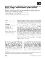
Fig. 1 Immunohistochemical staining of IGF1R expression in normal and neoplastic canine mammary glands. IGF1R (Insulin-like growth
factor type 1 receptor) expression was scored according to the intensity of the membrane staining in accordance with the HER-2 scoring
system. a Hair follicle positive for IGF1R expression, b Normal mammary gland with a score 2+ for IGF1R, c Invasive ductal mammary
carcinoma with a score 0 for IGF1R, d Invasive ductal mammary carcinoma with a score 1+ for IGF1R, (E) Invasive ductal mammary
carcinoma with a score 2+ for IGF1R, f Invasive ductal mammary carcinoma with a score 3+ for IGF1R (Immunohistochemical staining,
original magnification × 400). Bar = 50 micrometers
Jaillardon et al. BMC Cancer (2015) 15:664
Page 5 of 13
surrounding the carcinoma for ER and PR), as stated
in Table 1. For HER2 IHC, the pathway HER2 4-in-1
control slides (Roche Diagnostics) were chosen because they allow the quality of staining for each
HER2 score (0, 1+, 2+, 3+) to be assessed.
Photographs of slides were taken using an Eclipse 50i
microscope and a Nikon DS Fi-1 digital camera (Nikon
Instruments Europe B.V.).
Statistical analysis
The Statview (Statview 5 SAS Institute Inc.) and R (R 3.1.1
GUI 1.65) softwares were used for statistical analyses. Results are given as median and range unless otherwise indicated. Non-parametric tests were used after checking for
normality and independence of the data by KolmogorovSmirnov test and graphic assessment. The correlation between IGF1R expression and categorical variables (age
groups, histological grade, clinical stage, nodal stage,
hormone receptor status, and immunophenotype) was
analyzed using the Pearson chi-square test or the
Fisher exact test. Correlations between numeric variables
were determined by Spearman’s test. The Kaplan-Meier
non-parametric method was used for univariate survival
analysis and the log-rank test was used to assess differences among groups. Cox proportional-hazard regression
model was used to examine all factors found to be
predictive of survival in univariate analysis simultaneously.
A p-value of less than 0.05 was considered significant.
Results
Clinicopathological findings
The study population consisted in 117 intact and 33
spayed female dogs. Age at surgery ranged from 5.1 to
16.3 years (median 10.9 years). The 150 invasive carcinomas were classified as Luminal and Triple Negative
according to ER, PR and HER2 expressions [4, 5]: 47
(31.3 %) were of Luminal subtype (ERα ≥ 10 % and/or
PR ≥ 10 %), of which 17 were Luminal-A (Ki-67 < 20 %)
and 30 were Luminal-B (Ki-67 ≥ 20 %), and 103 (68.7 %)
were classified as Triple Negative (ERα < 10 %, PR < 10 %,
HER2 score other than 3+), of which 70 were basal-like
(Cytokeratin-CK 5/6 and/or Epidermal Growth Factor
Receptor-EGFR positive), and 33 were non-basal-like (CK
5/6 and EGFR negative). No carcinoma was HER2 overexpressing, although immunohistochemical scores 3+ were
obtained with the positive controls (human breast cancer
lines, control slides provided by Roche Diagnostics). The
main clinicopathological findings are summarized in
Table 1.
The median follow-up period was 36.3 months. In
total, 130 dogs (86.7 %) died. The median time between the date of diagnosis and the date of death was
8.4 months [2 days–60.3 months]. The median DFI
was 22.5 months with a 2-year recurrence and/or metastasis rate of 42 %. The median SS was 28.1 months
with a 2-year cancer-related mortality rate of 39.3 %.
The median OS was 11.0 months with a 2-year mortality rate of 68.7 %.
Table 2 Significant associations between IGF1R expression and clinicopathological features of the 150 canine mammary carcinomas
Parameters
Fisher’s exact
test
IGF1R score 2+
IGF1R score 3+
p-value
OR
95 % CI
p-value
OR
95 % CI
Grade I or II
-
1.00
-
-
1.00
-
Grade III
0.007
3.86
1.49–10.99
<0.001
6.54
2.57–18.53
-
1.00
-
-
1.00
-
0.87
1.08
0.44–2.68
0.01
3.11
1.32–7.62
Histological grade
LVI
<0.001
0.006
Absent
Present
ER expression
0.004
Positive (≥ 10 %)
-
1.00
-
-
1.00
-
Negative (< 10 %)
0.003
4.54
1.69–13.01
0.01
3.29
1.32–8.46
-
1.00
-
-
1.00
-
0.07
2.88
0.93–9.47
0.02
4.10
1.29–14.54
PR expression
0.04
Positive (≥ 10 %)
Negative (< 10 %)
Immunophenotype
0.03
Luminal
-
1.00
-
-
1.00
-
Triple Negative
0.02
2.86
1.16–7.20
0.02
2.88
1.20–7.04
IGF1R score 0–1+ is considered as the reference for each parameter
IGF1R Insulin-like growth factor type 1 receptor, LVI Lymphovascular Invasion, ER Estrogen Receptor, PR Progesterone Receptor, OR Odds Ratio, 95 % CI 95 %
Confidence Interval
Jaillardon et al. BMC Cancer (2015) 15:664
Page 6 of 13
IGF1R expression
The IGF1R staining was exclusively observed in the
plasma membrane with cytoplasmic blush only observed
when IGF1R was strongly expressed. Only membrane
immunoreactivity was taken into account for scoring
IGF1R expression. IGF1R was strongly expressed in epithelial cells of the hair follicles, hyperplastic and dysplastic mammary tissues adjacent to the tumors (Fig. 1). The
number of cases with IGF1R score 0-1+ was 34 (22.7 %,
of which 11 (7.3 %) score 0 and 23 (15.4 %) score 1+),
54 cases (36.0 %) were IGF1R score 2+ and 62 (41.3 %)
were IGF1R score 3+ (Fig. 1). Considering the luminal and triple negative immunophenotypes separately, the IGF1R 0–1+, 2+ and 3+ scores occurred in
17 (36.2 %), 14 (29.8 %) and 16 (34.0 %) luminal canine mammary carcinomas and in 17 (16.5 %), 40
(38.8 %) and 46 (44.7 %) triple-negative canine mammary carcinomas respectively.
Association of IGF1R expression and clinicopathological
features
IGF1R overexpression (IHC score 3+) was significantly
associated with aggressive features including lymphovascular invasion, histological grade III, absent or low ER
and PR expression, and the TN immunophenotype
(Table 2). In the Luminal subtype, IGF1R overexpression
was also significantly correlated with aggressive features
including high histological grade (OR = 7.78 [1.71–45.30],
p = 0.01) and lymphovascular invasion (OR = 5.42 [1.27–
27.20], p = 0.03), except for dermal infiltration for which
IGF1R score 2+ (OR = 0.07 [0.03–0.46], p = 0.02) and 3+
(OR = 0.13 [0.02–0.64], p = 0.02) were associated with an
absence of dermal infiltration (Additional file 2: Table S2).
In the TN subtype, IGF1R overexpression was only significantly related to a high histological grade (OR = 5.54
[1.67–22.25], p = 0.02).
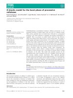
Prognostic value of IGF1R expression
By univariate analysis, IGF1R overexpression was associated with a poor outcome in terms of disease-free interval (p = 0.04), overall (p < 0.001) and specific (p = 0.001)
survival (Fig. 2). Univariate analyses revealed that other
factors were associated with a poor prognosis (DFI, OS
and SS), including multifocality of the mammary carcinoma, nodal stage at diagnosis, histological grade, surgical
margin status, lymphovascular invasion, ER expression
and immunophenotype (Tables 3, 4 and 5). Multivariate
analysis using Cox proportional-hazard regression was
then carried out. When several significant prognostic
factors were overlapping (for example nodal stage at
mastectomy and lymphovascular invasion or immunophenotype and ER/PR expression), only one was selected
as a covariate in the model.
Fig. 2 Kaplan-Meier analysis of OS, SS and DFI in 150 canine invasive
mammary carcinomas according to IGF1R expression. IGF1R: Insulin-like
growth factor type 1 receptor
For overall survival, IGF1R overexpression appeared to
be a strong and independent prognostic factor associated
with a poor outcome, as well as an age of more than 11
years, lymphovascular invasion, positive margin status of
the surgical sample and the presence of a peritumoral
inflammation (Table 3). With regard to specific survival,
IGF1R overexpression, lymphovascular invasion, and the
presence of central necrosis showed a significant independent prognostic value (Table 4). By multivariate analysis for disease-free interval, IGF1R overexpression was
Jaillardon et al. BMC Cancer (2015) 15:664
Page 7 of 13
Table 3 Factors associated with overall survival (OS) in canine invasive mammary carcinomas (n = 150)
Criteria
OS: Univariate analysis
OS: Multivariate analysis
(log-rank test) N = 150
(Cox regression model) N = 150
HR
95 % CI
<11 yrs
1.00
-
≥11 yrs
1.66
1.17–2.37
Age
p-value
HR
95 % CI
1.00
-
1.79
1.24–2.60
0.005
Multifocality
0.002
0.04
0.96
Unifocal
1.00
-
1.00
-
Multicentric
1.89
1.01–3.54
1.02
0.51–2.04
N0
1.00
-
-
-
N1
3.53
1.79–6.93
Lymph node status
p-value
<0.001
Histological grade
0.006
-
0.53
Grade I
1.00
-
-
1.00
-
-
Grade II
1.67
0.93–2.99
0.09
1.33
0.70–2.53
0.38
Grade III
2.37
1.35–4.18
0.003
1.42
0.77–2.62
0.26
Lymphovascular invasion
<0.001
0.01
No LVI
1.00
-
1.00
-
LVI
2.53
1.78–3.60
1.71
1.14–2.56
Complete excision
1.00
-
1.00
-
Incomplete excision
2.32
1.62–3.33
1.81
1.18–2.75
Surgical margins
<0.001
Muscle infiltration
0.006
0.001
0.77
No
1.00
-
1.00
-
Yes
1.86
1.27–2.71
1.07
0.70–1.63
No
1.00
-
1.00
-
Yes
1.53
1.08–2.17
1.53
1.03–2.29
-
-
1.00
-
1.34
0.88–2.04
Peritumoral Inflammation
0.02
ER
0.04
0.03
≥ 10 %
1.00
-
< 10 %
1.48
1.04–2.11
Luminal
1.00
-
Triple negative
1.54
1.05–2.26
Immunophenotype
0.03
IGF1R
-
0.17
<0.001
0.002
weak (0–1+)
1.00
-
-
1.00
-
-
moderate (2+)
1.31
0.81–2.10
0.27
1.40
0.85–2.33
0.19
strong (3+)
2.62
1.63–4.20
<0.001
2.74
1.63–4.62
0.002
Univariate (log rank test) and multivariate survival analyses (Cox proportional hazard regression)
HR Hazard Ratio, 95 % CI 95 % Confidence Interval, ER Estrogen Receptor, IGF1R Insulin-like Growth Factor type 1 Receptor, LVI Lymphovascular Invasion
When several significant prognostic factors overlapped, only one was selected for the multivariate analysis (LVI was chosen between lymph node status and LVI
because it could have been determined in all cases and immunophenotype was preferred to ER expression)
no longer significantly associated with an earlier recurrence, new primary tumor and/or lymph node and distant metastasis (p = 0.13) (Table 5).
The prognostic impact of IGF1R was also assessed separately in the luminal and the TN immunophenotypes.
In the luminal subtype (n = 47), IGF1R overexpression
was associated with a shorter OS (HR = 3.13 [1.41–6.96];
p = 0.005) and SS (HR = 4.72 [1.42–15.77]; p = 0.01)
by univariate analysis (Additional file 3: Tables S3 and
Additional file 4: Table S4). By multivariate analysis,
Jaillardon et al. BMC Cancer (2015) 15:664
Page 8 of 13
Table 4 Factors associated with specific survival (SS) in canine invasive mammary carcinomas (n = 150)
Criteria
SS: Univariate analysis
SS: Multivariate analysis
(log-rank test) N = 150
(Cox regression model) N = 150
HR
95 % CI
> 10 kgs
1.00
-
≤ 10 kgs
1.68
1.01–2.78
Body size
p-value
HR
95 % CI
0.04
Multifocality
0.63
1.00
1.16
0.64–2.09
0.02
0.85
Unifocal
1.00
-
1.00
-
Multicentric
2.36
1.12–4.97
1.09
0.44–2.72
N0
1.00
-
-
-
N1
5.07
1.88–13.67
Lymph node status
p-value
0.001
Histological grade
0.01
-
0.44
Grade I
1.00
-
1.00
-
-
Grade II
2.52
0.97–6.59
0.06
2.02
0.66–6.23
0.22
Grade III
3.74
1.47–9.55
0.006
2.03
0.67–6.19
0.21
Lymphovascular invasion
<0.001
0.002
No LVI
1.00
-
1.00
-
LVI
4.48
2.68–7.51
2.66
1.43–4.94
Complete excision
1.00
-
1.00
-
Incomplete excision
2.34
1.43–3.83
1.46
0.77–2.78
Surgical margins
<0.001
Muscle infiltration
0.24
0.01
0.76
No
1.00
-
1.00
-
Yes
1.88
1.14–3.11
1.10
0.58–2.11
No
1.00
-
1.00
-
Yes
1.64
1.02–2.63
1.74
0.96–3.15
Peritumoral Inflammation
0.04
Central necrosis
0.07
0.03
0.005
No
1.00
-
1.00
-
Yes
0.57
0.34–0.96
0.43
0.24–0.78
≥ 10 %
1.00
-
-
-
< 10 %
1.91
1.02–3.56
ER
0.04
Ki-67
0.01
< 20 %
1.00
≥ 20 %
2.65
1.21–5.80
Luminal
1.00
-
Triple negative
2.35
1.31–4.22
Immunophenotype
IGF1R
0.29
1.00
-
1.70
0.64–4.51
1.00
-
1.94
0.96–3.88
0.004
0.001
-
0.06
0.03
Jaillardon et al. BMC Cancer (2015) 15:664
Page 9 of 13
Table 4 Factors associated with specific survival (SS) in canine invasive mammary carcinomas (n = 150) (Continued)
weak (0–1+)
1.00
-
-
1.00
-
-
moderate (2+)
1.68
0.82–3.44
0.15
1.66
0.72–3.85
0.24
strong (3+)
3.36
1.68–6.72
<0.001
2.81
1.25–6.31
0.01
Univariate (log rank test) and multivariate survival analyses (Cox proportional hazard regression)
HR Hazard Ratio, 95 % CI 95 % Confidence Interval, ER Estrogen Receptor, IGF1R Insulin-like Growth Factor type 1 Receptor, LVI Lymphovascular Invasion
When several significant prognostic factors overlapped, only one was selected for the multivariate analysis (LVI was chosen between lymph node status and LVI
because it could have been determined in all cases and immunophenotype was preferred to ER expression)
IGF1R overexpression was also a significant strong
and independent prognostic factor associated with a
poor outcome in terms of OS and SS, as well as an
age of more than 11 years.
In the TN subtype (n = 103), IGF1R overexpression was
also associated with a shorter OS (HR = 2.24 [1.23–4.10];
p = 0.009) and SS (HR = 2.49 [1.07–5.81]; p = 0.03) by
univariate analysis. IGF1R expression retained a significant
and independent prognostic value for OS by multivariate
analysis, as well as the age of the dog at neutering, occurrence of a new primary mammary tumor, histological
grade, surgical margin status and presence of central necrosis (Additional file 5: Table S5). Finally, IGF1R was also
a significant and independent prognostic factor for SS in
the TN immunophenotype, with lymphovascular invasion
and central necrosis (HR = 0.47 [0.24–0.93]; p = 0.03) as
covariates (Additional file 6: Table S6).
IGF1R expression did not show any prognostic value
in terms of DFI either in the luminal or TN subgroup.
Discussion
The objective of this study was to investigate IGF1R expression in a large cohort of canine invasive carcinomas,
focusing on its relationship with the clinicopathological
features and prognosis, in terms of overall, specific and
disease-free survivals, in order to evaluate the similarities
between the role of IGF1R in the canine species and
those previously reported in human breast cancer. We
found that IGF1R was frequently expressed in canine invasive mammary carcinoma, as more than 90 % showed
at least a weak membrane staining for IGF1R. This result
is in accordance with the previous human studies, as
usually more than 80 % of the invasive breast cancer
cells are positive for IGF1R [18, 35, 38]. In human breast
cancer, few studies take into account both membrane
and cytoplasmic IGF1R expression [18, 39, 40]. We only
considered membrane staining for scoring IGF1R expression, as cytoplasmic blush was only observed when
IGF1R was strongly expressed. Methods used for IGF1R
scoring depend on the study, but most of the published
results consider that a score of 3+ by immunohistochemistry (mostly defined as complete and intense
membrane staining in more than 10 % of the cells, as for
HER2 scoring) defined IGF1R overexpression [19, 35].
Thus, we chose to score IGF1R in accordance with the
scoring of HER2 in breast cancer and then grouped the
negative scores (complete absence of membrane staining
or the presence of weak membrane staining in less than
10 % of the cells) and 1+ (incomplete membrane staining in more than 10 % of the cells), compared with the
positive scores 2+ (complete and weak to moderate
membrane staining in more than 10 % of the cells) and
3+ (complete and intense membrane staining in more
than 10 % of the cells) as Shin et al. previously did in
human breast cancer [19]. However, the grouping of the
score 0 and 1+ is questionable, as the normal canine
mammary gland [22] (Fig. 1), like the human breast [41,
42], naturally shows a weak (1+) to moderate (2+) IGF1R
expression, implying that the absence of expression is
abnormal and not necessarily a good prognostic factor.
Indeed, some studies showed that IGF1R negativity and
down-regulation was associated with a worse prognosis
[43] in tamoxifen-treated postmenopausal breast cancer
and correlated with aggressive features such as poor differentiation and high proliferation [44]. The number of
cases in the present study with a score 0 for IGF1R expression was too small (n = 11) to analyze this group
separately, implying that this is a rare condition that requires more cases for definitive conclusions.
When luminal and triple-negative subtypes were
assessed separately, IGF1R overexpression (score 3+) was
comparable in frequency to that reported in human breast
cancer in which more than 45 % of the triple-negative
breast carcinomas show strong expression of IGF1R
[18, 19, 40, 41]. In human breast cancer and canine mammary carcinoma, several studies have shown that IGF1R
expression parallels ER expression [18, 20, 39, 41], but we
found that IGF1R overexpression was correlated with the
negativity for ER and PR in the total cohort as Law et al.
showed for phosphorylated IGF1R/IR expression in human breast cancer [45]. This contradictory result could be
due to a biological difference concerning IGF1R and ER
between dogs and humans. The fact that IGF1R parallels
ER expression in canine mammary carcinoma in the study
of Queiroga et al. [20] is also controversial: the cohort was
small (40 mammary carcinomas) and unlike the present
study, the invasive nature of the mammary carcinomas
was not assessed. In our luminal subgroup, no correlation
was found between hormonal receptor (ER and PR) and
IGF1R expression. Nevertheless, this result has to be
Jaillardon et al. BMC Cancer (2015) 15:664
Page 10 of 13
Table 5 Factors associated with disease-free interval (DFI) in canine invasive mammary carcinomas (n = 150)
Criteria
DFI: Univariate analysis
DFI: Multivariate analysis
(log-rank test) N = 150
(Cox regression model) N = 150
HR
95 % CI
> 10 kgs
1.00
-
≤ 10 kgs
1.99
1.23–3.21
Body size
p-value
HR
95 % CI
0.005
Age
0.07
1.00
1.66
0.95–2.90
0.007
0.02
<11 yrs
1.00
-
1.00
-
≥11 yrs
1.88
1.19–2.98
1.91
1.13–3.24
Unifocal
1.00
-
1.00
-
Multicentric
2.56
1.22–5.38
0.86
0.32–2.29
Multifocality
0.01
Histological grade
p-value
0.76
0.06
0.46
Grade I
1.00
-
-
1.00
-
Grade II
1.90
0.89–4.04
0.10
1.76
0.67–4.62
0.25
Grade III
2.43
1.17–5.06
0.02
1.83
0.69–4.86
0.22
Lymphovascular invasion
<0.001
0.04
No LVI
1.00
-
1.00
-
LVI
2.86
1.81–4.51
1.91
1.02–3.58
Complete excision
1.00
-
1.00
-
Incomplete excision
1.94
1.22–3.07
1.57
0.85–2.88
Surgical margins
0.005
Muscle infiltration
0.15
0.005
0.99
No
1.00
-
1.00
-
Yes
2.00
1.23–3.28
1.00
0.54–1.86
No
1.00
-
1.00
-
Yes
1.57
1.00–2.47
1.69
0.95–2.99
Peritumoral Inflammation
0.04
Central necrosis
0.07
0.03
0.04
No
1.00
-
1.00
-
Yes
0.56
0.34–0.93
0.52
0.28–0.97
Luminal
1.00
-
1.00
-
Triple negative
1.80
1.08–3.00
1.54
0.82–2.86
Immunophenotype
0.02
CK5/6
0.18
0.01
0.09
< 10 %
1.00
-
1.00
-
≥ 10 %
0.55
0.35–0.87
0.61
0.34–1.08
weak (0–1+)
1.00
-
-
1.00
-
-
moderate (2+)
1.22
0.68–2.19
0.51
1.15
0.58–2.28
0.70
strong (3+)
2.09
1.14–3.83
0.02
1.74
0.86–3.55
0.13
IGF1R
0.04
0.23
Univariate (log rank test) and multivariate survival analyses (Cox proportional hazard regression)
HR Hazard Ratio, 95 % CI 95 % Confidence Interval, CK5/6 Cytokeratin 5/6, IGF1R Insulin-like Growth Factor type 1 Receptor, LVI Lymphovascular Invasion
Jaillardon et al. BMC Cancer (2015) 15:664
confirmed on a larger cohort of luminal canine mammary
carcinomas.
IGF1R expression was also correlated with other aggressive features in both luminal and TN subtypes (such
as high histological grades or presence of lymphovascular invasion). These results are in accordance with previously published studies in canine mammary carcinoma,
as IGF-1 and IGF1R expression were respectively related
to tumor malignancy [20] and histological types with
worse prognosis [21]. This finding is in line with the fact
that IGF1R is considered as a real oncogene closely involved in survival, proliferation, tumor growth, invasion
and metastasis as it was demonstrated in canine
osteosarcoma-derived cell lines [23]. In human breast
cancer, results are controversial and generally depend on
the ER status of the carcinomas. Indeed, extensive crosstalk between ER and IGF1R is now well-established from
several in vitro studies, which demonstrate a synergistic
effect of IGF1R and ER on the proliferation of human
breast cancer cells [46, 47]. Even if some studies did not
find any significant results [35, 48, 49], IGF1R positivity
was generally related to favorable prognostic features in
ER-positive breast cancer, including low histological
grade [19]. On the contrary, strong IGF1R expression
was associated with aggressive features in triple negative breast cancer, such as high histological grade
[40]. However, no study to date has investigated the
crosstalk between IGF1R and ER in canine mammary
cell lines. A difference of receptor biology between
Human and Dog cannot be excluded and should thus
be further investigated.
Some studies show that the complete negativity or
low expression of IGF1R is related to a worse prognosis [43, 44]. On the contrary, rare studies reveal
that high IGF1R mRNA [50] and phosphorylated
IGF1R/IR [45] are associated with a poor prognosis,
whatever the molecular subtype of breast cancer. In
addition, even if human studies show contradictory
results, it seems that the IGF1R prognostic value also
depends on the tumor ER status: in ER-positive mammary
carcinomas, IGF1R overexpression is related to a favorable
prognosis [18, 19] as opposed to the triple-negative subtype, in which IGF1R overexpression is associated with a
poor outcome [18, 19, 40]. In the present study, no difference was found between the luminal and triple-negative
subtypes of canine mammary carcinoma according to the
prognostic value of IGF1R expression: IGF1R overexpression was associated with a poor prognosis in both luminal
and triple-negative canine mammary carcinomas. The fact
that none of the dogs of this study received adjuvant
endocrine therapy is however a major difference between
humans and dogs after a diagnosis of luminal mammary
carcinoma, and this difference is likely to interfere with
prognosis. Furthermore, only 47 luminal mammary
Page 11 of 13
carcinomas were included in this study and further investigations with a higher number of luminal mammary carcinomas are needed to confirm this result. Nonetheless,
the expression and prognostic value of IGF1R overexpression is of particular interest in the triple negative subtype
since it is associated with a poor prognosis, particularly in
young women for which this type is more frequent [51].
Indeed, there is a lack of effective treatment for triple
negative breast cancer and the search for relevant therapeutic targets is of major concern [52]. IGF1R could be a
good candidate [53] with a translational approach based
on clinical trials in dogs.
Conclusions
IGF1R overexpression is common in canine mammary
carcinoma and related to a poor clinical outcome, particularly in the triple negative subtype. The Dog appears
to be a relevant naturally-occurring model of IGF1R
overexpressing triple-negative breast cancer, opening the
way for possible translational perspectives in the search
for new therapeutic opportunities, including anti-IGF1R
therapies.
Additional files
Additional file 1: Table S1. Primary antibodies and
immunohistochemical protocols (Benchmark XT Ventana, Roche
Diagnostics). All dilutions were performed using a commercially available
diluent (Ventana Medical Systems). aUltraview and bOptiview Universal
DAB detection kit: multimer-technology based detection system. cIView
Universal DAB detection kit: biotin streptavidin system. ERα: Estrogen
Receptor alpha, PR: Progesterone Receptor, HER2: Epidermal Growth
Factor type 2 Receptor, CK5/6: Cytokeratin 5/6, EGFR: Epidermal Growth
Factor type 1 Receptor, IGF1R: Insulin-like growth factor type 1 receptor.
CC1: Cell Conditioning 1. (DOC 33 kb)
Additional file 2: Table S2. Significant associations between IGF1R
expression and clinicopathological features of 47 luminal canine
mammary carcinomas. IGF1R score 0–1+ is considered as the reference
for each parameter. IGF1R Insulin-like Growth Factor type 1 Receptor. LVI:
Lymphovascular Invasion. OR: Odd Ratio. 95 % CI: 95 % Confidence Interval.
(DOC 30 kb)
Additional file 3: Table S3. Factors associated with overall survival (OS)
in 47 Luminal canine invasive mammary carcinomas. Univariate (log rank
test) and multivariate survival analyses (Cox proportional hazard regression).
HR: Hazard Ratio, 95 % CI: 95 % Confidence Interval, HER2: Epidermal
Growth Factor type 2 Receptor, CK5/6: Cytokeratin 5/6, EGFR: Epidermal
Growth Factor type 1 Receptor, IGF1R: Insulin-like Growth Factor type 1
Receptor. (DOC 35 kb)
Additional file 4: Table S4. Factors associated with specific survival (SS)
in 47 Luminal canine invasive mammary carcinomas. Univariate (log rank
test) and multivariate survival analyses (Cox proportional hazard
regression). HR: Hazard Ratio, 95 % CI: 95 % Confidence Interval, HER2:
Epidermal Growth Factor type 2 Receptor, IGF1R: Insulin-like Growth
Factor type 1 Receptor, LVI: Lymphovascular Invasion. (DOC 33 kb)
Additional file 5: Table S5. Factors associated with overall survival (OS)
in 103 Triple-negative canine invasive mammary carcinomas. Univariate
(log rank test) and multivariate survival analyses (Cox proportional hazard
regression). HR: Hazard Ratio, 95 % CI: 95 % Confidence Interval, IGF1R:
Insulin-like Growth Factor type 1 Receptor, LVI: Lymphovascular Invasion.
When several significant prognostic factors overlapped, only one was
selected for the multivariate analysis (LVI was chosen between lymph
Jaillardon et al. BMC Cancer (2015) 15:664
Page 12 of 13
node status and LVI because it could have been determined in all cases).
(DOC 38 kb)
Laënnec, Boulevard Jacques Monod, Saint Herblain-Nantes cedex, Nantes
F-44093, France.
Additional file 6: Table S6. Factors associated with specific survival (SS)
in 103 Triple-negative canine invasive mammary carcinomas. Univariate
(log rank test) and multivariate survival analyses (Cox proportional hazard
regression). HR: Hazard Ratio, 95 % CI: 95 % Confidence Interval, IGF1R:
Insulin-like Growth Factor type 1 Receptor, LVI: Lymphovascular Invasion.
When several significant prognostic factors overlapped, only one was
selected for the multivariate analysis (LVI was chosen between lymph
node status and LVI because it could have been determined in all
cases). (DOC 35 kb)
Received: 25 April 2015 Accepted: 1 October 2015
Abbreviations
CK5/6: Cytokeratin 5/6; CMC: Canine mammary carcinoma; DFI: Disease-free
interval; EGFR: Epidermal growth factor receptor; ER: Estrogen receptor;
HE: Hematoxylin and eosin; HER2: Human epidermal growth factor receptor
2; IGF: Insulin-like growth factor; IGF1R: Insulin like growth factor type 1
receptor; IHC: Immunohistochemistry; IR: Insulin receptor; OS: Overall survival;
PR: Progesterone receptor; SS: Specific survival; TN: Triple negative.
Competing interests
None of the authors of this paper has a financial or personal relationship
with other people or organizations that could inappropriately influence or
bias the content of the paper.
Authors’ contributions
LJ carried out the design of the study, the analysis and interpretation of the
data, participated in the immunophenotype of the mammary carcinomas,
drafted the work and wrote the manuscript. JA carried out the histological
analysis, the immunophenotype of the mammary carcinomas and revised
the manuscript. TG contributed to the acquisition of the data, participated in
the follow-up of the dogs and contributed to the survival study. DL participated
in the histological and immunophenotype analysis of the mammary carcinomas
in relation to breast cancer classification. MC and BS participated in the design
of the study, drafted and revised the manuscript. FN carried out the histological
analysis and complete immunophenotype of the mammary carcinomas,
participated in the design of the study, contributed to the analysis and
interpretation of the data and help to draft the work. All authors read
and approved the final manuscript.
Acknowledgements
The authors thank Dr Claire Hanzenne, Dr Ingrid Bemelmans, Dr Catherine
Ibisch, Dr Floriane Morio, and Dr Clotilde de Brito, who helped in the collection
of clinical and follow-up data of the dogs. We are deeply indebted to Pr Laura
Pena (Veterinary School, University of Madrid, Spain) and Pr Adelina Gama
(University of Vila Real, Portugal) for their expertise in canine mammary
carcinomas and their involvement in the classification, grading, and
determination of immunophenotypes. The authors also thank the veterinary
pathologists (Dr Jean-Loïc Le Net, Dr Virginie Théau, Dr Pierre Lagourette, Dr
Olivier Albaric and Dr Sophie Labrut) who performed the initial diagnoses, as
well as the technicians in histopathology (Mr. Bernard Fernandez, Mrs Florence
Lezin, and Catherine Guéreaud). Finally, we thank the referring veterinarians and
the owners of the dogs included in this study, who gave us the clinical and
follow-up data.
Financial support
This work was supported by the French National Cancer Institute (INCa,
Institut National du Cancer) with a grant for PhD students on translational
research (Grant N°201108; 2011). This work was partly financially sustained by
Roche diagnostics for the immunophenotype of the carcinomas.
Author details
1
Oniris, Université Nantes-Angers-Le Mans, Department of Human Health,
Biomedical Research and Animal Models, AMaROC Unit and LDHvet
laboratory, Nantes Atlantic College of Veterinary Medicine, Food Science and
Engineering, Site de la Chantrerie, Route de Gachet, Nantes F-44307, France.
2
Institut de Cancérologie de l’Ouest, Boulevard Jacques Monod Saint
Herblain-Nantes cedex, Centre de Recherche du Cancer Nantes-Angers,
UMR-INSERM U892/CNRS 6299, Nantes F-44805, France. 3Hopital G&R
References
1. Ranieri G, Gadaleta CD, Patruno R, Zizzo N, Daidone MG, Hansson MG, et al.
A model of study for human cancer: spontaneous occurring tumors in
dogs. Biological features and translation for new anticancer therapies. Crit
Rev Oncol Hematol. 2013;88:187–97.
2. Lindblad-Toh K, Wade CM, Mikkelsen TS, Karlsson EK, Jaffe DB, Kamal M,
et al. Genome sequence, comparative analysis and haplotype structure of
the domestic dog. Nature. 2005;438:803–19.
3. Pinho SS, Carvalho S, Cabral J, Reis CA, Gärtner F. Canine tumors: a spontaneous
animal model of human carcinogenesis. Transl Res. 2012;159:165–72.
4. Gama A, Alves A, Schmitt F. Identification of molecular phenotypes in
canine mammary carcinomas with clinical implications: application of the
human classification. Virchows Arch. 2008;453:123–32.
5. Sassi F, Benazzi C, Castellani G, Sarli G. Molecular-based tumour subtypes of
canine mammary carcinomas assessed by immunohistochemistry. BMC Vet
Res. 2010;6:5.
6. Liu D, Xiong H, Ellis AE, Northrup NC, Rodriguez CO, O’Regan RM, et al.
Molecular homology and difference between spontaneous canine
mammary cancer and human breast cancer. Cancer Res. 2014;74:5045–56.
7. Abadie J, Nguyen F, Loussouarn D, Bemelmans I, Catherine C, Albaric O,
et al. Spontaneous canine mammary carcinoma as a model of human
triple-negative breast cancer. J Comp Pathol. 2012;146:79.
8. Kim NH, Lim HY, Im KS, Kim JH, Sur J-H. Identification of triple-negative and
basal-like canine mammary carcinomas using four basal markers. J Comp
Pathol. 2013;148:298–306.
9. Khandwala HM, McCutcheon IE, Flyvbjerg A, Friend KE. The effects of
insulin-like growth factors on tumorigenesis and neoplastic growth. Endocr
Rev. 2000;21:215–44.
10. Kleinberg DL, Wood TL, Furth PA, Lee AV. Growth hormone and insulin-like
growth factor-I in the transition from normal mammary development to
preneoplastic mammary lesions. Endocr Rev. 2009;30:51–74.
11. Samani AA, Yakar S, LeRoith D, Brodt P. The role of the IGF system in cancer
growth and metastasis: overview and recent insights. Endocr Rev. 2007;28:20–47.
12. Werner H, Bruchim I. The insulin-like growth factor-I receptor as an
oncogene. Arch Physiol Biochem. 2009;115:58–71.
13. Davison Z, de Blacquière GE, Westley BR, May FEB. Insulin-like growth factordependent proliferation and survival of triple-negative breast cancer cells:
implications for therapy. Neoplasia. 2011;13:504–15.
14. Litzenburger BC, Creighton CJ, Tsimelzon A, Chan BT, Hilsenbeck SG, Wang
T, et al. High IGF-IR activity in triple-negative breast cancer cell lines and
tumorgrafts correlates with sensitivity to anti-IGF-IR therapy. Clin Cancer Res.
2011;17:2314–27.
15. Jones RL, Kim ES, Nava-Parada P, Alam S, Johnson FM, Stephens AW, et al.
Phase I study of intermittent oral dosing of the insulin-like growth factor-1
and insulin receptors inhibitor OSI-906 in patients with advanced solid
tumors. Clin Cancer Res. 2014;21:693–700.
16. Robertson JF, Ferrero J-M, Bourgeois H, Kennecke H, de Boer RH, Jacot W, et al.
Ganitumab with either exemestane or fulvestrant for postmenopausal women
with advanced, hormone-receptor-positive breast cancer: a randomised,
controlled, double-blind, phase 2 trial. Lancet Oncol. 2013;14:228–35.
17. Ma CX, Suman VJ, Goetz M, Haluska P, Moynihan T, Nanda R, et al. A phase I
trial of the IGF-1R antibody Cixutumumab in combination with temsirolimus
in patients with metastatic breast cancer. Breast Cancer Res Treat.
2013;139:145–53.
18. Hartog H, Horlings HM, Van Der Vegt B, Kreike B, Ajouaou A, Van De Vijver
MJ, et al. Divergent effects of insulin-like growth factor-1 receptor
expression on prognosis of estrogen receptor positive versus triple negative
invasive ductal breast carcinoma. Breast Cancer Res Treat. 2011;129:725–36.
19. Shin S-J, Gong G, Lee HJ, Kang J, Bae YK, Lee A, et al. Positive expression of
insulin-like growth factor-1 receptor is associated with a positive hormone
receptor status and a favorable prognosis in breast cancer. J Breast Cancer.
2014;17:113–20.
20. Queiroga FL, Pérez-Alenza MD, Silvan G, Peña L, Lopes CS, Illera JC. Crosstalk
between GH/IGF-I axis and steroid hormones (progesterone, 17beta-estradiol)
in canine mammary tumours. J Steroid Biochem Mol Biol. 2008;110:76–82.
Jaillardon et al. BMC Cancer (2015) 15:664
21. Dolka I, Motyl T, Malicka E, Sapierzynski R, Fabisiak M. Relationship between
receptors for insulin-like growth factor - I, steroid hormones and apoptosisassociated proteins in canine mammary tumors. Pol J Vet Sci. 2011;14:245–51.
22. Klopfleisch R, Hvid H, Klose P, da Costa A, Gruber AD. Insulin receptor is
expressed in normal canine mammary gland and benign adenomas but
decreased in metastatic canine mammary carcinomas similar to human
breast cancer. Vet Comp Oncol. 2010;8:293–301.
23. MacEwen EG, Pastor J, Kutzke J, Tsan R, Kurzman ID, Thamm DH, et al. IGF-1
receptor contributes to the malignant phenotype in human and canine
osteosarcoma. J Cell Biochem. 2004;92:77–91.
24. Maniscalco L, Iussich S, Morello E, Martano M, Gattino F, Miretti S, et al.
Increased expression of insulin-like growth factor-1 receptor is correlated
with worse survival in canine appendicular osteosarcoma. Vet J. 2014.
25. Thamm DH, Huelsmeyer MK, Mitzey AM, Qurollo B, Rose BJ, Kurzman ID. RTPCR-based tyrosine kinase display profiling of canine melanoma: IGF-1
receptor as a potential therapeutic target. Melanoma Res. 2010;20:35–42.
26. Peters MAJ, Mol JA, van Wolferen ME, Oosterlaken-Dijksterhuis MA, Teerds KJ, van
Sluijs FJ. Expression of the insulin-like growth factor (IGF) system and
steroidogenic enzymes in canine testis tumors. Reprod Biol Endocrinol. 2003;1:22.
27. Shamloula MM, El-Shorbagy SH, Saied EME. P63 and cytokeratin8/18
expression in breast, atypical ductal hyperplasia, ductal carcinoma in situ
and invasive duct carcinoma. J Egypt Natl Canc Inst. 2007;19:202–10.
28. Moriya T, Kanomata N, Kozuka Y, Fukumoto M, Iwachido N, Hata S, et al.
Usefulness of immunohistochemistry for differential diagnosis between
benign and malignant breast lesions. Breast Cancer. 2009;16:173–8.
29. Misdrop W, Else RW, Hellmen E LT. Histological classification of mammary
tumors of the dog and the cat. In World Health Organization International
Histological Classification of Tumors of Domestic Animals. 2nd edition.
Edited by Armed Forces Institute of Pathology. Washington DC; 1999:1–59
30. Goldschmidt M, Peña L, Rasotto R, Zappulli V. Classification and grading of
canine mammary tumors. Vet Pathol. 2011;48:117–31.
31. Elston CW, Ellis IO. Pathological prognostic factors in breast cancer. I. The
value of histological grade in breast cancer: experience from a large study
with long-term follow-up. Histopathology. 1991;19:403–10.
32. Peña L, Gama A, Goldschmidt MH, Abadie J, Benazzi C, Castagnaro M, et al.
Canine mammary tumors: a review and consensus of standard guidelines
on epithelial and myoepithelial phenotype markers, HER2, and hormone
receptor assessment using immunohistochemistry. Vet Pathol. 2014;51:127–45.
33. Senkus E, Kyriakides S, Penault-Llorca F, Poortmans P, Thompson A,
Zackrisson S, et al. Primary breast cancer: ESMO clinical practice guidelines
for diagnosis, treatment and follow-up. Ann Oncol. 2013;24 Suppl 6:vi7–23.
34. Wolff AC, Hammond MEH, Hicks DG, Dowsett M, McShane LM, Allison KH,
et al. Recommendations for human epidermal growth factor receptor 2
testing in breast cancer: American society of clinical oncology/College of
American Pathologists Clinical Practice Guideline Update. J Clin Oncol.
2013;31:3997–4013.
35. Shimizu C, Hasegawa T, Tani Y, Takahashi F, Takeuchi M, Watanabe T, et al.
Expression of insulin-like growth factor 1 receptor in primary breast cancer:
immunohistochemical analysis. Hum Pathol. 2004;35:1537–42.
36. Gama A, Gärtner F, Alves A, Schmitt F. Immunohistochemical expression of
Epidermal Growth Factor Receptor (EGFR) in canine mammary tissues. Res
Vet Sci. 2009;87:432–7.
37. Rakha EA, El-Sayed ME, Green AR, Paish EC, Lee AHS, Ellis IO. Breast
carcinoma with basal differentiation: a proposal for pathology definition
based on basal cytokeratin expression. Histopathology. 2007;50:434–8.
38. Papa V, Gliozzo B, Clark GM, McGuire WL, Moore D, Fujita-Yamaguchi Y,
et al. Insulin-like growth factor-I receptors are overexpressed and predict a
low risk in human breast cancer. Cancer Res. 1993;53:3736–40.
39. Happerfield LC, Miles DW, Barnes DM, Thomsen LL, Smith P, Hanby A. The
localization of the insulin-like growth factor receptor 1 (IGFR-1) in benign
and malignant breast tissue. J Pathol. 1997;183:412–7.
40. Iqbal J, Thike AA, Cheok PY, Tse GM-K, Tan PH. Insulin growth factor
receptor-1 expression and loss of PTEN protein predict early recurrence in
triple-negative breast cancer. Histopathology. 2012;61:652–9.
41. Bhargava R, Beriwal S, McManus K, Dabbs DJ. Insulin-like growth factor
receptor-1 (IGF-1R) expression in normal breast, proliferative breast lesions,
and breast carcinoma. Appl Immunohistochem Mol Morphol AIMM Off Publ
Soc Appl Immunohistochem. 2011;19:218–25.
42. Tamimi RM, Colditz GA, Wang Y, Collins LC, Hu R, Rosner B, et al. Expression
of IGF1R in normal breast tissue and subsequent risk of breast cancer. Breast
Cancer Res Treat. 2011;128:243–50.
Page 13 of 13
43. Aaltonen KE, Rosendahl AH, Olsson H, Malmström P, Hartman L, Fernö M.
Association between insulin-like growth factor-1 receptor (IGF1R) negativity
and poor prognosis in a cohort of women with primary breast cancer. BMC
Cancer. 2014;14:794.
44. Schnarr B, Strunz K, Ohsam J, Benner A, Wacker J, Mayer D. Down-regulation
of insulin-like growth factor-I receptor and insulin receptor substrate-1
expression in advanced human breast cancer. Int J Cancer. 2000;89:506–13.
45. Law JH, Habibi G, Hu K, Masoudi H, Wang MYC, Stratford AL, et al.
Phosphorylated insulin-like growth factor-i/insulin receptor is present in all
breast cancer subtypes and is related to poor survival. Cancer Res.
2008;68:10238–46.
46. Hamelers IHL, Steenbergh PH. Interactions between estrogen and insulinlike growth factor signaling pathways in human breast tumor cells. Endocr
Relat Cancer. 2003;10:331–45.
47. Surmacz E, Bartucci M. Role of estrogen receptor alpha in modulating IGF-I
receptor signaling and function in breast cancer. J Exp Clin Cancer Res.
2004;23:385–94.
48. Chong KYM, Subramanian A, Mokbel K, Sharma AK. The prognostic
significance of the insulin-like growth factor-1 ligand and receptor
expression in breast cancer tissue. J Surg Oncol. 2011;104:228–35.
49. Maor S, Yosepovich A, Papa MZ, Yarden RI, Mayer D, Friedman E, et al.
Elevated insulin-like growth factor-I receptor (IGF-IR) levels in primary breast
tumors associated with BRCA1 mutations. Cancer Lett. 2007;257:236–43.
50. Peiró G, Adrover E, Sánchez-Tejada L, Lerma E, Planelles M, Sánchez-Payá J,
et al. Increased insulin-like growth factor-1 receptor mRNA expression
predicts poor survival in immunophenotypes of early breast carcinoma.
Mod Pathol an Off J United States Can Acad Pathol Inc. 2011;24:201–8.
51. Boyle P. Triple-negative breast cancer: epidemiological considerations and
recommendations. Ann Oncol. 2012;23 Suppl 6:vi7–12.
52. Engebraaten O, Vollan HKM, Børresen-Dale A-L. Triple-negative breast cancer
and the need for new therapeutic targets. Am J Pathol. 2013;183:1064–74.
53. Beckwith H, Yee D. Insulin-like growth factors, insulin, and growth hormone
signaling in breast cancer: implications for targeted therapy. Endocr Pract.
2014;1–18.
Submit your next manuscript to BioMed Central
and take full advantage of:
• Convenient online submission
• Thorough peer review
• No space constraints or color figure charges
• Immediate publication on acceptance
• Inclusion in PubMed, CAS, Scopus and Google Scholar
• Research which is freely available for redistribution
Submit your manuscript at
www.biomedcentral.com/submit