The different role of intratumoral and peritumoral lymphangiogenesis in gastric cancer progression and prognosis
Bạn đang xem bản rút gọn của tài liệu. Xem và tải ngay bản đầy đủ của tài liệu tại đây (1.5 MB, 8 trang )
Pak et al. BMC Cancer (2015) 15:498
DOI 10.1186/s12885-015-1501-9
RESEARCH ARTICLE
Open Access
The different role of intratumoral and
peritumoral lymphangiogenesis in gastric
cancer progression and prognosis
Kyung Ho Pak1, Ara Jo4,6, Hye Ji Choi4,6, Younghee Choi2, Hyunki Kim3 and Jae-Ho Cheong4,5,6*
Abstract
Background: Tumor-induced lymphangiogenesis plays a crucial role in metastasis and tumor progression.
However, the significance of intratumoral lymphovascular density (I-LVD) and peritumoral lymphovascular density
(P-LVD) has been controversial in gastric cancer. The purpose of this study was to investigate the differences of
clinicopathologic characteristics with respect to I-LVD and P-LVD in gastric cancer.
Methods: Samples of I-LVD and P-LVD from 66 patients who had undergone radical gastrectomy for gastric cancer
were assessed after staining with D2-40, an immunostaining marker for lymphatic endothelium. The mean number
of lymphatic vessels in three hotspots was calculated in intratumoral and peritumoral areas.
Results: The peritumoral lymphatics were enlarged with dilated lumens compared to the intratumoral lymphatics.
I-LVD was positively correlated with diffuse gastric cancer subtype, tumor stage, lymphovascular invasion, tumor
node metastasis stage, and overall survival (P <0.05). P-LVD was associated with lymphovascular invasion, node
stage, and disease-free survival (P <0.05).
Conclusions: We conclude that P-LVD had an important role in lymph node metastasis, while I-LVD was more
associated with depth of tumor invasion. However, both LVDs contributed to gastric cancer progression and
prognosis.
Keywords: Lymphangiogenesis, Lymphovascular density (LVD), Gastric cancer
Background
In 2012, gastric cancer was responsible for 723,000 deaths
and was ranked as the world’s third leading cause of cancer
mortality [1]. Gastric cancer was also the second most
common malignancy in Korea [2]. Regional lymph nodes
are the most common site of tumor spread and lymph node
metastasis is a major prognostic factor in gastric carcinomas. Thus, understanding the mechanism of lymphatic
metastasis represents a crucial step that could result in a
new therapeutic target in the treatment of gastric cancer.
Recent studies have suggested that lymphangiogenesis,
the formation of new lymphatic vessels induced by tumors,
is directly correlated with the extent of metastasis of solid
* Correspondence:
4
Depatment of Surgery, Yonsei University College of Medicine, 50-1
Yonsei-ro, Seodaemun-gu, 120-752 Seoul, South Korea
5
Department of Biochemistry & Molecular Biology, Yonsei University College
of Medicine, 50-1 Yonsei-ro, Seodaemun-gu, 120-752 Seoul, South Korea
Full list of author information is available at the end of the article
tumors in lymph nodes [3–8]. Lymphatic vessel density
(LVD) is a quantitative measure of tumor lymphangiogenesis and is measured by directly counting lymphatic vessels.
It has been reported that high LVD in gastric cancer
correlates with regional lymph node metastasis and poor
prognosis [9–11]. However, in these studies, the effect of
lymphangiogenesis associated with intratumoral lymphatics
or peritoumoral lymphatics was not evaluated.
There is considerable debate about the roles of intratumoral versus peritumoral lymphatics in the pathology of
the primary tumor [3, 5, 8, 12–16]. It is well established
that peritumoral lymphatics are predominantly responsible for providing access to cancer cells during metastasis [16]. Peritumoral-LVD (P-LVD) was associated with
lymph node metastases of tumors, for example, of the
breast, prostate, and uterine cervix [3, 5, 8]. In contrast,
intratumoral-LVD (I-LVD) was predictive of lymphatic
metastasis in cancers of other organs, including papillary
© 2015 Pak et al. This is an Open Access article distributed under the terms of the Creative Commons Attribution License
( which permits unrestricted use, distribution, and reproduction in any medium,
provided the original work is properly credited. The Creative Commons Public Domain Dedication waiver (http://
creativecommons.org/publicdomain/zero/1.0/) applies to the data made available in this article, unless otherwise stated.
Pak et al. BMC Cancer (2015) 15:498
carcinoma of the thyroid and squamous cell carcinoma
of the head, neck, esophagus, and oral cavity [12–15].
Nonetheless, the role of different lymphatics in gastric
cancer is unclear and remains controversial. Some researchers have concluded that P-LVD is more important
than I-LVD [17–19], while others present conflicting
results [20–23].
Here, we investigated the clinical significance of I-LVD
and P-LVD in gastric cancer to evaluate their role as risk
factors for lymph node metastasis, disease recurrence,
and overall survival.
Methods
Patients and specimens
Three hundred and sixty-five patients with gastric cancer underwent curative radical gastrectomy at Severance
Hospital of Yonsei University, Seoul, Korea during the
period of June 2005 to December 2005. This is the longest follow-up period that we are aware of in which
complete data, including lymphovascular invasion (LVI)
status, was collected. For preliminary data to test a correlation between the location of lymphatics and clinicopathologic parameters, we selected 66 cases from the
365 cases with their Tumor, Node, Metastasis (TNM)
stages according to Lauren’s classification. None of the
patients these specimens came from had received preoperative chemotherapy or radiotherapy. The study
population consisted of 47 men (71 %) and 19 women
(19 %). The patients’ ages ranged from 23 to 81 years
and the average age at diagnosis was 59 years. There
were 18 cases of early gastric cancer and 48 cases of advanced gastric cancer. The histological stage was based
on classification system cited in the American Joint
Committee on Cancer’s Staging Manual, 7th edition.
Other clinical features assessed and are summarized in
Table 1. Patients were followed up clinically for at least
five years after surgery, except in mortality cases. The
follow-up time ranged from 3 to 69 months, with an
average follow-up time of 55 months. The current study
was approved by the Institutional Review Board of
Severance Hospital, Yonsei University (4-2012-0427).
Page 2 of 8
antibody, an immunostaining marker for lymphatic
endothelium. Non-specific antibody binding was blocked
with normal rabbit serum. The slides were incubated
with primary antibodies for D2-40 (1:100 diluted; DAKO
Cytomation, Glostrup, Denmark). Antigen-antibody reaction was visualized using 3-amino-9-ethylcarbazole
substrate (AEC, NeoMarkers, Fremont, CA, USA) as
chromogen. The slides were counterstained with Mayer’s
hematoxylin and mounted. The immune-staining of all
66 selected formalin fixed paraffin embedded tissues
were performed in a way under the same conditions.
I-LVD was measured at the tumor center while P-LVD
was measured in the periphery within 2 mm of tumors
adjacent to the invasion front. The two values were
assessed separately. LVD was detected by immunostaining with D2-40. First, we selected five hot spot areas
with highly D2-40 positive vessels in peritumoral sections and intratumoral sections were identified by scanning the sections at 40 X magnification. Next, the
number of D2-40 positive vessels was counted in randomly selected three fields in each hot spot area at 200
X magnification [24]. The mean value for the three fields
was taken as the LVD. The 66 cases were divided into
two groups according to the mean LVD level, either
I-LVD or P-LVD group. Scoring and counting were performed independently by two pathologists without
knowledge of clinicopathological data or survival of
patients.
Statistical analysis
Analyses were performed using the SPSS statistical software
program for Windows version 21 (SPSS Inc., Chicago, IL,
USA). Comparison of the means was performed using
Student’s t-test and one-way ANOVA, followed by Tukey’s
multiple comparison test for continuous variables. The
survival data for both groups were analyzed by means of
the Kaplan-Meier method and the log-rank test was used
for the assessment of the difference between the two
groups. Two-sided P <0.05 was considered a statistically
significant difference.
Results
Immunohistochemistry and assessment of LVD
Tumor specimens were prospectively collected at the tissue bank of the hospital after operation. Immunohistochemical staining was performed on 4-μm thick samples
that had been fixed in 10 % formalin and embedded in
paraffin. The paraffin was solubilized and removed with
xylene and the sections were rehydrated. Endogenous
peroxidase was blocked with 3 % hydrogen peroxide for
10 min. Immunoperoxidase staining was performed
using a streptavidin-biotin peroxidase method. Antigen
retrieval was performed using 0.01 M sodium citrate
buffer through microwave processing for D2-40
Intratumoral and peritumoral lymphatic characteristics in
gastric cancer
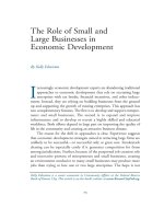
The D2-40-positive lymphatic vessels had irregular morphology and thin-walled lumens. Lymphatic vessels in gastric
tissues were mostly located in the layer of submucosa.
Intratumoral lymphatic vessels were usually collapsed,
small, and irregular in all intestinal and diffuse types (Fig. 1a
and b), but some non-collapsed lymphatics had open
lumens and occasionally contained invading tumor-cell
clusters (Fig. 2). The peritumoral lymphatic vessels were
enlarged with dilated lymphatic cavities, regardless of their
Lauren classification (Fig. 3a and b). Overall, the mean
Pak et al. BMC Cancer (2015) 15:498
Page 3 of 8
Table 1 Correlation of LVD with clinicopathological parameters and survival
Factors (N)
I-LVD
Age
Sex
Extent of resection
Tumor location
Tumor size
Lauren classification
Differentiation
Tumor stage (AJCC 7th ed.)
LVI
Node stage (AJCC 7th ed.)
th
Tumor node metastasis stage (AJCC 7 ed.)
DFS (month)
OS (month)
P-LVD
mean ± SD
P
mean ± SD
P
<60 (33)
12.60 ± 5.32
N.S.
10.52 ± 3.22
N.S.
>60 (33)
11.99 ± 3.15
Male (47)
12.31 ± 4.79
Female (19)
12.24 ± 3.11
Total gastrectomy (16)
13.78 ± 5.62
Subtotal gastrectomy (50)
11.82 ± 3.81
Upper 1/3 (12)
12.01 ± 3.30
Middle 1/3 (21)
13.49 ± 5.18
11.26 ± 3.63
Lower 1/3 (32)
12.33 ± 4.37
10.63 ± 2.92
<5cm (36)
12.20 ± 4.98
>5cm (30)
12.40 ± 3.53
Intestinal type (31)
11.20 ± 3.06
Diffuse type (35)
13.26 ± 5.09
Differentiated (31)
11.03 ± 2.80
Undifferentiated (35)
13.41 ± 5.15
11.53 ± 3.99
N.S.
11.06 ± 3.91
10.88 ± 2.83
N.S.
12.36 ± 5.30
N.S.
11.74 ± 5.40
10.55 ± 3.61
0.048*
10.10 ± 2.74
0.021*
10.49 ± 2.74
11.70 ± 3.64
T3 (26)
12.23 ± 3.95
11.05 ± 3.74
T4 (11)
15.16 ± 5.37
12.18 ± 3.37
0.024*
0.004*
9.87 ± 3.55
10.18 ± 3.71
N0 (36)
11.26 ± 3.84
3.34 ± 2.20
10.25 ± 0.55
N2 (7)
13.51 ± 3.79
12.38 ± 1.84
N3 (20)
14.16 ± 5.00
12.67 ± 4.38
I (20)
10.45 ± 3.93
12.27 ± 3.58
III (18)
14.17 ± 5.14
LVD-Low (38)
57.82 ± 3.07
LVD-High (28)
51.45 ± 4.84
LVD-Low (38)
61.70 ± 2.62
LVD-High (28)
52.60 ± 4.44
N.S.
N.S.
0.028*
12.12 ± 3.28
N1 (3)
II (30)
N.S.
11.47 ± 4.25
10.22 ± 3.53
10.96 ± 3.80
N.S.
11.55 ± 4.23
12.95 ± 4.09
13.99 ± 4.48
N.S.
11.56 ± 3.62
T1 (18)
Negative (37)
N.S.
10.59 ± 2.87
N.S.
T2 (11))
Positive (29)
N.S.
0.052
0.029*
10.00 ± 2.77
9.81 ± 3.02
0.040*
0.061
10.88 ± 3.18
12.56 ± 4.48
N.S.
59.31 ± 2.77
0.037*
51.21 ± 4.81
0.031*
60.06 ± 2.70
0.088
55.96 ± 4.14
LVI, lympho-vascular invasion; DFS, disease free survival; OS, overall survival
*, P <0.05; N.S., not significant; data are expressed as means ± SD
I-LVD was higher than the mean P-LVD (12.29 ± 4.35 vs.
11.01 ± 3.62 [±SD throughout], P = 0.025). In addition, the
mean I-LVD of the intestinal and diffuse subtypes was significantly different (11.20 ± 3.06 vs. 13.27 ± 5.09, respectively, P = 0.048). However, the P-LVD of the two subtypes
was not significantly different..
Correlations of I-LVD and P-LVD with clinicopathological
parameters and prognosis
The correlations of I-LVD and P-LVD with clinicopathological parameters are shown in Table 1. Neither I-LVD
nor P-LVD correlated with age, sex, extent of resection,
tumor location, or tumor size. In addition to the association of I-LVD with the diffuse type of cancer, it was also
significantly associated with the undifferentiated type
(P = 0.021), tumor stage (T-stage; P = 0.024), LVI
(P = 0.004), tumor node metastasis (TNM) stage
(P = 0.029), and poor overall survival (P = 0.031; refer
also to Fig. 4 a and b). Although it failed to reach statistical significance, the data showed a trend for I-LVD to
associate with node (N)-stage (P = 0.052). In comparison,
P-LVD had a significant correlation with LVI (P = 0.028),
Pak et al. BMC Cancer (2015) 15:498
Page 4 of 8
Fig. 1 Intratumoral lympho-vascular density (I-LVD). a intestinal type, b diffuse type. Arrows indicate lymphatics
N-stage (P = 0.040), poor disease-free survival (P = 0.037;
also refer to Fig. 5a), and a tendency for association with
TNM-stage (P = 0.061) and overall survival (P = 0.088,
Fig. 5b). Lauren classification, differentiation, and
T-stage were not correlated with P-LVD.
We suggest that these results are consistent with different biological roles for I-LVD and P-LVD in gastric
cancer. I-LVD was more closely correlated with depth of
invasion than it was with lymph node metastasis, while
P-LVD had a strong relationship with lymph node metastasis rather than depth of invasion. Both parameters
correlated with TNM stage and oncological long-term
survival. Thus, both intratumoral lymphatics and peritumoral lymphatics may contribute to gastric cancer progression and prognosis but in different ways.
Discussion
A study by Padera and coworkers, with an in vivo animal
model, has conclusively established that peritumoral
lymphatics are predominantly responsible for the uptake
ability of cancer cells during metastasis [16]. However,
the role of intratumoral versus peritumoral lymphatics
Fig. 2 Intratumoral lymphatics containing tumor (Lymphovascular invasion)
Pak et al. BMC Cancer (2015) 15:498
Page 5 of 8
Fig. 3 Peritumoral lympho-vascular density (P-LVD). a intestinal type, b diffuse type. Arrows indicate lymphatics
in the pathology of primary human tumors [3, 5, 8, 12–15]
has not been so convincingly demonstrated. To date, there
have been seven studies comparing the values of I-LVD and
P-LVD in lymph node metastasis. In three of them, P-LVD
was more highly correlated with lymph node metastasis
and a poor prognosis than was I-LVD [17–19], although
other studies do not confirm the correlation. Lee et al. [25]
reported that I-LVD in early gastric cancer was related to
lymph node metastasis, while P-LVD was not, in either
early or late stages of the disease. Gao et al. [26] found that
both I-LVD and P-LVD were associated with lymph node
metastasis in early gastric cancer, but only P-LVD was correlated with it in advanced stages. Raica et al. [21] reported
that both I- and P-LVD were related to lymph node metastasis and a poor prognosis, but in yet another study, there
was no correlation between LVD in either location and
lymph node metastasis or prognosis [22]. Based on our results, we suggest that P-LVD is more important in lymph
node metastasis than is I-LVD. Some association of I-LVD
with lymph node metastasis was indicated and although it
failed to reach statistical significance (P = 0.052), the data
do not exclude the possibility that I-LVD influences lymph
node metastasis.
The distribution of lymphatic channels in tumors also
appears to vary by the organ affected. Few lymphatic
channels in breast cancers have been identified in intratumoral areas, with the majority located around the
tumor periphery [27]. However, increased LVD within
the tumor and in peritumoral areas has been observed
in cutaneous melanoma [28]. In our study, I-LVD was
generally higher than P-LVD, in agreement with Lee
et al. [20]. In addition, I-LVD was positively correlated
with the depth of tumor invasion. These findings
indicate that intratumoral lymphatic channels are the
product of neovascularization by tumor cells rather than
simple entrapment of pre-existing, normal lymphatic
channels. LVD in both locations was correlated with
TNM stage and led to a poor prognosis. This may
Fig. 4 Survival curve according to the intratumoral lympho-vascular density (I-LVD). a Disease-free survival, b overall survival
Pak et al. BMC Cancer (2015) 15:498
Page 6 of 8
Fig. 5 Survival curve according to the peritumoral lympho-vascular density (P-LVD). a Disease-free survival, b overall survival
indicate that high LVD in either location contributes to
gastric cancer progression, but that they act in different
manners.
The results indicate a correlation between P-LVD and
TNM stage although they did not quite reach significance (P = 0.061). Likewise, we observed some clear
trends in our analyses of long-term survival, but the statistical analysis requires that they should be interpreted
cautiously. Nonetheless, the results are intriguing and
we believe the failure to detect clear differences is due to
the small sample size and that both I-LVD and P-LVD
are important in cancer progression and the poor prognosis of gastric cancer patients.
Angiogenesis inhibitor (Avastin®, bevacizumab) has
been used as a molecular targeting agent in the treatment of colon cancer and renal cell carcinoma [29, 30].
Although it was ineffective in the treatment of gastric
cancer in the AVAGAST trial, the results cannot be
considered conclusive because the study was analyzed
without the biomarker’s classification [31]. In addition,
the REGARD trial showed that the VEGFR-2 inhibitor
prolonged the survival of patients with advanced gastric
cancer [32]. Similar to the growing awareness of the
importance of angiogenesis, awareness of the importance
of lymphangiogenesis is emerging. In particular, in the
case of tumors in which prognosis is dependent on
lymph node status and in which lymph node metastasis
is a major biological process leading to distant tumor
propagation, lymphangiogenesis inhibition is of utmost
clinical importance. Thus, continued research on
lymphangiogenesis is critical.
We have some limitations to draw a solid conclusion
in this study. One of them is a selection bias. We
selected 66 cases arbitrarily from 365 patients for IHC
staining, although we matched TNM stages according to
Lauren’s classification to remove the effect of tumor
aggressiveness. Small samples must be one of limitations
too. However, our results could suggest some conceptual
important points. In this study, we used D2-40 for staining of lymphatics. D2-40 is a commercially available
monoclonal antibody directed against human podoplanin a transmembrane mucoprotein that is expressed in
lymphatic endothelial cells. Many studies indicate that
D2-40 is specific for lymphatic invasion and LVD in
human cancers, including gastric cancer [33–37]. However, some studies reported that D2-40 could be
expressed in other tissues such as seminoma, epithelioid,
adrenal cortical tumor, Kaposi sarcoma, adnexal tumors
of the skin [38–43]. D2-40 immunoreactivity is restricted
to lymphatic endothelium, not blood vessels. Therefore,
the conjunction with specific vascular marker, such as
CD31 or CD34, which highlights both blood vessel and
lymphatic endothelium was suggested to detect
lymphatics specifically [44]. Wang et al. performed
immune-staining of lymphatics with D2-40 and CD31 in
gastric cancer tissue and showed that D2-40 showed exclusively stained lymphatic endothelium [18]. Consistently, our
study also demonstrated that D2-40 was a good lymphatic
endothelial maker in gastric cancer tissue.
In conclusion, although we should be cautious due to
the small sample size, P-LVD might have a more important role in lymph node metastasis than I-LVD, while
I-LVD was associated with depth of tumor invasion.
Collectively, high LVD in both locations contributed to
gastric cancer progression and prognosis; thus, lymphangiogenesis inhibition should be considered an important
target of therapy for treatment of gastric cancer.
Conclusions
We conclude that P-LVD was significantly associated
with lymph node metastasis, while I-LVD was more
associated with depth of tumor invasion. However, both
LVDs contributed to gastric cancer progression and
prognosis.
Pak et al. BMC Cancer (2015) 15:498
Abbreviations
I-LVD: Intratumoral lymphatic vessel density; P-LVD: Peritumoral lymphatic
vessel density; LVD: Lymphatic vessel density; LVI: Lymphatic vascular
invasion.
Competing interests
There is no disclosure of any commercial interest that the authors may have
in the subject of study or the source of any financial or material support.
Authors’ contributions
JC was the guarantor of integrity of the entire study, designed the research.
KHP analyzed the data and drafted the manuscript. AJ and HJC participated
in the experiments of immunohistochemical staining. YC and HK counted
lympho-vascular density. All authors have read and approved in final
manuscript.
Page 7 of 8
10.
11.
12.
13.
14.
Acknowledgements
This study was supported by Grant No. HURF-2013-30 from the Hallym
University Research Fund, by a grant from the National R&D Program for
Cancer Control, Ministry of Health and Welfare, Republic of Korea (1020390)
and by the National Research Foundation of Korea (NRF) grant funded by
the Korea government (MSIP) (No. NRF-2011-0030705).
15.
16.
Author details
1
Department of Surgery, Hallym University Medical Center, Hwasung, South
Korea. 2Department of Pathology, Hallym University Medical Center,
Hwasung, South Korea. 3Department of Pathology, Yonsei University College
of Medicine, 50-1 Yonsei-ro, Seodaemun-gu, 120-752 Seoul, South Korea.
4
Depatment of Surgery, Yonsei University College of Medicine, 50-1
Yonsei-ro, Seodaemun-gu, 120-752 Seoul, South Korea. 5Department of
Biochemistry & Molecular Biology, Yonsei University College of Medicine,
50-1 Yonsei-ro, Seodaemun-gu, 120-752 Seoul, South Korea. 6Brain Korea 21
PLUS Project for Medical Science, Yonsei University College of Medicine,
Seoul, South Korea.
17.
18.
19.
20.
Received: 16 May 2015 Accepted: 19 June 2015
21.
References
1. Ferlay J, Soerjomataram I, Dikshit R, Eser S, Mathers C, Rebelo M, Parkin DM,
Forman D, Bray F. Cancer incidence and mortality worldwide: sources,
methods and major patterns in GLOBOCAN 2012. Int J Cancer. 2014.
2. Jung KW, Won YJ, Kong HJ, Oh CM, Cho H, Lee DH, et al. Cancer statistics in
Korea: Incidence, mortality, survival, and prevalence in 2012. Cancer Res
Treat. 2015;47:127-141.
3. Gombos Z, Xu X, Chu CS, Zhang PJ, Acs G. Peritumoral lymphatic vessel
density and vascular endothelial growth factor C expression in early-stage
squamous cell carcinoma of the uterine cervix. Clin Cancer Res.
2005;11(23):8364–71.
4. Liang P, Hong JW, Ubukata H, Liu HR, Watanabe Y, Katano M, et al.
Increased density and diameter of lymphatic microvessels correlate with
lymph node metastasis in early stage invasive colorectal carcinoma.
Virchows Arch. 2006;448(5):570–5.
5. Roma AA, Magi-Galluzzi C, Kral MA, Jin TT, Klein EA, Zhou M. Peritumoral
lymphatic invasion is associated with regional lymph node metastases in
prostate adenocarcinoma. Mod Pathol. 2006;19(3):392–8.
6. Kaneko I, Tanaka S, Oka S, Kawamura T, Hiyama T, Ito M, et al. Lymphatic
vessel density at the site of deepest penetration as a predictor of lymph
node metastasis in submucosal colorectal cancer. Dis Colon Rectum.
2007;50(1):13–21.
7. Longatto-Filho A, Pinheiro C, Pereira SM, Etlinger D, Moreira MA, Jube LF,
et al. Lymphatic vessel density and epithelial D2-40 immunoreactivity in
pre-invasive and invasive lesions of the uterine cervix. Gynecol Oncol.
2007;107(1):45–51.
8. El-Gohary YM, Metwally G, Saad RS, Robinson MJ, Mesko T, Poppiti RJ.
Prognostic significance of intratumoral and peritumoral lymphatic density
and blood vessel density in invasive breast carcinomas. Am J Clin Pathol.
2008;129(4):578–86.
9. Cao F, Hu YW, Li P, Liu Y, Wang K, Ma L, et al. Lymphangiogenic and
angiogenic microvessel density in chinese patients with gastric carcinoma:
22.
23.
24.
25.
26.
27.
28.
29.
30.
correlation with clinicopathologic parameters and prognosis. Asian Pac J
Cancer Prev. 2013;14(8):4549–52.
Yang LP, Fu LC, Guo H, Xie LX. Expression of vascular endothelial growth
factor C correlates with lymphatic vessel density and prognosis in human
gastroesophageal junction carcinoma. Onkologie. 2012;35(3):88–93.
Yu JW, Wu JG, Tajima Y, Li XQ, Du GY, Zheng LH, et al. Study on lymph
node metastasis correlated to lymphangiogenesis, lymphatic vessel
invasion, and lymph node micrometastasis in gastric cancer. J Surg Res.
2011;168(2):188–96.
Hall FT, Freeman JL, Asa SL, Jackson DG, Beasley NJ. Intratumoral lymphatics
and lymph node metastases in papillary thyroid carcinoma. Arch
Otolaryngol Head Neck Surg. 2003;129(7):716–9.
Maula SM, Luukkaa M, Grenman R, Jackson D, Jalkanen S, Ristamaki R.
Intratumoral lymphatics are essential for the metastatic spread and
prognosis in squamous cell carcinomas of the head and neck region.
Cancer Res. 2003;63(8):1920–6.
Inoue A, Moriya H, Katada N, Tanabe S, Kobayashi N, Watanabe M, et al.
Intratumoral lymphangiogenesis of esophageal squamous cell carcinoma
and relationship with regulatory factors and prognosis. Pathol Int.
2008;58(10):611–9.
Zhao D, Pan J, Li XQ, Wang XY, Tang C, Xuan M. Intratumoral
lymphangiogenesis in oral squamous cell carcinoma and its
clinicopathological significance. J Oral Pathol Med. 2008;37(10):616–25.
Padera TP, Kadambi A, di Tomaso E, Carreira CM, Brown EB, Boucher Y,
et al. Lymphatic metastasis in the absence of functional intratumor
lymphatics. Science. 2002;296(5574):1883–6.
Gou HF, Chen XC, Zhu J, Jiang M, Yang Y, Cao D, et al. Expressions of
COX-2 and VEGF-C in gastric cancer: correlations with lymphangiogenesis
and prognostic implications. J Exp Clin Cancer Res. 2011;30:14.
Wang XL, Fang JP, Tang RY, Chen XM. Different significance between
intratumoral and peritumoral lymphatic vessel density in gastric cancer: a
retrospective study of 123 cases. BMC Cancer. 2010;10:299.
Coskun U, Akyurek N, Dursun A, Yamac D. Peritumoral lymphatic
microvessel density associated with tumor progression and poor prognosis
in gastric carcinoma. J Surg Res. 2010;164(1):110–5.
Lee K, Park do J, Choe G, Kim HH, Kim WH, Lee HS. Increased intratumoral
lymphatic vessel density correlates with lymph node metastasis in early
gastric carcinoma. Ann Surg Oncol. 2010;17(1):73–80.
Raica M, Ribatti D, Mogoanta L, Cimpean AM, Ioanovici S. Podoplanin
expression in advanced-stage gastric carcinoma and prognostic value of
lymphatic microvessel density. Neoplasma. 2008;55(5):455–60.
Rudno-Rudzinska J, Kielan W, Grzebieniak Z, Dziegiel P, Donizy P, Mazur G,
et al. High density of peritumoral lymphatic vessels measured by D2-40/
podoplanin and LYVE-1 expression in gastric cancer patients: an excellent
prognostic indicator or a false friend? Gastric Cancer. 2013;16(4):513–20.
Gao P, Zhou GY, Zhang QH, Su ZX, Zhang TG, Xiang L, et al.
Lymphangiogenesis in gastric carcinoma correlates with prognosis. J Pathol.
2009;218(2):192–200.
Weidner N, Semple JP, Welch WR, Folkman J. Tumor angiogenesis and
metastasis–correlation in invasive breast carcinoma. N Engl J Med.
1991;324(1):1–8.
Lee SJ, Kim JG, Sohn SK, Chae YS, Moon JH, Kim SN, et al. No association of
vascular endothelial growth factor-A (VEGF-A) and VEGF-C expression with
survival in patients with gastric cancer. Cancer Res Treat. 2009;41(4):218–23.
Gao P, Zhou GY, Zhang QH, Xiang L, Zhang SL, Li C, et al.
Clinicopathological significance of peritumoral lymphatic vessel density in
gastric carcinoma. Cancer Lett. 2008;263(2):223–30.
Williams CS, Leek RD, Robson AM, Banerji S, Prevo R, Harris AL, et al.
Absence of lymphangiogenesis and intratumoural lymph vessels in human
metastatic breast cancer. J Pathol. 2003;200(2):195–206.
Straume O, Jackson DG, Akslen LA. Independent prognostic impact of
lymphatic vessel density and presence of low-grade lymphangiogenesis in
cutaneous melanoma. Clin Cancer Res. 2003;9(1):250–6.
de Gramont A, Van Cutsem E, Schmoll HJ, Tabernero J, Clarke S, Moore MJ, et al.
Bevacizumab plus oxaliplatin-based chemotherapy as adjuvant treatment for
colon cancer (AVANT): a phase 3 randomised controlled trial. Lancet Oncol.
2012;13(12):1225–33.
Escudier B, Pluzanska A, Koralewski P, Ravaud A, Bracarda S, Szczylik C, et al.
Bevacizumab plus interferon alfa-2a for treatment of metastatic renal cell
carcinoma: a randomised, double-blind phase III trial. Lancet.
2007;370(9605):2103–11.
Pak et al. BMC Cancer (2015) 15:498
Page 8 of 8
31. Ohtsu A, Shah MA, Van Cutsem E, Rha SY, Sawaki A, Park SR, et al.
Bevacizumab in combination with chemotherapy as first-line therapy in
advanced gastric cancer: a randomized, double-blind, placebo-controlled
phase III study. J Clin Oncol. 2011;29(30):3968–76.
32. Fuchs CS, Tomasek J, Yong CJ, Dumitru F, Passalacqua R, Goswami C, et al.
Ramucirumab monotherapy for previously treated advanced gastric or
gastro-oesophageal junction adenocarcinoma (REGARD): an international,
randomised, multicentre, placebo-controlled, phase 3 trial. Lancet.
2014;383(9911):31–9.
33. Evangelou E, Kyzas PA, Trikalinos TA. Comparison of the diagnostic accuracy
of lymphatic endothelium markers: Bayesian approach. Mod Pathol.
2005;18(11):1490–7.
34. Walgenbach-Bruenagel G, Tolba RH, Varnai AD, Bollmann M, Hirner A,
Walgenbach KJ. Detection of lymphatic invasion in early stage primary
colorectal cancer with the monoclonal antibody D2-40. Eur Surg Res.
2006;38(5):438–44.
35. Niakosari F, Kahn HJ, Marks A, From L. Detection of lymphatic invasion in
primary melanoma with monoclonal antibody D2-40: a new selective
immunohistochemical marker of lymphatic endothelium. Arch Dermatol.
2005;141(4):440–4.
36. Marinho VF, Metze K, Sanches FS, Rocha GF, Gobbi H. Lymph vascular
invasion in invasive mammary carcinomas identified by the endothelial
lymphatic marker D2-40 is associated with other indicators of poor
prognosis. BMC Cancer. 2008;8:64.
37. Arigami T, Natsugoe S, Uenosono Y, Arima H, Mataki Y, Ehi K, et al.
Lymphatic invasion using D2-40 monoclonal antibody and its relationship
to lymph node micrometastasis in pN0 gastric cancer. Br J Cancer.
2005;93(6):688–93.
38. Schacht V, Dadras SS, Johnson LA, Jackson DG, Hong YK, Detmar M.
Up-regulation of the lymphatic marker podoplanin, a mucin-type transmembrane
glycoprotein, in human squamous cell carcinomas and germ cell tumors. The
American journal of pathology. 2005;166(3):913–21.
39. Ordonez NG. D2-40 and podoplanin are highly specific and sensitive
immunohistochemical markers of epithelioid malignant mesothelioma.
Hum Pathol. 2005;36(4):372–80.
40. Browning L, Bailey D, Parker A. D2-40 is a sensitive and specific marker in
differentiating primary adrenal cortical tumours from both metastatic clear
cell renal cell carcinoma and phaeochromocytoma. J Clin Pathol.
2008;61(3):293–6.
41. Kahn HJ, Bailey D, Marks A. Monoclonal antibody D2-40, a new marker of
lymphatic endothelium, reacts with Kaposi’s sarcoma and a subset of
angiosarcomas. Mod Pathol. 2002;15(4):434–40.
42. Liang H, Wu H, Giorgadze TA, Sariya D, Bellucci KS, Veerappan R, et al.
Podoplanin is a highly sensitive and specific marker to distinguish primary
skin adnexal carcinomas from adenocarcinomas metastatic to skin. Am J
Surg Pathol. 2007;31(2):304–10.
43. Rabban JT, Chen YY. D2-40 expression by breast myoepithelium: potential
pitfalls in distinguishing intralymphatic carcinoma from in situ carcinoma.
Hum Pathol. 2008;39(2):175–83.
44. Kalof AN, Cooper K. D2-40 immunohistochemistry–so far! Adv Anat Pathol.
2009;16(1):62–4.
Submit your next manuscript to BioMed Central
and take full advantage of:
• Convenient online submission
• Thorough peer review
• No space constraints or color figure charges
• Immediate publication on acceptance
• Inclusion in PubMed, CAS, Scopus and Google Scholar
• Research which is freely available for redistribution
Submit your manuscript at
www.biomedcentral.com/submit