TRAIL-receptor preferences in pancreatic cancer cells revisited: Both TRAIL-R1 and TRAIL-R2 have a licence to kill
Bạn đang xem bản rút gọn của tài liệu. Xem và tải ngay bản đầy đủ của tài liệu tại đây (1.85 MB, 11 trang )
Mohr et al. BMC Cancer (2015) 15:494
DOI 10.1186/s12885-015-1508-2
RESEARCH ARTICLE
Open Access
TRAIL-receptor preferences in pancreatic
cancer cells revisited: Both TRAIL-R1 and
TRAIL-R2 have a licence to kill
Andrea Mohr1†, Rui Yu2† and Ralf M. Zwacka1*
Abstract
Background: TRAIL is a potent and specific inducer of apoptosis in tumour cells and therefore is a possible new
cancer treatment. It triggers apoptosis by binding to its cognate, death-inducing receptors, TRAIL-R1 and TRAIL-R2.
In order to increase its activity, receptor-specific ligands and agonistic antibodies have been developed and some
cancer types, including pancreatic cancer, have been reported to respond preferentially to TRAIL-R1 triggering. The
aim of the present study was to examine an array of TRAIL-receptor specific variants on a number of pancreatic
cancer cells and test the generality of the concept of TRAIL-R1 preference in these cells.
Methods: TRAIL-R1 and TRAIL-R2 specific sTRAIL variants were designed and tested on a number of pancreatic
cancer cells for their TRAIL-receptor preference. These sTRAIL variants were produced in HEK293 cells and were
secreted into the medium. After having measured and normalised the different sTRAIL variant concentrations, they
were applied to pancreatic and control cancer cells. Twenty-four hours later apoptosis was measured by DNA
hypodiploidy assays. Furthermore, the specificities of the sTRAIL variants were validated in HCT116 cells that were
silenced either for TRAIL-R1 or TRAIL-R2.
Results: Our results show that some pancreatic cancer cells use TRAIL-R1 to induce cell death, whereas other
pancreatic carcinoma cells such as AsPC-1 and BxPC-3 cells trigger apoptosis via TRAIL-R2. This observation extended
to cells that were naturally TRAIL-resistant and had to be sensitised by silencing of XIAP (Panc1 cells). The measurement
of TRAIL-receptor expression by FACS revealed no correlation between receptor preferences and the relative levels of
TRAIL-R1 and TRAIL-R2 on the cellular surface.
Conclusions: These results demonstrate that TRAIL-receptor preferences in pancreatic cancer cells are variable and that
predictions according to cancer type are difficult and that determining factors to inform the optimal TRAIL-based
treatments still have to be identified.
Keywords: TRAIL, Pancreatic cancer, DR4 specific TRAIL variant, DR5 specific TRAIL variant, Apoptosis, TRAIL receptor
Background
Pancreatic cancers are one of the most serious oncological diseases, for which novel treatment options are
urgently needed. TRAIL is a cytokine that is involved in
natural tumour surveillance mechanisms and as recombinant protein has been shown to exert specific antitumour effects by induction of apoptosis in cancer cells
[1–5]. Apoptosis is triggered after binding of TRAIL to
* Correspondence:
†
Equal contributors
1
School of Biological Sciences, University of Essex, Wivenhoe Park, Colchester
CO4 3SQ, United Kingdom
Full list of author information is available at the end of the article
one of its two receptors, TRAIL-receptor 1 (TRAIL-R1)
or TRAIL-receptor 2 (TRAIL-R2), also known as DR4
and DR5, respectively [6–8]. Binding of TRAIL to these
two receptors stimulates the formation of a protein complex called the death-inducing signaling complex (DISC).
It consists of TRAIL-R1 and/or TRAIL-R2, the adaptor
protein Fas-associated death domain (FADD) and
procaspase-8. At the DISC, caspase-8 is activated by a
mechanism that involves dimerisation and proteolytic
cleavage [9, 10]. Active caspase-8 can then, either directly, or indirectly via the BH3-only protein Bid, activate
effector caspases, such as caspase-3, which in turn cleave
© 2015 Mohr et al. This is an Open Access article distributed under the terms of the Creative Commons Attribution
License ( which permits unrestricted use, distribution, and reproduction in
any medium, provided the original work is properly credited. The Creative Commons Public Domain Dedication waiver
( applies to the data made available in this article, unless otherwise stated.
Mohr et al. BMC Cancer (2015) 15:494
Page 2 of 11
many cellular substrates resulting in the biochemical
and morphological features characteristic of apoptosis
[11]. Aside from the two death domain (DD)-containing,
apoptosis-inducing receptors, TRAIL-R1 and TRAIL-R2,
three additional decoy receptors exist, TRAIL-R3
(DcR1), TRAIL-R4 (DcR2) and Osteoprogerin (OPG)
[6, 7, 12–14]. These decoy receptors can inhibit the
apoptosis-inducing function of TRAIL [15]. To address
this issue, agonistic antibodies against either TRAIL-R1 or
TRAIL-R2 have been developed and have been tested in
pre-clinical and as well as clinical studies [16–21].
In addition, engineered variants of TRAIL, containing
specific amino acid changes leading to specific targeting
of TRAIL-R1 or TRAIL-R2 have been designed and have
shown improved anti-tumour effects in-vitro and in-vivo
when compared to wild-type TRAIL [22–27]. Such
TRAIL-receptor variants have been studied in the context of various specific cancer types as well as in the
context of combination treatments [28–32]. TRAIL variants might hold important advantages over TRAILreceptor specific antibodies as they are smaller than
antibodies and might therefore be better able to reach
and infiltrate growing tumours. In addition, such proteins can be further optimised to increase activity, specificity and stability and they can be used as part of gene
and cell therapeutic approaches [31, 33–38]. This way of
potentially improving the therapeutic efficacy of TRAIL
by using TRAIL-receptor specific agents is of particular
interest for pancreatic cancer, as previous studies have
shown that pancreatic tumour cells preferentially use
TRAIL-R1 to execute TRAIL-induced apoptosis [39, 40].
Thus, agonistic TRAIL-R1 specific antibodies or TRAILR1 targeting variants of TRAIL were regarded as having
a higher therapeutic potential than normal TRAIL in the
treatment of pancreatic carcinoma.
We wondered, given the molecular heterogeneity of
tumours, how such a uniform TRAIL response with respect to receptor preferences could be possible. Therefore, we set out to examine an array of pancreatic cancer
cells for their TRAIL-receptor preferences. We found
that a number of pancreatic cancer cells used TRAIL-R2
rather than TRAIL-R1 to initiate apoptosis signalling.
These results demonstrate that, while TRAIL-receptor
specific variants constitute a potentially substantial improvement to conventional TRAIL therapies, generalised
predictions according to cancer type are difficult. Therefore, additional research is needed to identify factors that
determine the optimal TRAIL variant (or antibody) on a
case-by-case basis for each individual tumour.
cell lines Panc1 and PancTu1, the human embryonic
kidney cell line HEK293, the human colon cancer cell
line Colo205 and the human cervix carcinoma cell line
HeLa were maintained in Dulbecco’s modified Eagle’s
medium (DMEM). The human pancreatic cancer cell
lines AsPC-1, BxPC-3 and Colo357 were cultured in
RPMI-1640 medium. The human colorectal cancer cell
line HCT116 was cultured in McCoy’s medium and the
human prostate cancer PC-3 cells were grown in Ham’s
F12 medium. All media were supplemented with 10 %
FBS, 100 U/ml penicillin and 100 μg/ml streptomycin.
Cells were cultured in a humidified incubator at 37 °C
and 5 % CO2.
Methods
Transfection of HEK293 cells
Reagents and cell culture
HEK293 cells were transfected using the Calciumphosphate method. Briefly, before transfection, fresh 2 %
FBS containing medium was added to the cells. For each
All reagents were purchased from Sigma (St. Louis, MO)
unless otherwise stated. The human pancreatic cancer
Generation of sTRAIL constructs
Generation of sTRAIL constructs and site-directed mutagenesis have been previously described [31]. Briefly, the
soluble portion of human TRAIL (amino acids 114–281)
was first subcloned into the NheI/NotI sites of a pcDNA3
plasmid (Invitrogen) giving rise to pcDNA3.sTRAIL. Then
an exogenous signal peptide sequence of the human
fibrillin protein, the Furin cleavage site (Furin CS) and
Isoleucine-zipper sequence (ILZ) cassette was cloned
into the BamHI/NheI sites of the pcDNA3.sTRAIL vector. The resulting vector was termed sTRAILwt. The two
sTRAILDR5 and three sTRAILDR4 constructs were generated using the Quick-Change site-directed mutagenesis kit (Stratagene, La Jolla, CA) and confirmed by
DNA sequencing.
TRAIL Enzyme-linked Immunosorbent Assay (ELISA)
TRAIL concentrations were measured by a human
TRAIL/TNFSF10 Quantikine ELISA Kit as recommended
by the manufacturer (R&D Systems, Minneapolis, MN).
Before the measurement the medium supernatants were
pipetted off the respective HEK293 producer cells and
then centrifuged to clear them of any cellular debris.
TRAIL receptor surface stain
For the TRAIL receptor stain we used monoclonal antiTRAIL-R1 (DJR1) and anti-TRAIL-R2 (DJR2-4) antibodies (1 μg/106 cells; BioLegend, San Diego, CA) that
were conjugated to Phycoerythrin (PE). The isotype control antibody (MOPC-21) (1 μg/106 cells) was also purchased from BioLegend. The surface expression of
TRAIL receptors was measured by incubating cells with
the PE-conjugated mouse anti-human TRAIL-R1 and
mouse anti-human TRAIL-R2 antibodies as described
previously [41].
Mohr et al. BMC Cancer (2015) 15:494
well of a 6-well plate, 0.5 ml HBS were aliquoted into a
sterile 1.5 ml Eppendorf tube. In a separate tube 5 μg of
plasmid DNA were mixed with 250 μl CaCl2 (2.5 mM)
and sterile water added to 0.5 ml. The CaCl2/DNA mix
was then added to the HBS in a drop-wise fashion and
constant vortexing at slow speed. After 45 min of incubation at room temperature, the mixture was slowly
added to the cells. After 4 h, the medium was removed
and the cells were washed with PBS and fresh growth
medium added.
Apoptosis assay
Apoptosis was measured according to Nicoletti et al.
(DNA hypoploidy assay) and has been described
previously [42, 43]. Trypsinised cells including the supernatant medium and PBS wash-solution were directly
transferred into FACS tubes and centrifuged at 1,300 rpm
for 7 min at 4 °C. After washing the cell pellet with PBS,
Nicoletti buffer (Sodium citrate 0.1 % (w/v) supplemented
with 0.1 % Triton X-100 (w/v) and propidium iodide at
50 μg/ml) was added. Then the tubes were vortexed for
10 s at medium speed and left for 5 h in a refrigerator.
The fluorescence intensity was then measured by flow cytometry and analysed using the Venturi One software
package (Applied Cytometry, Sheffield, UK). Where specified, untreated cells were taken as reference to calculate
specific apoptosis by subtraction of the basal cell death
values from the apoptosis levels of treated cells.
RNAi knock-down constructs and stable cell line
generation
The following small hairpin (sh) RNA motifs were used
to silence: DR5 (5′-GCTAGAAGGTAATGCAGACTCT
GCCATGTC -3’), DR4 (5′-GCTGTTCTTTGACAAGT
TGC-3’) and XIAP (5′-GTGGTAGTCCTGTTTCAGC-3’).
Sense and antisense oligos containing the sh-sequence and
a 5’ overhang representing a restricted BbsI site and EcoRI
site on the 3’ side were hybridised to generate doublestranded DNA fragments. These fragments were then
cloned into a BbsI/EcoRI opened up pU6.ENTR plasmid (Life Technologies, Carlsbad, CA). The resulting
pU6.ENTR plasmids (pU6.ENTR.shDR5, pU6.ENTR.shDR4,
pU6.ENTR.shXIAP) were used to generate the pBlockiT.shDR5, pBlock-iT.shDR4 and pBlock-iT.shXIAP
plasmids using the pBLOCK-iT6-DEST vector (Life
Technologies) and LR Clonase II. This was used to generate
stable DR5 and DR4 knock-down clones of HCT116 cells
and stable XIAP knock-down clones of PancTu1 and Panc1
cells. For this, the pBlock-iT.shDR5, pBlock-iT.shDR4 and
pBlock-iT.shXIAP plasmids were FuGeneHD-transfected
(Roche, Basle, Switzerland) into HCT116, PancTu1 and
Panc1 cells, respectively. Three days later, the transfected cells were split into Blasticidin containing selection medium. Clones were then picked, transferred to
Page 3 of 11
24 well-plates and analysed for DR5, DR4 and XIAP
knock-down, respectively. Clones that did not show a
knock-down were used as controls and labelled
PancTu1.shctrl and Panc1.shctrl, respectively. These
control clones were tested and shown to behave like
parental cells.
Statistical analysis
Experimental values are expressed as mean value ± standard error (SEM). For significance analyses, analysis of variance (ANOVA) between groups was used and P < 0.05 (*)
was considered significant.
Results
Expression and specificity of DR4- and DR5-specific TRAIL
variants
We used soluble TRAIL (sTRAIL) expression constructs
that we described previously [31, 36] to address the
TRAIL-receptor preference in pancreatic cancer. These
constructs contain an exogenous signal peptide sequence
from the human fibrillin-1 gene, a cleavage site for the
ubiquitous protease Furin, an Isoleucine Zipper domain
and the ectodomain of TRAIL (aa114-aa281). In addition
to the wild-type TRAIL construct (sTRAILwt), we engineered constructs expressing three different DR4-specific
sTRAIL variants (termed sTRAILDR4–1, sTRAILDR4–2 and
sTRAILDR4–3) and two specific for DR5 (labelled
sTRAILDR5–1 and sTRAILDR5–2). These sTRAILDR4 and
sTRAILDR5 variants contained various amino acid changes
(Fig. 1a) [26, 44]. Following transfection of HEK293 cells
we could demonstrate that all TRAIL variants were
expressed and secreted to comparable levels (Fig. 1b).
TRAIL receptor specificity was confirmed in HCT116
cells silenced for TRAIL-R1 and TRAIL-R2, respectively.
We chose HCT116 cells, because of their relatively balanced TRAIL-receptor preference and expression levels
(Fig. 1c). Cells with knocked-down TRAIL-R1 showed decreased apoptosis with all three sTRAILDR4 variants, but
elevated levels with sTRAILDR5 as compared to sTRAILwt
(Fig. 1d). In contrast, cells with silenced TRAIL-R2 exhibited markedly reduced apoptosis in response to sTRAILDR5,
whereas the levels of cell death were increased with
the sTRAILDR4 variants, in particular with sTRAILDR4–3
(Fig. 1d). The likely reason for this observation is that in
TRAIL-R1 and TRAIL-R2 silenced cells the chance of
homotrimer formation is increased with sTRAIL variants
when compared to sTRAILwt and parental cells resulting
in higher apoptosis levels [24, 44].
Induction of apoptosis by TRAIL variants in pancreatic
cancer cells
Next, we tested several pancreatic cancer cells with the
array of TRAIL variants. In parallel, we analysed cancer
cells for which TRAIL-receptor preferences have been
Mohr et al. BMC Cancer (2015) 15:494
Page 4 of 11
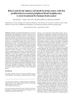
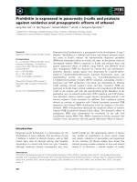
Fig. 1 Design, expression and specificity of sTRAIL specific variants. a Schematic drawing of sTRAIL constructs, all of which contain a heterologous
signal peptide sequence from the human fibrillin-1 gene (hFIB) ligated to a Furin cleavage site (Furin CS), an Isoleucine Zipper (ILZ) domain and
the soluble part of TRAIL (aa114-aa281). The expression was driven by a conventional CMV promoter/enhancer element (CMV). The mutations in
sTRAILwt leading to the two sTRAILDR5 (TRAIL-R2 specific) and three sTRAILDR4 (TRAIL-R1 specific) variants are shown in the respective sTRAIL
segments. b Results of ELISA analyses for TRAIL showing the levels of secreted sTRAILwt (yellow), sTRAILDR5–1 (dark green), sTRAILDR5–2 (light
green), sTRAILDR4–1 (dark blue), sTRAILDR4–2 (light blue) and sTRAILDR4–3 (blue-grey) into the supernatant of HEK293 cells that were transfected with
the described constructs. Results for cells transfected with an EGFP control expression construct (ctrl; grey) are also shown. c FACS histogram of
HCT116 cells showing membrane expression levels of TRAIL-R1 (red) and TRAIL-R2 (blue). The FACS profile of the isotype control is shown as filled
black. d Supernatants from HEK293 cells transfected with either an EGFP control expression plasmid (grey), sTRAILwt (yellow), sTRAILDR5–1 (dark
green), sTRAILDR5–2 (light green), sTRAILDR4–1 (dark blue), sTRAILDR4–2 (light blue) or sTRAILDR4–3 (blue-grey) were normalised to 2 ng/ml TRAIL (the
EGFP control was diluted 1:2 in fresh medium) and then applied to HCT116 (left), HCT.shDR4 (centre) and HCT.shDR5 cells (right), respectively,
before apoptosis was measured 24 h later
clearly documented, namely HeLa cells (TRAIL-R1) and
Colo205 cells (TRAIL-R2). Next, we applied the sTRAIL
variants to the pancreatic cancer cells Colo357, BxPC-3
and AsPC-1. After 24 h exposure to the sTRAIL variants
we measured apoptosis and found that HeLa (Fig. 2a)
and Colo205 (Fig. 2b) cells showed higher cell death
levels with sTRAILDR4 and sTRAILDR5, respectively.
However, while Colo357 pancreatic cancer cells exhibited elevated cell death rates with sTRAILDR4 (Fig. 2c) as
reported previously [45], BxPC-3 (Fig. 2d) and AsPC-1
(Fig. 2e) cells responded with higher apoptosis levels to
sTRAILDR5.
TRAIL-receptor expression profile is not associated with
receptor preferences
Next, we analysed whether the observed preferences for
either TRAIL-R1 (HeLa, Colo357) or TRAIL-R2 (Colo205,
BxPC-3, AsPC-1) could be linked to the surface expression
levels of the two receptors. Using PE-conjugated antibodies
against TRAIL-R1 and TRAIL-R2 and the appropriate isotype control, we found that HeLa cells harboured robust
levels of TRAIL-R1 on their cell surface (MFI ratio:
4.12 +/− 0.05), whereas on Colo357 cells, we could
detect only comparably low levels of TRAIL-R1 (MFI
ratio: 2.51 +/− 0.43) (Fig. 3a). TRAIL-R2 levels in both
HeLa and Colo357 cells are slightly higher than
TRAIL-R1 (TRAIL-R2 MFI ratios: HeLa: 6.24 +/− 1.49
and Colo357: 3.42 +/− 0.55) (Fig. 3a). In the group of
cells that preferentially responded to sTRAILDR5 all
cells showed higher levels of TRAIL-R2 (MFI ratios:
Colo205: 7.33 +/− 0.14, AsPC-1: 10.42 +/− 2.43, BxPC-3:
4.54 +/− 0.75) than TRAIL-R1 (MFI ratios: Colo205: 2.90
+/− 0.04, AsPC-1: 4.02 +/− 0.96, BxPC-3: 2.31 +/− 0.55)
(Fig. 3b), with all of them expressing levels that are not
distinguishable from the group of cells reacting better to
sTRAILDR4. Thus, there is no straightforward correlation
between the levels of TRAIL-R1 and TRAIL-R2 and
TRAIL-receptor preference in TRAIL-induced apoptosis.
Mohr et al. BMC Cancer (2015) 15:494
Page 5 of 11
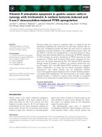
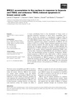
Fig. 2 The TRAIL receptor preference for apoptosis induction is variable in pancreatic cancer cells. a-e Supernatants from HEK293 cells that were
transfected with the EGFP control expression construct (ctrl; grey; 1:2 diluted), sTRAILwt (yellow; 2 ng/ml), sTRAILDR5–1 (dark green; 2 ng/ml),
sTRAILDR5–2 (light green; 2 ng/ml), sTRAILDR4–1 (dark blue; 2 ng/ml), sTRAILDR4–2 (light blue; 2 ng/ml) or sTRAILDR4–3 (blue-grey; 2 ng/ml) were then
transferred onto (a) HeLa cells (prototypic DR4 specific cell type), (b) Colo205 cells (prototypic DR5 specific cell type), (c) Colo357 pancreatic
cancer cells, (d) BxPC-3 pancreatic cancer cells and (e) AsPC-1 pancreatic cancer cells. After 24 h apoptosis was measured
Induction of apoptosis in sensitised TRAIL resistant
pancreatic cancer cells
It is well known that some pancreatic cancer cells are resistant to TRAIL (Fig. 4a). Therefore, in order to examine
the TRAIL receptor preference in such cells, we silenced
the anti-apoptotic protein XIAP in PancTu1 (PancTu1.shXIAP) and Panc1 (Panc1.shXIAP) cells and treated them
with sTRAIL variants. The results show that knocking
down of XIAP sensitised the cells to TRAIL-induced
apoptosis, with sTRAILDR4 having a significantly better effect in PancTu1.shXIAP (Fig. 4b), as previously described,
but sTRAILDR5 leading to more apoptosis in Panc1.shXIAP (Fig. 4c). Thus not all pancreatic cancer cells possess
a preference for the TRAIL-R1 apoptosis pathway as reported previously [39, 40]. Instead, a group of pancreatic
cancer cells have a higher propensity to undergo TRAILinduced apoptosis via TRAIL-R2.
TRAIL-receptor expression profile is not associated with
receptor preferences in XIAP-silenced pancreatic cancer cells
Next, we also measured TRAIL-R1 and TRAIL-R2 expression on the surface of both Panc1.shctrl and
PancTu1.shctrl cells as well as their XIAP-silenced counterparts, Panc1.shXIAP and PancTu1.shXIAP cells. We
found that the profiles of TRAIL-receptor expression
did not differ between the control cells (Panc1.shctrl and
PancTu1.shctrl) and the corresponding XIAP knockdown clones (Panc1.shXIAP and PancTu1.shXIAP)
(Fig. 5a and b). TRAIL-R1 expression was almost undetectable in Panc1.shctrl and Panc1.shXIAP (MFI ratios: Panc1.shctrl: 1.81 +/− 0.44 and Panc1.shXIAP:
1.74 +/− 0.30), whereas TRAIL-R2 expression was
readily detectable (MFI ratios: Panc1.shctrl: 3.33 +/−
0.57 and Panc1.shXIAP: 3.2 +/− 0.60). In PancTu1.shctrl and PancTu1.shXIAP both TRAIL-R1 and
TRAIL-R2 were expressed at robust levels (TRAIL-R1
MFI ratios: PancTu1.shctrl: 3.15 +/− 0.17 and PancTu1.shXIAP: 2.52 +/− 0.10; TRAIL-R2 MFI ratios:
PancTu1.shctrl: 5.67 +/− 0.13 and PancTu1.shXIAP:
5.90 +/− 0.08). This comparison of TRAIL-receptor
levels in TRAIL resistant pancreatic cells also does not
show a clear correlation between TRAIL-receptor expression levels and TRAIL-receptor preference after
XIAP sensitisation.
Mohr et al. BMC Cancer (2015) 15:494
Page 6 of 11
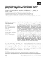
Fig. 3 TRAIL-receptor surface expression profiles of pancreatic cancer cells and control cell lines. a FACS histograms of HeLa and Colo357 cells
showing membrane expression levels of TRAIL-R1 (black) and TRAIL-R2 (red). The FACS profile of the isotype control is shown as filled grey.
b FACS histograms of Colo205, AsPC-1 and BxPC-3 cells showing membrane expression levels of TRAIL-R1 (black) and TRAIL-R2 (red). The FACS
profile of the isotype control is shown as filled grey. c Quantification of the FACS results for TRAIL-R1 (black) and TRAIL-R2 (red) for HeLa, Colo357,
Colo205, AsPC-1 and BxPC-3 cells. The surface expression levels of the two receptors are expressed as MFI ratios
Discussion
Initially it was thought that TRAIL-R2 is the main
apoptosis-inducing receptor for the death ligand TRAIL
[27]. This led to the development and testing of
agonistic antibodies against this receptor as potential
anti-cancer agents [16, 18, 46, 47]. However, more recently reports showed that TRAIL-R1 has a more prominent role, than first thought, in specific types of cancer
Mohr et al. BMC Cancer (2015) 15:494
Page 7 of 11
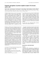
Fig. 4 TRAIL receptor preference is also variable in apoptosis-sensitised pancreatic cancer cells. a TRAIL-sensitive control cells (PC-3), Panc1, Panc1.shctrl,
PancTu1 and PancTu1.shctrl were treated with 10 ng/ml rTRAIL for 24 h, before apoptosis was measured. b PancTu1.shXIAP cells were treated with
supernatants from HEK293 cells that were transfected with the EGFP control expression construct (ctrl; grey), sTRAILwt (yellow), sTRAILDR5–2 (light green)
or sTRAILDR4–3 (blue-grey). After 24 h apoptosis was measured. c Panc1.shXIAP cells were treated with supernatants from HEK293 cells that were
transfected with the EGFP control expression construct (ctrl; grey), sTRAILwt (yellow), sTRAILDR5–2 (light green) or sTRAILDR4–3 (blue-grey). After 24 h
apoptosis was measured
such as lymphoid malignancies [29] and leukaemic cells
[30, 48]. Additionally, it was suggested that pancreatic
cancer cells also trigger TRAIL-induced apoptosis
mainly through TRAIL-R1 [39, 40]. However, when we
analysed a wider array of pancreatic cancer cell lines we
found that 2 out of 3 pancreatic cancer cells preferred
the TRAIL-R2 pathway in response to TRAIL. In
addition, Panc1 cells also showed higher apoptosis levels
when treated with sTRAILDR5 and XIAP was silenced
concomitantly (Table 1).
While these results appear to contrast the two afore
mentioned publications [39, 40], it is important to point
out that we used, at least in part, different cell lines and
sTRAIL variant proteins instead of agonistic antibodies.
Interestingly, the results in one of the reports indicate
that both TRAIL-R1 and TRAIL-R2 agonistic antibodies
can trigger apoptosis in pancreatic cells and that the
TRAIL-R1 preference was only detected when one of
the two receptors was inhibited by blocking antibodies
followed by treatment with TRAIL [39]. In contrast, the
second study found clear differences between the
apoptosis-inducing activities of the two agonistic antibodies, with a clear preference for TRAIL-R1. It is therefore possible that sTRAIL variant proteins and TRAILreceptor specific antibodies have distinct effects owing
to their different modes of action with regard to their
Mohr et al. BMC Cancer (2015) 15:494
Page 8 of 11
Fig. 5 TRAIL-receptor surface expression profiles of TRAIL resistant pancreatic cancer cells and their XIAP silenced counterparts. a FACS
histograms of Panc1.shctrl and Panc1.shXIAP cells showing membrane expression levels of TRAIL-R1 (black) and TRAIL-R2 (red). The FACS profile
of the isotype control is shown as filled grey. b FACS histograms of PancTu1.shctrl and PancTu1.shXIAP cells showing membrane expression levels
of TRAIL-R1 (black) and TRAIL-R2 (red). The FACS profile of the isotype control is shown as filled grey. c Quantification of the FACS results for
TRAIL-R1 (black) and TRAIL-R2 (red) for Panc1.shctrl, Panc1.shXIAP, PancTu1.shctrl and PancTu1.shXIAP cells. The surface expression levels of the
two receptors are expressed as MFI ratios
Mohr et al. BMC Cancer (2015) 15:494
Page 9 of 11
Table 1 TRAIL-R preference of different cancer cell types
Cell line
Cancer cell type
TRAIL-receptor preference
HCT116
colorectal carcinoma
DR5
Colo205
colorectal carcinoma
DR5
HeLa
cervical carcinoma
DR4
Colo357
pancreatic carcinoma
DR4
BxPC-3
pancreatic carcinoma
DR5
AsPC-1
pancreatic carcinoma
DR5
Panc1
pancreatic carcinoma
DR5
PancTu1
pancreatic carcinoma
DR4
receptor engagement. Notwithstanding, the notion that
pancreatic cancer cells and possibly other tumour types
have a general TRAIL receptor preference needs to be
re-visited, re-examined and possibly refined. Furthermore, we tested whether the expression profile of
TRAIL-R1 and TRAIL-R2 could determine receptor
preference, but failed to observe any clear correlation.
These findings are generally in line with results reported
earlier [39]. Thus, other factors and mechanisms than
surface expression levels of the TRAIL-receptors must
determine their apoptosis-inducing function.
Potential molecular mechanisms that could determine
whether a receptor can be activated are O-glycosylation
of both receptors [49] as well as S-palmitoylation, Snitrosylation, N-glycosylation and ubiquitination of
TRAIL-R1 [50–53]. Thus, despite being present on the
cell surface a receptor might be relatively inactive,
making it impossible to determine receptor preferences based solely on expression levels.
An area where specific TRAIL variants and/or agonistic antibodies can be used with good predictability is in
combination treatments, in which up-regulation of
either TRAIL-R1 or TRAIL-R2 can be targeted by the
respective variant. For example, pre-treatment with the
anti-cancer drug doxorubicin gave rise to significantly
increased cell death when treated with the agonistic
TRAIL-R2 antibody lexatumumab [54]. In addition,
combined treatment of colorectal tumours with lexatumumab and radiotherapy had similar sensitising effects
[55]. Soluble TRAILDR5 also showed better apoptosis
inducing effects after priming with 5-Fluorouracil as
compared to sTRAILwt or sTRAILDR4, because the drug
caused p53-independent upregulation of TRAIL-R2 [31].
In contrast, HDAC inhibition has been shown to result
in sensitisation to TRAIL-R1 specific apoptosis [48, 56].
Of note in this context is that the individual activation of TRAIL-R1 and -R2 could be an advantage,
since it was shown that combined exposure to
DR4- and DR5-selective TRAIL variants in cells,
sensitive for both receptors, was more potent in
triggering apoptosis when compared to single agent
treatment [22]. Other factors that can influence TRAIL receptor preferences are so called non-canonical pathways
including the activation of NF-κB, p38 and JNK [57]. The
issue with these pathways is that they have been reported
to have opposing effects and different apoptosis factor requirements depending on cell type and cellular context
[57]. For example, TRAIL-induced JNK activation has
been reported to be caspase-dependent in HeLa human
cervical cancer cells, but caspase-independent in the human rhabdomyosarcoma Kym-1 cell line [58]. These findings illustrate that the TRAIL receptors have varying, cell
type-specific and in parts receptor specific capabilities to
recruit different signalling complexes to their intracellular
domain. These complexes and their individual constituents might have an impact on the apoptosis-inducing
function of the receptors and thereby may contribute to
TRAIL-receptor preferences in TRAIL-triggered cell
death.
Consequently, further research is needed to better
understand potential differences between TRAIL agonistic antibodies and recombinant TRAIL proteins and
variants. Additionally, it is important to elucidate the
molecular components that determine TRAIL-receptor
preferences in order to be able to select the best TRAIL
agents to potentially treat pancreatic cancer and other
tumour types in the future.
Conclusions
We discovered that not all pancreatic cancer cells favour
the TRAIL-R1 pathway to induce apoptosis and that
no clear and direct correlation exists between the surface expression levels of TRAIL-R1 and TRAIL-R2 and
their preference for one of the two receptors. AsPC-1,
BxPC-3 and Panc1 cells elicit apoptosis via TRAIL-R2,
whereas Colo357 cells and PancTu1 cells preferred
TRAIL-R1 to induce cell death. Thus, claims of general
cancer type specific TRAIL receptor preference should
be taken with a pinch of salt.
Abbreviations
ANOVA: Analysis of variance between groups; CMV: Cytomegalie virus;
DISC: Death-inducing signaling complex; DMEM: Dulbecco’s modified Eagle’s
medium; EGFP: Enhanced green fluorescent protein; ELISA: Enzyme-linked
Immunosorbent Assay; FACS: Fluorescence-activated Cell Sorting; FADD:
Fas-associated death domain; FBS: Fetal bovine serum; FIB: Fibrillin; Furin
CS: Furin cleavage site; HBS: Hepes-buffered saline; ILZ: Isoleucine zipper;
JNK: c-Jun N-terminal kinase; NF-κB: Nuclear factor kappa-light-chain-enhancer
of activated B cells; OPG: Osteoprogerin; PBS: Phosphate-buffered saline; PE:
R-Phycoerythrin; RNAi: RNA interference; RPMI 1640 medium: Roswell Park
Memorial Institute 1640 medium; SEM: Standard error of the mean; TRAIL:
TNF-related apoptosis-inducing ligand; sTRAIL: soluble TNF-related apoptosisinducing ligand; TRAIL-R1/DR4: TRAIL-receptor 1/Death-receptor 4; TRAIL-R2/
DR5: TRAIL-receptor 2/ Death-receptor 5; TRAIL-R3/DcR1: TRAIL-receptor 3/
Decoy-receptor 1; TRAIL-R4/DcR2: TRAIL-receptor 4/Decoy-receptor 2; XIAP:
X-linked Inhibitor of apoptosis protein.
Competing interests
The authors declare that they have no competing interests.
Mohr et al. BMC Cancer (2015) 15:494
Authors’ contributions
AM and RY designed the study; performed experiments; analysed and
interpreted data; wrote the manuscript. RMZ conceived and designed this
study; analysed and interpreted data; wrote the manuscript. All authors read
and approved the final manuscript.
Acknowledgements
The work was supported by an EU-FP6-STREP (TRIDENT) award. The work
was also supported by an EU-FP6 Marie-Curie Excellence Team Award (MIST)
and by an EU-RTN Award (ApopTrain) (to R. M. Z).
Author details
1
School of Biological Sciences, University of Essex, Wivenhoe Park, Colchester
CO4 3SQ, United Kingdom. 2School of Medicine, Ningbo University, Ningbo,
Zhejiang 315211, P.R. China.
Received: 17 April 2015 Accepted: 19 June 2015
References
1. Duiker EW, Mom CH, de Jong S, Willemse PH, Gietema JA, van der Zee AG,
et al. The clinical trail of TRAIL. Eur J Cancer. 2006;42(14):2233–40.
2. Lemke J, von Karstedt S, Zinngrebe J, Walczak H. Getting TRAIL back on
track for cancer therapy. Cell Death Differ. 2014;21(9):1350–64.
3. Wiley SR, Schooley K, Smolak PJ, Din WS, Huang CP, Nicholl JK, et al.
Identification and characterization of a new member of the TNF family that
induces apoptosis. Immunity. 1995;3(6):673–82.
4. Wu GS. TRAIL as a target in anti-cancer therapy. Cancer Lett.
2009;285(1):1–5.
5. Micheau O, Shirley S, Dufour F. Death receptors as targets in cancer. Br J
Pharmacol. 2013;169(8):1723–44.
6. Chaudhary PM, Eby M, Jasmin A, Bookwalter A, Murray J, Hood L. Death
receptor 5, a new member of the TNFR family, and DR4 induce
FADD- dependent apoptosis and activate the NF-kappaB pathway.
Immunity. 1997;7(6):821–30.
7. Schneider P, Thome M, Burns K, Bodmer JL, Hofmann K, Kataoka T, et al.
TRAIL receptors 1 (DR4) and 2 (DR5) signal FADD-dependent apoptosis and
activate NF-kappaB. Immunity. 1997;7(6):831–6.
8. Mahalingam D, Szegezdi E, Keane M, de Jong S, Samali A. TRAIL receptor
signalling and modulation: Are we on the right TRAIL? Cancer Treat Rev.
2009;35(3):280–8.
9. Sprick MR, Weigand MA, Rieser E, Rauch CT, Juo P, Blenis J, et al. FADD/
MORT1 and caspase-8 are recruited to TRAIL receptors 1 and 2 and are
essential for apoptosis mediated by TRAIL receptor 2. Immunity.
2000;12(6):599–609.
10. Hellwig CT, Rehm M. TRAIL signaling and synergy mechanisms used in
TRAIL-based combination therapies. Mol Cancer Ther. 2012;11(1):3–13.
11. Bratton SB, MacFarlane M, Cain K, Cohen GM. Protein complexes activate
distinct caspase cascades in death receptor and stress-induced apoptosis.
Exp Cell Res. 2000;256(1):27–33.
12. Degli-Esposti MA, Dougall WC, Smolak PJ, Waugh JY, Smith CA, Goodwin
RG. The novel receptor TRAIL-R4 induces NF-kappaB and protects against
TRAIL-mediated apoptosis, yet retains an incomplete death domain.
Immunity. 1997;7(6):813–20.
13. Degli-Esposti MA, Smolak PJ, Walczak H, Waugh J, Huang CP, DuBose RF,
et al. Cloning and Characterization of TRAIL-R3, a Novel Member of the
Emerging TRAIL Receptor Family. J Exp Med. 1997;186(7):1165–70.
14. Emery JG, McDonnell P, Burke MB, Deen KC, Lyn S, Silverman C, et al.
Osteoprotegerin is a receptor for the cytotoxic ligand TRAIL. J Biol Chem.
1998;273(23):14363–7.
15. LeBlanc HN, Ashkenazi A. Apo2L/TRAIL and its death and decoy receptors.
Cell Death Differ. 2003;10(1):66–75.
16. Camidge DR, Herbst RS, Gordon MS, Eckhardt SG, Kurzrock R, Durbin B, et al.
A phase I safety and pharmacokinetic study of the death receptor 5
agonistic antibody PRO95780 in patients with advanced malignancies. Clin
Cancer Res. 2010;16(4):1256–63.
17. Chuntharapai A, Dodge K, Grimmer K, Schroeder K, Marsters SA, Koeppen H,
et al. Isotype-dependent inhibition of tumor growth in vivo by monoclonal
antibodies to death receptor 4. J Immunol. 2001;166(8):4891–8.
Page 10 of 11
18. Ichikawa K, Liu W, Zhao L, Wang Z, Liu D, Ohtsuka T, et al. Tumoricidal
activity of a novel anti-human DR5 monoclonal antibody without
hepatocyte cytotoxicity. Nat Med. 2001;7(8):954–60.
19. Trarbach T, Moehler M, Heinemann V, Kohne CH, Przyborek M, Schulz C,
et al. Phase II trial of mapatumumab, a fully human agonistic monoclonal
antibody that targets and activates the tumour necrosis factor
apoptosis-inducing ligand receptor-1 (TRAIL-R1), in patients with
refractory colorectal cancer. Br J Cancer. 2010;102(3):506–12.
20. van Geelen CM, Pennarun B, Le PT, de Vries EG, de Jong S. Modulation of
TRAIL resistance in colon carcinoma cells: different contributions of DR4 and
DR5. BMC Cancer. 2011;11:39.
21. den Hollander MW, Gietema JA, de Jong S, Walenkamp AM, Reyners AK,
Oldenhuis CN, et al. Translating TRAIL-receptor targeting agents to the
clinic. Cancer Lett. 2013;332(2):194–201.
22. Reis CR, van der Sloot AM, Natoni A, Szegezdi E, Setroikromo R, Meijer M,
et al. Rapid and efficient cancer cell killing mediated by high-affinity death
receptor homotrimerizing TRAIL variants. Cell Death Dis. 2010;1, e83.
23. Reis CR, van der Sloot AM, Szegezdi E, Natoni A, Tur V, Cool RH, et al.
Enhancement of antitumor properties of rhTRAIL by affinity increase toward
its death receptors. Biochemistry (Mosc). 2009;48(10):2180–91.
24. Szegezdi E, van der Sloot AM, Mahalingam D, O’Leary L, Cool RH, Munoz IG,
et al. Kinetics in signal transduction pathways involving promiscuous
oligomerizing receptors can be determined by receptor specificity:
apoptosis induction by TRAIL. Mol Cell Proteomics. 2012;11(3):M111 013730.
25. Tur V, van der Sloot AM, Reis CR, Szegezdi E, Cool RH, Samali A, et al.
DR4-selective tumor necrosis factor-related apoptosis-inducing ligand
(TRAIL) variants obtained by structure-based design. J Biol Chem.
2008;283(29):20560–8.
26. van der Sloot AM, Tur V, Szegezdi E, Mullally MM, Cool RH, Samali A, et al.
Designed tumor necrosis factor-related apoptosis-inducing ligand variants
initiating apoptosis exclusively via the DR5 receptor. Proc Natl Acad Sci U S A.
2006;103(23):8634–9.
27. Kelley RF, Totpal K, Lindstrom SH, Mathieu M, Billeci K, DeForge L, et al.
Receptor-selective mutants of apoptosis-inducing ligand 2/tumor necrosis
factor-related apoptosis-inducing ligand reveal a greater contribution of
death receptor (DR) 5 than DR4 to apoptosis signaling. J Biol Chem.
2005;280(3):2205–12.
28. Duiker EW, de Vries EG, Mahalingam D, Meersma GJ, Boersma-van
Ek W, Hollema H, et al. Enhanced antitumor efficacy of a
DR5-specific TRAIL variant over recombinant human TRAIL in a
bioluminescent ovarian cancer xenograft model. Clin Cancer Res.
2009;15(6):2048–57.
29. MacFarlane M, Kohlhaas SL, Sutcliffe MJ, Dyer MJ, Cohen GM. TRAIL
receptor-selective mutants signal to apoptosis via TRAIL-R1 in primary
lymphoid malignancies. Cancer Res. 2005;65(24):11265–70.
30. Szegezdi E, Reis CR, van der Sloot AM, Natoni A, O’Reilly A, Reeve J, et al.
Targeting AML through DR4 with a novel variant of rhTRAIL. J Cell Mol Med.
2011;15(10):2216–31.
31. Yu R, Deedigan L, Albarenque SM, Mohr A, Zwacka RM. Delivery of
sTRAIL variants by MSCs in combination with cytotoxic drug treatment
leads to p53-independent enhanced antitumor effects. Cell Death Dis.
2013;4, e503.
32. Meijer A, Kruyt FA, van der Zee AG, Hollema H, Le P, ten Hoor KA, et al.
Nutlin-3 preferentially sensitises wild-type p53-expressing cancer cells to
DR5-selective TRAIL over rhTRAIL. Br J Cancer. 2013;109(10):2685–95.
33. Kim CY, Jeong M, Mushiake H, Kim BM, Kim WB, Ko JP, et al. Cancer gene
therapy using a novel secretable trimeric TRAIL. Gene Ther.
2006;13(4):330–8.
34. Kim SM, Lim JY, Park SI, Jeong CH, Oh JH, Jeong M, et al. Gene therapy
using TRAIL-secreting human umbilical cord blood-derived mesenchymal
stem cells against intracranial glioma. Cancer Res. 2008;68(23):9614–23.
35. Menon LG, Kelly K, Yang HW, Kim SK, Black PM, Carroll RS. Human bone
marrow-derived mesenchymal stromal cells expressing S-TRAIL as a cellular
delivery vehicle for human glioma therapy. Stem Cells. 2009;27(9):2320–30.
36. Mohr A, Albarenque SM, Deedigan L, Yu R, Reidy M, Fulda S, et al. Targeting
of XIAP Combined with Systemic Mesenchymal Stem Cell-Mediated Delivery
of sTRAIL Ligand Inhibits Metastatic Growth of Pancreatic Carcinoma Cells.
Stem Cells. 2010;28(11):2109–20.
37. Mohr A, Henderson G, Dudus L, Herr I, Kuerschner T, Debatin KM, et al.
AAV-encoded expression of TRAIL in experimental human colorectal cancer
leads to tumor regression. Gene Ther. 2004;11(6):534–43.
Mohr et al. BMC Cancer (2015) 15:494
38. Mohr A, Lyons M, Deedigan L, Harte T, Shaw G, Howard L, et al.
Mesenchymal stem cells expressing TRAIL lead to tumour growth inhibition
in an experimental lung cancer model. J Cell Mol Med. 2008;12(6B):2628–43.
39. Lemke J, Noack A, Adam D, Tchikov V, Bertsch U, Roder C, et al. TRAIL
signaling is mediated by DR4 in pancreatic tumor cells despite the
expression of functional DR5. J Mol Med. 2010;88(7):729–40.
40. Stadel D, Mohr A, Ref C, MacFarlane M, Zhou S, Humphreys R, et al.
TRAIL-induced apoptosis is preferentially mediated via TRAIL receptor 1 in
pancreatic carcinoma cells and profoundly enhanced by XIAP inhibitors.
Clin Cancer Res. 2010;16(23):5734–49.
41. Buneker CK, Yu R, Deedigan L, Mohr A, Zwacka RM. IFN-gamma combined
with targeting of XIAP leads to increased apoptosis-sensitisation of TRAIL
resistant pancreatic carcinoma cells. Cancer Lett. 2012;316(2):168–77.
42. Mohr A, Buneker C, Gough RP, Zwacka RM. MnSOD protects colorectal
cancer cells from TRAIL-induced apoptosis by inhibition of Smac/DIABLO
release. Oncogene. 2008;27(6):763–74.
43. Nicoletti I, Migliorati G, Pagliacci MC, Grignani F, Riccardi C. A rapid and
simple method for measuring thymocyte apoptosis by propidium iodide
staining and flow cytometry. J Immunol Methods. 1991;139(2):271–9.
44. Reis CR, van Assen AH, Quax WJ, Cool RH. Unraveling the binding
mechanism of trivalent tumor necrosis factor ligands and their receptors.
Mol Cell Proteomics. 2011;10(1):M110 002808.
45. Yu R, Albarenque SM, Cool RH, Quax WJ, Mohr A, Zwacka RM. DR4 specific
TRAIL variants are more efficacious than wild-type TRAIL in pancreatic
cancer. Cancer Biol Ther. 2014;15(12):1658–66.
46. Buchsbaum DJ, Zhou T, Grizzle WE, Oliver PG, Hammond CJ, Zhang S, et al.
Antitumor efficacy of TRA-8 anti-DR5 monoclonal antibody alone or in
combination with chemotherapy and/or radiation therapy in a human
breast cancer model. Clin Cancer Res. 2003;9(10 Pt 1):3731–41.
47. Muhlenbeck F, Schneider P, Bodmer JL, Schwenzer R, Hauser A, Schubert G,
et al. The tumor necrosis factor-related apoptosis-inducing ligand receptors
TRAIL-R1 and TRAIL-R2 have distinct cross-linking requirements for initiation
of apoptosis and are non-redundant in JNK activation. J Biol Chem.
2000;275(41):32208–13.
48. MacFarlane M, Inoue S, Kohlhaas SL, Majid A, Harper N, Kennedy DB, et al.
Chronic lymphocytic leukemic cells exhibit apoptotic signaling via TRAIL-R1.
Cell Death Differ. 2005;12(7):773–82.
49. Wagner KW, Punnoose EA, Januario T, Lawrence DA, Pitti RM, Lancaster K,
et al. Death-receptor O-glycosylation controls tumor-cell sensitivity to the
proapoptotic ligand Apo2L/TRAIL. Nat Med. 2007;13(9):1070–7.
50. Rossin A, Derouet M, Abdel-Sater F, Hueber AO. Palmitoylation of the TRAIL
receptor DR4 confers an efficient TRAIL-induced cell death signalling.
Biochem J. 2009;419(1):185–92. 2 p following 92.
51. Tang Z, Bauer JA, Morrison B, Lindner DJ. Nitrosylcobalamin promotes cell
death via S nitrosylation of Apo2L/TRAIL receptor DR4. Mol Cell Biol.
2006;26(15):5588–94.
52. Yoshida T, Shiraishi T, Horinaka M, Wakada M, Sakai T. Glycosylation
modulates TRAIL-R1/death receptor 4 protein: different regulations of two
pro-apoptotic receptors for TRAIL by tunicamycin. Oncol Rep.
2007;18(5):1239–42.
53. van de Kooij B, Verbrugge I, de Vries E, Gijsen M, Montserrat V, Maas C, et al.
Ubiquitination by the membrane-associated RING-CH-8 (MARCH-8) ligase
controls steady-state cell surface expression of tumor necrosis factor-related
apoptosis inducing ligand (TRAIL) receptor 1. J Biol Chem.
2013;288(9):6617–28.
54. Wu XX, Jin XH, Zeng Y, El Hamed AM, Kakehi Y. Low concentrations of
doxorubicin sensitizes human solid cancer cells to tumor necrosis factorrelated apoptosis-inducing ligand (TRAIL)-receptor (R) 2-mediated apoptosis
by inducing TRAIL-R2 expression. Cancer Sci. 2007;98(12):1969–76.
55. Marini P, Denzinger S, Schiller D, Kauder S, Welz S, Humphreys R, et al.
Combined treatment of colorectal tumours with agonistic TRAIL receptor
antibodies HGS-ETR1 and HGS-ETR2 and radiotherapy: enhanced effects in
vitro and dose-dependent growth delay in vivo. Oncogene.
2006;25(37):5145–54.
56. Natoni A, MacFarlane M, Inoue S, Walewska R, Majid A, Knee D, et al. TRAIL
signals to apoptosis in chronic lymphocytic leukaemia cells primarily
through TRAIL-R1 whereas cross-linked agonistic TRAIL-R2 antibodies
facilitate signalling via TRAIL-R2. Br J Haematol. 2007;139(4):568–77.
Page 11 of 11
57. Azijli K, Weyhenmeyer B, Peters GJ, de Jong S, Kruyt FA. Non-canonical
kinase signaling by the death ligand TRAIL in cancer cells: discord in the
death receptor family. Cell Death Differ. 2013;20(7):858–68.
58. Muhlenbeck F, Haas E, Schwenzer R, Schubert G, Grell M, Smith C, et al.
TRAIL/Apo2L activates c-Jun NH2-terminal kinase (JNK) via caspasedependent and caspase-independent pathways. J Biol Chem.
1998;273(49):33091–8.
Submit your next manuscript to BioMed Central
and take full advantage of:
• Convenient online submission
• Thorough peer review
• No space constraints or color figure charges
• Immediate publication on acceptance
• Inclusion in PubMed, CAS, Scopus and Google Scholar
• Research which is freely available for redistribution
Submit your manuscript at
www.biomedcentral.com/submit