Nrf2 is a potential prognostic marker and promotes proliferation and invasion in human hepatocellular carcinoma
Bạn đang xem bản rút gọn của tài liệu. Xem và tải ngay bản đầy đủ của tài liệu tại đây (3.5 MB, 12 trang )
Zhang et al. BMC Cancer (2015) 15:531
DOI 10.1186/s12885-015-1541-1
RESEARCH ARTICLE
Open Access
Nrf2 is a potential prognostic marker and
promotes proliferation and invasion in
human hepatocellular carcinoma
Mingxin Zhang1†, Chao Zhang1†, Lingmin Zhang2†, Qi Yang1, Suna Zhou3, Qinsheng Wen1 and Jingjie Wang1*
Abstract
Background: Nuclear factor E2-related factor 2 (Nrf2 or NFE2L2) is abundantly expressed in cancer cells and
relates to proliferation, invasion, and chemoresistance. Our early observations also found that expression of Nrf2
was up-regulated in kinds of cancer including human hepatocellular carcinoma (HCC) cells. But there are limited
reports about the expression, significance, function of Nrf2 in HCC.
Methods: First, Nrf2 expression was analyzed in HCC cell lines and tumor samples. Then, the relationship of Nrf2
with clinicopathological factors and survival were analyzed. Further, the effect of Nrf2 on cell proliferation,
apoptosis, and metastasis was examined in vitro by modulating expression of Nrf2 through specific shRNA or
expression plasmid. Last, the potential mechanisms were also investigated.
Results: Nrf2 was up-regulated in HCC, and expression of Nrf2 was correlated with tumor differentiation, metastasis,
and tumor size. Univariate and multivariate analyses indicated that high Nrf2 expression might be a poor prognostic
factor. Further studies demonstrated that inhibition of Nrf2 expression inhibited proliferation by inducing apoptosis and
repressed invasion, and up-regulation of Nrf2 expression resulted in opposite phenotypes. Moreover, there are positive
correlation between Nrf2 expression and that of Bcl-xL and MMP-9.
Conclusion: Nrf2 is a potential prognostic marker and promotes proliferation and invasion in human hepatocellular
carcinoma partly through regulating expression of Bcl-xL and MMP-9.
Keywords: Nuclear factor E2-related factor 2, Human hepatocellular carcinoma, Prognostic marker, Proliferation,
Invasion
Background
Hepatocellular carcinoma (HCC) is one of the most common malignancies worldwide, especially in Asia [1]. The
mortality rate of HCC has been increasing in China since
the 1990s, and HCC has become the second leading cause
of cancer death [2]. Although there have been significant
improvements in surgical techniques and diagnostic methods in recent years, long-term prognosis is still unsatisfactory largely due to the high recurrence and invasion
rates even after resection (50 % to 70 % at 5 y) [3, 4]. Multiple risk factors have been associated with the initiation
and development of HCC, including chronic infection of
* Correspondence:
†
Equal contributors
1
Department of Gastroenterology, Tangdu Hospital, Fourth Military Medical
University, Xi’an 710038, Shaanxi Province, China
Full list of author information is available at the end of the article
hepatitis viruses (B, C, or D), aflatoxin, alcohol abuse, hereditary metabolic liver diseases, and diabetes mellitus [5].
However, little is known regarding the molecular mechanisms underlying this aggressive behavior. Therefore, a
reliable prognostic biomarker may help clinicians predict
the characteristics of the malignancy and decrease the rate
of unfavorable outcomes in a high-risk population.
Nuclear factor E2-related factor 2 (Nrf2 or NFE2L2) is a
key transcription regulator for antioxidant and detoxification enzymes [6]. Nrf2 activation is observed in nonparenchymal cells including hepatic stellate cells and Kupffer
cells as well as in parenchymal hepatocytes [7, 8]. Moreover,
many kinds of Nrf2 target genes are also expressed in the
liver. Nrf2 plays protective roles in hepatic inflammation,
fibrosis, hepatocarcinogenesis, and regeneration via its target gene induction [9]. However, recent studies found that
© 2015 Zhang et al. This is an Open Access article distributed under the terms of the Creative Commons Attribution License
( which permits unrestricted use, distribution, and reproduction in any medium,
provided the original work is properly credited. The Creative Commons Public Domain Dedication waiver (http://
creativecommons.org/publicdomain/zero/1.0/) applies to the data made available in this article, unless otherwise stated.
Zhang et al. BMC Cancer (2015) 15:531
Nrf2 is abundantly expressed in cancer cells including
HCC and relates to proliferation, invasion, and chemoresistance [10–12]. Our early observations also found that
expression of Nrf2 was up-regulated in kinds of cancer including HCC [13–18]. But there are limited reports about
the expression, significance, function of Nrf2 in HCC.
In this study, we investigated whether expression of Nrf2
level has prognostic significance in HCC. Immunohistochemical expression of Nrf2 was examined in a total of 65
HCC patients who underwent a surgical resection without
any neoadjuvant or adjuvant chemotherapy. We also investigated whether the expression levels of Nrf2 correlate with
malignant behaviors of HCC including proliferation, apoptosis, and invasion through modulation of Nrf2 expression
by RNA interference and expression plasmid.
Page 2 of 12
Table 1 Clinicopathologic variables and the expression status of
Nrf2
Variables
Total
Nrf2
High
Patients
We chose80 patients received resection for HCC at
Tangdu Hospital, Fourth Military Medical University and
First Affiliated Hospital, Medical School, Xi’an JiaoTong
University between January 2005 and December 2009.
Of these, staging or clinicopathologic information was
incomplete for 10 patients, and either specimen blocks
or follow-up records were not available for 5 patients.
As a result, 65 patients were retrospectively reviewed.
None of these 65 patients received neoadjuvant or
adjuvant chemotherapy before operation. Patients were
followed closely until December 31, 2011 for more
than 6 months, and the mean duration of follow-up
was 16.6 months (±9.2 months). Tumor stage was defined
according to tumor-node-metastasis (TNM) classification
of the American Joint Committee on International Union
against Cancer. Tumor differentiation was assessed according to Edmonson and Steiner grading system. The
clinicopathological features of patients are shown in
Table 1. Our study was approved by the ethics committee
of the Fourth Military Medical University and written
informed consents were obtained from all the patients.
Cell culture
HCC cell lines (Hep3B, Bel-7402, and HepG2) and
normal liver cell line L02 were obtained from the Type Culture Collection Cell Bank, Chinese Academy of Science
Committee (Shanghai, China). Cells were cultured in RPMI
1640 with 10 % of fetal bovine serum (FBS), 100 U/ml of
penicillin, and 100 U/ml of streptomycin at 37 °C in a 5 %
CO2 incubator.
Immunohistochemical staining and analysis
Tissue specimens were fixed in neutral buffered formalin
(10 % v/v formalin in water; pH 7.4) and embedded in
paraffin wax. Serial sections of 4-μm thickness were cut
and mounted on charged glass slides. Conditions for
P
0.828
0.381
1.092
0.398
15.023
<0.001
10.955
0.001
2.578
0.167
0.828
0.381
0.088
1.000
5.388
0.026
0.427
0.579
Low
Gender
Male
44
34
10
Female
21
14
7
<60
30
24
6
≥60
35
24
11
Age
Metastasis
Negative
35
19
16
Positive
30
29
1
Well + Moderate
35
20
15
Poor
30
28
2
Differentiation
Methods
χ2
HBV infection
Negative
14
8
6
Positive
51
40
11
No
21
14
7
Yes
44
34
10
Liver cirrhosis
AFP
≤400 μg/L
21
16
5
>400 μg/L
44
32
12
<5 cm
34
21
13
≥5 cm
31
27
4
Tumor size
Tumor number
Single
35
27
8
Multiple
30
21
9
Nrf2 were optimized and evaluated by two independent
pathologists. The rabbit polyclonal antibody against Nrf2
(Santa Cruz Biotechnology, Santa Cruz, CA) was used at
dilutions of 1:500. The Streptavidin-Peroxidase technique (Golden Bridge International, Beijing, China) was
used as described [13]. An irrelevant rabbit antiserum
served as a negative control. Sections were counterstained with Mayer’s hematoxylin.
Immunohistochemical analysis
Two observers who were blinded to clinical and followup data evaluated staining results independently and coobserved for a consensus when they were divergent. Each
slide was evaluated using a semiquantitative scoring system for both the intensity of the stain and the percentage
of positive malignant cells. Nrf2 immunoreactivity was
predominant in the nucleus. The percentage scoring of
Zhang et al. BMC Cancer (2015) 15:531
immunoreactive tumor cells was as follows: 0 (0 %), 1
(1-10 %), 2 (11-50 %) and 3 (>50 %). The staining intensity was visually scored and stratified as follows: 0
(negative), 1 (weak), 2 (moderate) and 3 (strong). A
final score was obtained for each case by multiplying
the percentage and the intensity score. Therefore, tumors with a multiplied score exceeding 4 (median of
total scores for Nrf2) were deemed to be high expressions of Nrf2; all other scores were considered to be
low expressions of Nrf2 [13].
Western blot analysis
Anti-Nrf2, anti-Bcl-xL, anti-MMP9, and anti-β-actin
antibodies were obtained from Santa Cruz Biotech
(Santa Cruz, CA, USA). For Western blot analyses,
20 μg of total protein were electrophoresed on a 10 %
SDS-PAGE gel, transferred onto to PVDF membrane,
blocked, and then incubated with primary antibody as
indicated above. Corresponding horseradish peroxidase
(HRP)-conjugated secondary antibody was then used on
them at room temperature for 2 h. After chemiluminescence reaction with enhanced ECL detection reagents
(Amersham, Little Chalfont, Buckinghamshire, England)
according to the manufacturer’s instructions, the membranes were visualized by exposure to X-ray film in dark.
Densitometric analysis was performed using Scion Image
software (Scion Corporation, Frederick, MD).
Quantitative real time polymerase chain reaction
(qRT-PCR)
qRT-PCR assay was carried out by a BioRad iQ5 RealTime PCR Detection System to analyze the mRNA levels
of Nrf2. The reverse transcription reaction was carried
out in a 20 μL volume with 1 μg total RNA. The reaction
was incubated at 37 °C for 15 min, then 85 °C for 5 s;
1 μL of the RT product was used in each PCR. The PCR
cycling began with template denaturation at 95 °C for
5 min, followed by 40 cycles of 95 °C for 10 s, 60 °C for
20 s, 72 °C for 20 s, and 78 °C for 20 s. Final PCR products
were resolved in agarose gel electrophoresis and a single
band of expected size indicated the specificity of the reaction. The PCR primer sets used for cDNA amplification
were as follows: Nrf2 sense 5′-ACACGGTCCACAGC
TCATC-3′, anti-sense 5′-TGCCTCCAAGTATGTCAA
TA -3′; and GAPDH sense 5′-ACCACAGTCCATGC
CATCAC-3′, anti-sense 5′-TCCACCACC CTGTTGC
TGTA-3′. Final PCR products were resolved in agarose
gel electrophoresis and a single band of expected size
indicated the specificity of the reaction. Relative quantification was performed using the 2-ΔΔCT, and data were
normalized by using GAPDH as an internal standard.
Each PCR amplification was performed in triplicate to
verify the results.
Page 3 of 12
Immunofluorescence assay
Cells (5 × 10⁴ cells/mL) were grown on coverslips in 24well plates and pretreated with different interventions.
The cells were washed with cold PBS, fixed in 4 % paraformaldehyde, permeabilized with 0.3 % Triton X-100,
blocked with 5 % bovine serum albumin (BSA), and incubated at 4 °C overnight with Nrf2 antibodies. After
washing with PBS, cells were incubated at 37 °C for 1 h
with FITC- conjugated secondary antibody, then stained
with the fluorochrome dye DAPI (1 μg/ml, Roche) to
visualize the cell nuclei, and observed under a fluorescence microscope (Olympus).
shRNA design, plasmid construction and transfection
The pGP U6-shRNA plasmids were constructed by cloning
the respective shRNAs into the pGPU6/GFP/Neo vector
(GenePharma, Shanghai, China) and renamed as shRNANrf2. shNC contained an unrelated shRNA sequence, with
no homology to any human gene, and was used as a negative control. The sequence targeting Nrf2 were described
before [16]. The primers for human Nrf2 cDNA were as
follows: forward 5′-CCGCTCGAGATGATGGACTTGGA
GCTGCC-3′, reverse 5′-GGGGTACCGTGTTTTTCTTA
ACATCTGGC-3′. Human Nrf2 cDNA was cloned into
the cloning site of the vector pEGFP-N1 (GeneChem,
Shanghai, China) using the standard recombinant DNA
technique as described before [17]. The new plasmid was
named as pEGFP-Nrf2. And a blank vector (pEGFP) was
used as negative control. Cells were seeded in a 24-well
plate at a concentration of 1 × 10⁵ cells per well. Lipofectamine 2000 (Invitrogen, Carlsbad, CA, USA) was used for
transfection according to the manufacturer’s instructions.
Fresh culture medium was changed 6 h after transfection,
and the cells were harvested 48 h after transfection for
analysis. The shNC was used as a negative control. For
verification of knock-down or up-regulation of Nrf2 in the
transient transfected cell line, qRT-PCR and western blot
analysis were performed, with Nrf2 expression normalized
to the control.
Cell viability assays
Cell viability was determined using an MTT assay according to the manufacturer’s protocol. The absorbance
of each well was measured using a multidetection microplate reader (BMG LABTECH, Durham, NC, USA) at a
wavelength of 570 nm. All experiments were performed
in quadruplicate.
Cell apoptosis assays
Cells were washed with PBS and resuspended in 500 μL
binding buffer containing 2.5 μL annexin V-phycoerythrin
(PE) and 5 μL propidine iodide (PI) to determine the phosphatidylserine (PS) exposure on the outer plasma membrane. After incubation, the samples were analyzed using
Zhang et al. BMC Cancer (2015) 15:531
Page 4 of 12
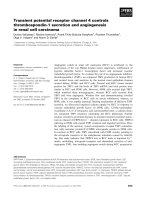
Fig. 1 Expression and significance of Nrf2 in hepatocellular carcinoma. a-d Typical immunohistological features of Nrf2 expression in
hepatocellular carcinoma (HCC). a Expression of Nrf2 in HCC with low differentiation. b Expression of Nrf2 in HCC with metastasis. c Expression of
Nrf2 in HCC with well differentiation. d Expression of Nrf2 in HCC without metastasis. Magnifications: a, c × 200, b, d × 400; e-f Negative staining
in hepatocellular carcinoma. Magnifications: e, × 200; f, × 400; g-h Kaplan-Meier survival analysis, P value was obtained using the log-rank test of
the difference. g Overall survival (OS) differences between patients with high and low levels of Nrf2 protein expression; h Disease free survival
(DFS) differences between patients with high and low levels of Nrf2 protein expression
Zhang et al. BMC Cancer (2015) 15:531
Page 5 of 12
flow cytometry (FACSCalibur, BD Biosciences, San Jose,
CA). The experiment was repeated three times.
crystal violet. The cells were quantified from five different fields under a light microscope. The experiment was
repeated in triplicate.
Cell invasion assay
Cell invasion was measured using transwell chambers
(Millipore, Billerica, USA) coated with Matrigel. After
transfection, the harvested cells were suspended in
serum free RPMI 1640 and were added into the upper
compartment of the chamber; conditioned RPMI 1640
medium with 20 % (v/v) FBS was used as a chemoattractant and placed in the bottom compartment of the
chamber. After incubation, the cells were removed from
the upper surface of the filter with a cotton swab. The
invaded cells were then fixed and stained using 0.1 %
Statistical analysis
Statistical analysis was done using the SPSS software
package (version 13.0, SPSS Institute). The association
between staining index and other categorical factors potentially predictive of prognosis was analyzed using the
Chi-square test and Fisher’s exact test. Overall survival
(OS) was defined as the time from the date of surgery to
the date of last follow-up or death from any case. Disease free survival (DFS) time was defined as the interval
between the date of surgery and the date of recurrence.
Table 2 Univariate analysis for overall survival and disease free survival
Variables
P
Overall survival
Median ± SE
95 % CI
Nrf2
P
Disease free survival
Median ± SE
95 % CI
24.43 ± 3.33
17.90-30.96
11.24 ± 0.76
9.75-12.73
<0.01
Low
30.40 ± 4.16
22.25-38.56
High
13.87 ± 0.95
12.02-15.73
Gender
<0.01
0.93
0.91
Male
17.56 ± 1.32
14.97-20.15
15.43 ± 2.91
9.72-21.13
Female
19.07 ± 3.64
11.94-26.20
14.15 ± 1.05
12.08-16.22
<60
18.98 ± 2.50
14.08-23.88
15.33 ± 1.99
11.43-19.23
≥60
18.43 ± 2.31
13.90-22.97
14.93 ± 1.86
11.29-18.56
Age
0.94
Metastasis
0.91
<0.01
<0.01
Negative
22.56 ± 2.48
17.69-27.42
18.18 ± 1.99
14.29-22.07
Positive
12.97 ± 1.18
10.66-15.29
10.47 ± 0.93
8.64-12.30
Well + Moderate
22.13 ± 2.52
17.20-27.07
17.81 ± 2.01
13.87-21.75
Poor
13.51 ± 1.22
11.12-15.89
10.94 ± 0.92
9.02-12.87
Differentiation
0.01
HBV infection
0.02
0.69
0.70
Negative
19.64 ± 3.90
12.00-27.29
15.89 ± 3.07
9.87-21.92
Positive
18.80 ± 2.02
14.83-22.77
15.24 ± 1.65
12.01-18.47
No
21.09 ± 2.22
16.73-25.44
17.09 ± 1.77
13.62-20.56
Yes
17.24 ± 2.13
13.07-21.40
13.90 ± 1.70
10.56-17.23
Liver cirrhosis
0.09
AFP
0.10
0.20
0.19
≤400 μg/L
20.84 ± 2.02
16.88-24.79
16.82 ± 1.62
13.65-19.99
>400 μg/L
17.78 ± 2.27
13.35-22.22
14.36 ± 1.81
10.80-17.91
<5 cm
21.98 ± 2.77
16.55-27.41
17.72 ± 2.22
13.38-22.06
≥5 cm
15.25 ± 1.56
12.18-18.31
12.35 ± 1.27
9.85-14.85
Tumor size
0.09
Tumor number
0.10
0.60
0.57
Single
20.21 ± 2.71
14.89-25.52
16.35 ± 2.19
12.06-20.63
multiple
16.83 ± 1.79
13.32-20.35
13.63 ± 1.43
10.84-16.43
Zhang et al. BMC Cancer (2015) 15:531
Page 6 of 12
Survival curve and median survival were estimated by the
Kaplan-Meier method. Their differences were verified by
log-rank test. Multivariate analysis was done using the
Cox proportional hazard regression analysis. Differences
between groups were assessed using an unpaired, twotailed Student’s t test; P < 0.05 was considered significant.
Result
Expression of Nrf2 in HCC tissues and its significance
Level of Nrf2 was evaluated by immunohistochemical
analysis. Fig. 1a and d shows representative expression
patterns of Nrf2 in HCC. Nrf2 was found nuclear and
cytoplasmic localization, but primarily in the nucleus.
And in HCC with poor differentiation or metastasis,
Nrf2 showed more nuclear localization compared to that
in HCC with well differentiation or no metastasis. There
were significant correlations between the high level of
Nrf2 expression and the tumor differentiation, metastasis, and tumor size,. However, the high level rates were
not significantly correlated with gender, age, HBV infection, liver cirrhosis. alpha-fetal protein (AFP) levels, and
tumor number (Table 1). Then, Kaplan-Meier analysis
was used to calculate the impact of classic clinicopathologic features and protein expression on survival (Table 2,
Fig. 1g and h). High expression of Nrf2, tumor differentiation, and metastasis were associated with decreased
survival (P < 0.05), whereas other clinicopathological variables were not significant. Cox regression analysis revealed a statistically significant correlation with Nrf2
expression (P < 0.05, Table 3).
detection of expression of Nrf2 by western blot, all HCC
cell lines (Hep3B, Bel-7402, and HepG2) had an overexpression of Nrf2 compared to normal liver cell line
L02 (Fig. 2a). Bel-7402 and HepG2, with highest or
lowest expression levels of Nrf2, were chose for further
experiments. Then, subcellular location of Nrf2 was
evaluated by immunofluorescence assay. In LO2 cells,
Nrf2 expression was present in the cytoplasm, while in
Bel-7402 cells, Nrf2 localization was found both in nucleus and cytoplasm, but mainly in nucleus (Fig. 2b).
The subcellular location of Nrf2 in Bel-7402 was consistent with that of immunohistochemical results.
Transient transfection effect on Nrf2 mRNA and protein
level
To knock down the endogenous expression of Nrf2 in Bel7402 cells, we applied a plasmid vector expressing specific
shRNA sequence targeting Nrf2 (shRNA-Nrf2). As a control, we stably transfected the Bel-7402 cells with the same
plasmid vector expressing a control shRNA sequence
(shNC) that did not target any known human gene.
Through mRNA and protein expression analysis, we found
that the shNC cells have a similar Nrf2 level as the parental Bel-7402 cells, which were significantly higher than the
level in the shNrf2 cells (Fig. 3a, b and c). We then applied
a expression plasmid named pEGFP-Nrf2 to up-regulate
expression of Nrf2 in HepG2. The mRNA and protein expression analysis confirmed that pEGFP-Nrf2 significantly
increased expression of Nrf2 in transfected HepG2 cells
(Fig. 3d, e and f).
Expression and subcellular location of Nrf2 in HCC cell
lines
Since high level of Nrf2 expression correlated with the
tumor differentiation, metastasis, and tumor size and
served as independent prognostic factor, we then investigate the expression of Nrf2 in HCC cell lines. After
Table 3 Multivariate Cox proportional hazards analysis for
overall survival and disease free survival
P
Overall survival
Nrf2
5.96 2.46-14.69 <0.01 5.84
2.37-14.39
<0.01
Gender
0.62 0.30-1.27
0.20
0.63
0.31-1.29
0.20
Age
0.85 0.23-3.14
0.81
0.86
0.23-3.17
0.82
Metastasis
0.96 0.23-4.07
0.96
1.08
0.27-4.32
0.92
RR
95 % CI
Disease free survival
P
Variables
RR
95 % CI
Differentiation
0.76 0.16-3.76
0.74
0.67
0.14-3.16
0.62
HBV infection
0.64 0.29-1.40
0.26
0.64
0.28-1.41
0.26
Liver cirrhosis
1.78 0.90-3.51
0.10
1.80
0.92-3.55
0.09
AFP
1.93 0.92-4.06
0.08
1.91
0.91-4.01
0.09
Tumor size
1.56 0.57-4.24
0.39
1.59
0.58-4.31
0.39
Tumor number 1.73 0.45-6.66
0.43
1.72
0.45-6.60
0.43
Nrf2 promotes cell proliferation by inhibiting apoptosis
To investigate whether Nrf2 modulates cell proliferation in
HCC cells, we assayed its effect on cell proliferation activity. The proliferation activities of Bel-7402 cells transfected
with shRNA-Nrf2 and HepG2 cells transected with
pEGFP-Nrf2 were determined using an MTT assay. As
shown in Fig. 4a and b, inhibition of Nrf2 expression had a
significant decrease in cell viability while increasing Nrf2
expression got the opposite results (P < 0.05). Following experiments demonstrated that shRNA-Nrf2 transfection induced apoptosis and pEGFP-Nrf2 transfection inhibited
apoptosis, showing that the cell proliferation inhibition
effect was partly due to the inhibition of apoptosis
(Fig. 4c to f ). We therefore assessed the expression of
Bcl-xL, an apoptosis related protein regulating death and
survival, in Bel-7402 cells transfected with shRNA-Nrf2
and HepG2 cells transected with pEGFP-Nrf2. Expression
of Bcl-xL was positively correlated with the expression of
Nrf2: inhibition of Nrf2 decreased the Bcl-xL expression
while up-regulation of Nrf2 increased the Bcl-xL expression (Fig. 4g to h).
Zhang et al. BMC Cancer (2015) 15:531
Page 7 of 12
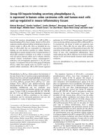
Fig. 2 Expression and Subcellular location of Nrf2 in hepatocellular carcinoma cell lines. a-b Expression of Nrf2 in different human hepatocellular
carcinoma cell lines (Hep3B, Bel-7402, and HepG2), with normal human liver cell line LO2 as control; c Subcellular location of Nrf2 was detected
by immunofluorescence assay. In LO2 cells, Nrf2 expression was present in the cytoplasm, while in Bel-7402 cells, Nrf2 localization was found both
in nucleus and cytoplasm, but mainly in nucleus. Magnifications: ×400. *P <0.05 compared with LO2
Nrf2 regulates cell invasion in vitro
Because there was a correlation between Nrf2 and metastasis, a transwell assay was performed to investigate the
role of Nrf2 on the invasion of HCC cells. Downregulation of Nrf2 expression repressed the cell invasion ability of Bel-7402 cells, and up-regulation of
Nrf2 expression promoted the cell invasion ability of
HepG2 cells (P < 0.05, Fig. 5a to d). These findings
suggest that Nrf2 regulates cell invasion of the HCC
cell lines in vitro. We therefore assessed the expression of
matrix metalloproteinases-9 (MMP-9), a protein regulating cell migration and invasion, in Bel-7402 cells
transfected with shRNA-Nrf2 and HepG2 cells transected with pEGFP-Nrf2. Expression of MMP-9 was
positively correlated with the expression of Nrf2: inhibition of Nrf2 decreased the MMP-9 expression
Zhang et al. BMC Cancer (2015) 15:531
Page 8 of 12
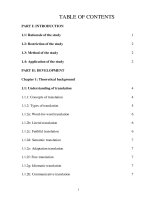
Fig. 3 Modulation of endogenous Nrf2 expression. a After transfected with Nrf2-shRNA (shRNA-867, shRNA-1118, shRNA-1757, or shRNA-2019) or
control shRNA (shNC), expression levels of Nrf2 mRNA in Bel-7402 cells were detected by qRT-PCR; b-c After transfected with Nrf2-shRNA
(shRNA-867, shRNA-1118, shRNA-1757, or shRNA-2019) or control shRNA (shNC), expression levels of Nrf2 protein in Bel-7402 cells were detected
by western blot; d After transfected with Nrf2 expression plasmid (pEFGP-Nrf2-1 or pEFGP-Nrf2-2) or mock pEGFP plasmid (pEGFP-NC), expression
levels of Nrf2 mRNA in HepG2 cells were detected by qRT-PCR; e-f After transfected with Nrf2 expression plasmid (pEFGP-Nrf2-1 or pEFGP-Nrf2-2)
or mock pEGFP plasmid (pEGFP-NC), expression levels of Nrf2 protein in HepG2 cells were detected by western blot. *P <0.05 compared with
control (Bel-7402 cells or HepG2 cells respectively) or shNC and pEGFP-NC
while up-regulation of Nrf2 increased the MMP-9
expression (Fig. 5e to f ).
Discussion
Nrf2, a key transcription factor, plays a pivotal role in endogenous protection against oxidative stress. Upon exposure of cells to oxidative stress or chemopreventive
compounds, Nrf2 translocates to the nucleus, forms a
heterodimer with its obligatory partner Maf, and binds to
the antioxidant response element (ARE) sequence to activate those encoding endogenous antioxidants, phase II
detoxifying enzymes, and transporters [19]. As a result, activation of the Nrf2 pathway confers protection against
subsequent toxic/carcinogenic exposure. Therefore, Nrf2
has been viewed as a “good” protein that protects humans
from genotoxic damage caused by carcinogens. Several in
Zhang et al. BMC Cancer (2015) 15:531
Fig. 4 (See legend on next page.)
Page 9 of 12
Zhang et al. BMC Cancer (2015) 15:531
Page 10 of 12
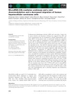
(See figure on previous page.)
Fig. 4 Effect of Nrf2 on cell proliferation and apoptosis. a After shRNA-Nrf2 (shRNA-1757 or shRNA-2019) or control shRNA (shNC) transduction,
the growth of Bel-7402 cells was analyzed at different time points using the MTT assay; b After Nrf2 expression plasmid (pEFGP-Nrf2-1 or
pEFGP-Nrf2-2) or mock pEGFP plasmid (pEGFP-NC) transduction, the growth of HepG2 cells was analyzed at different time points using
the MTT assay; c-d Flow cytometric analysis of the effect of Nrf2 on the apoptosis of Bel-7402 cells by down-regulation of expression of
Nrf2; e-f Flow cytometric analysis of the effect of Nrf2 on the apoptosis of HepG2 cells by up-regulation of expression of Nrf2. g After
shRNA-Nrf2 (shRNA-1757 or shRNA-2019) or control shRNA (shNC) transduction, expression of Bcl-xL were detected by western blot in Bel-7402
cells; h After Nrf2 expression plasmid (pEFGP-Nrf2-1 or pEFGP-Nrf2-2) or mock pEGFP plasmid (pEGFP-NC) transduction, expression of Bcl-xL were
detected by western blot in HepG2 cells. *P < 0.05 compared with control (Bel-7402 cells or HepG2 cells respectively) or shNC and pEGFP-NC
Fig. 5 Effect of Nrf2 on cell invasion in vitro. a Bel-7402 cells transfected with shRNA-Nrf2 (shRNA-1757 or shRNA-2019) or control shRNA (shNC)
were subjected to transwell invasion assays; b The invasive cell numbers are the average count of five random microscopic fields detected using
the transwell invasion assay; c HepG2 cells transfected with Nrf2 expression plasmid (pEFGP-Nrf2-1 or pEFGP-Nrf2-2) or mock pEGFP plasmid
(pEGFP-NC) were subjected to transwell invasion assays; d The invasive cell numbers are the average count of five random microscopic fields detected
using the transwell invasion assay. Each bar represents the mean ± SD of the counts. e After shRNA-Nrf2 (shRNA-1757 or shRNA-2019)) or control shRNA
(shNC) transduction, expression of MMP-9 were detected by western blot in Bel-7402 cells; f After Nrf2 expression plasmid (pEFGP-Nrf2-1 or pEFGP-Nrf2-2)
or mock pEGFP plasmid (pEGFP-NC) transduction, expression of MMP-9 were detected by western blot in HepG2 cells. *P < 0.05 compared with control
(Bel-7402 cells or HepG2 cells respectively) or shNC and pEGFP-NC
Zhang et al. BMC Cancer (2015) 15:531
vivo studies using Nrf2-null mice further verified the pivotal role of Nrf2 in cancer protection [20–22].
Interestingly, recent emerging data has revealed the
“dark” side of Nrf2. Nrf2 and its downstream genes are
over-expressed in many cancer cell lines and human cancer tissues, giving cancer cells an advantage for survival
and growth [23]. In cancer tissues and cells, loss of Keap1,
an Nrf2 negatively regulator, leads to nuclear localization
and constitutive activation of Nrf2 [24–27]. Furthermore,
Nrf2 is up-regulated in resistant cancer cells and is
thought to be responsible for acquired chemoresistance.
Observations including ours found that mutation or overexpression of Nrf2 in kinds of cancer [13–18]. Our previous studies suggest that Nrf2 confers chemoresistance of
HCC and inhibition of Nrf2 by sorafenib could sensitize
Bel-7402/5-FU cells to 5-FU [17]. But there are limited reports about the expression, significance, function of Nrf2
in HCC. Then, we will focus our attention on the oncogenic functions of Nrf2 in HCC in the certain research.
Our results showed that there were significant correlations between the expression of Nrf2 and metastasis, differentiation, and tumor size. Then, high expression level
of Nrf2 was an independent factor that indicated poor
prognosis in HCC patients. Furthermore, we detected
the expression and subcellular localization of Nrf2 in
HCC cell lines. Consistent with the immunohistochemical
results and other reports in lung cancer and cervical cancer, Nrf2 were over-expression and nuclear localization in
HCC, indicating Nrf2 was constitutive activated [26, 27].
This evidence suggests that over-expression of Nrf2 in
tumor cells may play roles in the development of HCC
and may have prognostic value.
Furthermore, to reveal the exact role of Nrf2 in HCC, we
tested the effect of Nrf2 on proliferation, apoptosis, and invasion by modulating the expression level of Nrf2 using
Nrf2-shRNA and pEGFP-Nrf2. The results suggested that
Nrf2 acted as an oncogene in HCC. First, Nrf2 could induce proliferation due to the regulation of apoptosis and
promote invasion in HCC cells. In our opinion, this invasion related ability could reveal the correlation between
Nrf2 and metastasis: over-expression of Nrf2 promoted the
metastasis of HCC cells. Then, we investigated the potential
mechanisms of Nrf2 in regulating proliferation, apoptosis,
and invasion. Bcl-2 family proteins are the prototypical
antiapoptotic proteins, and Bcl-xL was the first protein discovered with a similar function [28]. MMP9, which belongs
to the ECM-degrading enzyme family, is involved in migration and invasion of tumor cells [29]. Considering the role
of Bcl-xL and MMP-9 in cell survival and invasion respectively, we investigated their relationship with expression of
Nrf2. There are positive correlation between Nrf2 expression and that of Bcl-xL and MMP-9. Consistent with
previous studies, Nrf2 up-regulated expression of Bcl-xL
and MMP-9 in HCC cells resulted in cell proliferation,
Page 11 of 12
apoptosis inhibitation, and invasion [17, 30]. We will carry
out in vivo experiments further to confirm the role of Nrf2
and its target genes in HCC.
Conclusions
In conclusion, this was the first study to systemically
evaluate the oncogenic functions of the Nrf2 in HCC.
Our findings demonstrated that Nrf2 was up-regulated
in HCC, and expression of Nrf2 was correlated with
tumor differentiation metastasis, and tumor size. We
found that Nrf2 was an independent prognostic factor in
HCC patients. We also concluded that Nrf2 promoted
proliferation by inhibiting apoptosis and enhanced the
invasive ability of HCC cells partly through regulating
expression of Bcl-xL and MMP-9.
Abbreviations
Nrf2: Nuclear factor E2-related factor 2; HCC: Human hepatocellular
carcinoma; TNM: Tumor-nodemetastasis; HRP: Horseradish peroxidase;
qRT-PCR: Quantitative real time polymerase chain reaction; PI: Propidine
iodide; PS: Phosphatidylserine; AFP: Alpha-fetal protein.
Competing interests
The authors declare that they have no competing interests.
Authors’ contributions
MZ, CZ and LZ constructed the manuscript. MZ, CZ and LZ were responsible
for clinical data and evaluated clinical data; formed analysis of relation
between clinical data and survival data. QY and SZ carried out intro
experiments. QW and JW reviewed the manuscript. All authors read and
approval the final manuscript.
Authors’ information
Mingxin Zhang, Chao Zhang, Lingmin Zhang and Qi Yang are Co-first authors.
Acknowledgments
This work was supported by supported by the National Natural Science
Foundation of China (NSFC 81301922, 81270485 and 81302055).
Author details
1
Department of Gastroenterology, Tangdu Hospital, Fourth Military Medical
University, Xi’an 710038, Shaanxi Province, China. 2Department of
Anesthesiology, First Affiliated Hospital, Medical School, Xi’an Jiaotong
University, Xi’an 710061, Shaanxi Province, China. 3Department of
Radiotherapy, Tangdu Hospital, Fourth Military Medical University, Xi’an
710038, Shaanxi Province, China.
Received: 3 January 2015 Accepted: 13 July 2015
References
1. Schütte K, Bornschein J, Malfertheiner P. Hepatocellular carcinoma–
epidemiological trends and risk factors. Dig Dis. 2009;27(2):80–92.
2. He J, Gu D, Wu X, Reynolds K, Duan X, Yao C, et al. Major causes of death
among men and women in China. N Engl J Med. 2005;353(11):1124–34.
3. Cabrera R, Nelson DR. Review article: the management of hepatocellular
carcinoma. Aliment Pharmacol Ther. 2010;31(4):461–76.
4. Cormier JN, Thomas KT, Chari RS, Pinson CW. Management of
hepatocellular carcinoma. J Gastrointest Surg. 2006;10(5):761–80.
5. El-Serag HB. Hepatocellular carcinoma. N Engl J Med. 2011;365(12):1118–27.
6. Surh YJ, Kundu JK, Li MH, Na HK, Cha YN. Role of Nrf2-mediated heme
oxygenase-1 upregulation in adaptive survival response to nitrosative stress. Arch
Pharm Res. 2009;32(8):1163–76.
7. Vasiliou V, Qamar L, Pappa A, Sophos NA, Petersen DR. Involvement of the
electrophile responsive element and p53 in the activation of hepatic stellate
cells as a response to electrophile menadione. Arch Biochem Biophys.
2003;413(2):164–71.
Zhang et al. BMC Cancer (2015) 15:531
8.
9.
10.
11.
12.
13.
14.
15.
16.
17.
18.
19.
20.
21.
22.
23.
24.
25.
26.
27.
28.
29.
30.
Yeligar SM, Machida K, Kalra VK. Ethanol-induced HO-1 and NQO1 are
differentially regulated by HIF-1alpha and Nrf2 to attenuate inflammatory
cytokine expression. J Biol Chem. 2010;285(46):35359–73.
Shin SM, Yang JH, Ki SH. Role of the Nrf2-ARE pathway in liver diseases.
Oxid Med Cell Longev. 2013;2013:763257.
Gao AM, Ke ZP, Shi F, Sun GC, Chen H. Chrysin enhances sensitivity of
BEL-7402/ADM cells to doxorubicin by suppressing PI3K/Akt/Nrf2 and
ERK/Nrf2 pathway. Chem Biol Interact. 2013;206(1):100–8.
Gao AM, Ke ZP, Wang JN, Yang JY, Chen SY, Chen H. Apigenin sensitizes
doxorubicin-resistant hepatocellular carcinoma BEL-7402/ADM cells to
doxorubicin via inhibiting PI3K/Akt/Nrf2 pathway. Carcinogenesis.
2013;34(8):1806–14.
Lee SE, Yang H, Jeong SI, Jin YH, Park CS, Park YS. Induction of heme
oxygenase-1 inhibits cell death in crotonaldehyde-stimulated HepG2 cells
via the PKC-δ-p38-Nrf2 pathway. PLoS One. 2012;7(7), e41676.
Wang J, Zhang M, Zhang L, Cai H, Zhou S, Zhang J, et al. Correlation of
Nrf2, HO-1, and MRP3 in gallbladder cancer and their relationships to
clinicopathological features and survival. J Surg Res. 2010;164(1):e99–105.
Ma R, Zhang M, Wang J, Cai H, Yeer M, Duan X. Expression and distribution
of Nrf2 in several hepatocellular carcinoma cell lines. Xi Bao Yu Fen Zi Mian
Yi Xue Za Zhi. 2011;27(6):608–10.
Mao J, Tangsakar E, Shen H, Wang Z, Zhang M, Chen J, et al. Expression and
clinical significance of Nrf2 in esophageal squamous cell carcinoma. Xi Bao
Yu Fen Zi Mian Yi Xue Za Zhi. 2011;27(11):1231–3.
Zhang L, Wang N, Zhou S, Ye W, Jing G, Zhang M. Propofol induces
proliferation and invasion of gallbladder cancer cells through activation of
Nrf2. J Exp Clin Cancer Res. 2012;31:66.
Pan H, Wang H, Zhu L, Mao L, Qiao L, Su X. The role of Nrf2 in migration and
invasion of human glioma cell U251. World Neurosurg. 2013;80(3–4):363–70.
Zhou S, Ye W, Shao Q, Zhang M, Liang J. Nrf2 is a potential therapeutic
target in radioresistance in human cancer. Crit Rev Oncol Hematol.
2013;88(3):706–15.
Ma Q. Role of nrf2 in oxidative stress and toxicity. Annu Rev Pharmacol
Toxicol. 2013;53:401–26.
Kwak MK, Itoh K, Yamamoto M, Sutter TR, Kensler TW. Role of transcription
factor Nrf2 in the induction of hepatic phase 2 and antioxidative enzymes
in vivo by the cancer chemoprotective agent, 3H-1, 2-dimethiole-3-thione.
Mol Med. 2001;7(2):135–45.
Chan JY, Kwong M. Impaired expression of glutathione synthetic enzyme
genes in mice with targeted deletion of the Nrf2 basic-leucine zipper
protein. Biochim Biophys Acta. 2000;1517(1):19–26.
Chanas SA, Jiang Q, McMahon M, McWalter GK, McLellan LI, Elcombe CR, et
al. Loss of the Nrf2 transcription factor causes a marked reduction in
constitutive and inducible expression of the glutathione S-transferase Gsta1,
Gsta2, Gstm1, Gstm2, Gstm3 and Gstm4 genes in the livers of male and
female mice. Biochem J. 2002;365(Pt 2):405–16.
Zhou S, Ye W, Zhang M, Liang J. The effects of nrf2 on tumor angiogenesis:
a review of the possible mechanisms of action. Crit Rev Eukaryot Gene Expr.
2012;22(2):149–60.
Kim WD, Kim YW, Cho IJ, Lee CH, Kim SG. E-cadherin inhibits nuclear
accumulation of Nrf2: implications for chemoresistance of cancer cells. J Cell
Sci. 2012;125(Pt 5):1284–95.
Kawai Y, Garduño L, Theodore M, Yang J, Arinze IJ. Acetylation-deacetylation
of the transcription factor Nrf2 (nuclear factor erythroid 2-related factor 2)
regulates its transcriptional activity and nucleocytoplasmic localization. J Biol
Chem. 2011;286(9):7629–40.
Ohta T, Iijima K, Miyamoto M, Nakahara I, Tanaka H, Ohtsuji M, et al. Loss of
Keap1 function activates Nrf2 and provides advantages for lung cancer cell
growth. Cancer Res. 2008;68(5):1303–9.
Konstantinopoulos PA, Spentzos D, Fountzilas E, Francoeur N, Sanisetty S,
Grammatikos AP, et al. Keap1 mutations and Nrf2 pathway activation in epithelial
ovarian cancer. Cancer Res. 2011;71(15):5081–9.
Boise LH, González-García M, Postema CE, Ding L, Lindsten T, Turka LA, et al.
bcl-x, a bcl-2-related gene that functions as a dominant regulator of
apoptotic cell death. Cell. 1993;74(4):597–608.
Bauvois B. New facets of matrix metalloproteinases MMP-2 and MMP-9 as
cell surface transducers: outside-in signaling and relationship to tumor
progression. Biochim Biophys Acta. 2012;1825(1):29–36.
Niture SK, Jaiswal AK. Nrf2-induced antiapoptotic Bcl-xL protein enhances
cell survival and drug resistance. Free Radic Biol Med. 2013;57:119–31.
Page 12 of 12
Submit your next manuscript to BioMed Central
and take full advantage of:
• Convenient online submission
• Thorough peer review
• No space constraints or color figure charges
• Immediate publication on acceptance
• Inclusion in PubMed, CAS, Scopus and Google Scholar
• Research which is freely available for redistribution
Submit your manuscript at
www.biomedcentral.com/submit