The prognostic value of SUMO1/Sentrin specific peptidase 1 (SENP1) in prostate cancer is limited to ERG-fusion positive tumors lacking PTEN deletion
Bạn đang xem bản rút gọn của tài liệu. Xem và tải ngay bản đầy đủ của tài liệu tại đây (1.51 MB, 13 trang )
Burdelski et al. BMC Cancer (2015) 15:538
DOI 10.1186/s12885-015-1555-8
RESEARCH ARTICLE
Open Access
The prognostic value of SUMO1/Sentrin
specific peptidase 1 (SENP1) in prostate
cancer is limited to ERG-fusion positive
tumors lacking PTEN deletion
Christoph Burdelski1†, Devi Menan2†, Maria Christina Tsourlakis2, Martina Kluth2, Claudia Hube-Magg2,
Nathaniel Melling1, Sarah Minner2, Christina Koop2, Markus Graefen3, Hans Heinzer3, Corinna Wittmer2,
Guido Sauter2, Ronald Simon2, Thorsten Schlomm3,4, Stefan Steurer2 and Till Krech2*
Abstract
Background: Posttranscriptional protein modification by SUMOylation plays an important role in tumor development
and progression. In the current study we analyzed prevalence and prognostic impact of the de-SUMOylation enzyme
SENP1 in prostate cancer.
Methods: SENP1 expression was analyzed by immunohistochemistry on a tissue microarray containing more than
12,400 prostate cancer specimens. Results were compared to tumor phenotype, ERG status, genomic deletions of 3p,
5q, 6q and PTEN, and biochemical recurrence.
Results: SENP1 immunostaining was detectable in 34.5 % of 9,516 interpretable cancers and considered strong in
7.3 %, moderate in 14.9 % and weak in 12.3 % of cases. Strong SENP1 expression was linked to advanced pT stage
(p < 0.0001), high Gleason grade (p < 0.0001), positive lymph node status (p = 0.0019), high pre-operative PSA levels
(p = 0.0037), and PSA recurrence (p < 0.0001). SENP1 expression was strongly associated with positive ERG fusion status
as determined by both in situ hybridization (FISH) and immunohistochemistry as well as with PTEN deletions.
Detectable SENP1 immunostaining was found in 41 % of ERG positive and in 47 % of PTEN deleted cancers but in only
30 % of ERG negative and 30 % of PTEN non-deleted cancers (p < 0.0001 each). Deletions of 3p, 5q, and 6q were
unrelated to SENP1 expression. Subset analyses revealed that the prognostic impact of SENP1 expression was solely
driven by the subgroup of ERG positive, PTEN undeleted cancers. In this subgroup, the prognostic role of SENP1
expression was independent of the preoperative PSA level, tumor stage, Gleason grade, and the status of the resection
margin.
Conclusions: SENP1 expression has strong prognostic impact in a molecularly defined subset of cancers. This is per se
not surprising as the biologic impact of each individual molecular event is likely to be dependent on its cellular
environment. However, such findings challenge the concept of finding clinically relevant molecular signatures that are
equally applicable to all prostate cancers.
Keywords: Prostate cancer, ERG fusion, PTEN deletion, SENP1, SUMO, Immunohistochemistry, Tissue microarray
* Correspondence:
†
Equal contributors
2
Institute of Pathology, University Medical Center Hamburg-Eppendorf,
Hamburg, Germany
Full list of author information is available at the end of the article
© 2015 Burdelski et al. This is an Open Access article distributed under the terms of the Creative Commons Attribution License
( which permits unrestricted use, distribution, and reproduction in any medium,
provided the original work is properly credited. The Creative Commons Public Domain Dedication waiver (http://
creativecommons.org/publicdomain/zero/1.0/) applies to the data made available in this article, unless otherwise stated.
Burdelski et al. BMC Cancer (2015) 15:538
Background
Prostate cancer is the most prevalent cancer in men in
Western societies [1]. Although most prostate cancers
have a rather indolent clinical course, this disease still
represents the third most common cause of cancer related death in men. A reliable distinction between the
indolent and the aggressive forms of the disease is highly
desirable to enhance therapeutic decisions. Despite recent advances, the only established pretreatment prognostic parameters currently include Gleason grade and
tumor extent on biopsies, preoperative prostate-specific
antigen (PSA), and clinical stage. Because these data are
statistically powerful but not sufficient for optimal individual treatment decisions, it can be hoped that a better
understanding of disease biology will eventually lead to
the identification of clinically applicable molecular
markers that enable a more reliable prediction of prostate cancer aggressiveness.
SUMOylation is a revertible posttranscriptional protein modification involving the binding of small
ubiquitin-like modifiers (SUMOs) to target proteins.
SUMOs are structurally related to ubiquitin and are covalently attached to target proteins by a SUMOconjugating system resembling the ubiquitination machinery [2]. SUMOylation affects protein stability and
activity, and regulates a variety of cellular processes,
such as nuclear transport, transcription, and apoptosis
[3]. Several proteins control the balance between
SUMOylation and de-SUMOylation. A key protein for
de-SUMOylation is SUMO1/Sentrin specific peptidase 1
(SENP1) [4], which deconjugates SUMOs from a large
number of SUMOylated proteins [5]. Since important
target genes of SENP1 include histone deacetylases and
cell cycle regulators like cyclin D1, SENP1 is also involved in control of epigenetic transcription and cell
proliferation [6–10]. Consequently, overexpression of
SENP1 has been found in various cancer types [10], such
as colon cancer [11], bladder cancer [12], head & neck
cancer [13], and lung cancer [14], and has been linked to
poor clinical features in some of these [13, 15]. In the
prostate gland, SENP1 was shown to act as a transcriptional activator of androgen receptor (AR) signaling [7].
Two studies analyzing SENP1 in 115 and 150 Asian
prostate cancer patients suggested that SENP1 overexpression might be an independent marker of poor prognosis [16, 17].
These promising findings prompted us to study the
putative prognostic value of SENP1 expression measurement in a large cohort including more than 12,400 European prostate cancers that have been assembled in a
tissue microarray (TMA) format. The database attached
to our TMA contains pathological and clinical follow-up
data, as well molecular data of key molecular alterations
of this disease such as ERG fusion and genomic deletion
Page 2 of 13
of PTEN, 3p13, 5q21, and 6q15, which were used to establish associations between SENP1 expression and distinct phenotypic and molecular subsets of prostate
cancers.
Methods
Patients
Radical prostatectomy specimens were available from
12,427 patients, undergoing surgery between 1992 and
2012 at the Department of Urology and the Martini
Clinics at the University Medical Center HamburgEppendorf. Follow-up data were available for a total of
11,665 patients with a median follow-up of 36 months
(range: 1 to 241 months; Table 1). Prostate specific antigen (PSA) values were measured following surgery and
PSA recurrence was defined as a postoperative PSA of ≥
0.2 ng/ml confirmed by a second determination with a
serum PSA ≥ 0.2 ng/ml. All prostate specimens were analyzed according to a standard procedure, including a
complete embedding of the entire prostate for histological analysis [18].
The TMA manufacturing process was described earlier
in detail [19]. In short, one 0.6 mm core was taken from
a representative tissue block from each patient. The tissues were distributed among 27 TMA blocks, each containing 144 to 522 tumor samples. For internal controls,
each TMA block also contained various control tissues,
including normal prostate tissue. The molecular database attached to this TMA contained results on ERG expression in 10,711 [20], ERG break apart FISH analysis
in 7,122 (expanded from [21]) and deletion status of
5q21 (CHD1) in 7932 (expanded from [22]), 6q15
(MAP3K7) in 6,069 (expanded from [23]), 10q23 (PTEN)
in 6,704 (expanded from [24]) and 3p13 (FOXP1) in
7,081 (expanded from [25]) cancers. Immunohistochemical data on Ki67 labeling index (LI) were available from
7,010 cancers (expanded from [26]).
The usage of archived diagnostic left-over tissues for
manufacturing of tissue microarrays and their analysis
for research purposes as well as patient data analysis has
been approved by the local ethics committee (Ethics
commission Hamburg, WF-049/09 and PV3652). All
work has been carried out in compliance with the
Helsinki Declaration.
Usage of patient data and routinely archived formalin
fixed left-over patient tissue samples for research purposes
by the attending physician is approved by local laws and
does not require written consent (HmbKHG, §12,1).
Immunohistochemistry
Freshly cut TMA sections were immunostained on one
day and in one experiment. Slides were deparaffinized and
exposed to heat-induced antigen retrieval for 5 min in an
autoclave at 121 °C in pH 7.8 Tris-EDTA-Citrate buffer.
Burdelski et al. BMC Cancer (2015) 15:538
Page 3 of 13
Table 1 Pathological and clinical data of the arrayed prostate cancers. Percentage in the column “Study cohort on TMA” refers to
the fraction of samples across each category. Percentage in column “Biochemical relapse among categories” refers to the fraction of
samples with biochemical relapse within each parameter in the different categories. Numbers do not always add up to 12,427 in the
different categories because of cases with missing data. Abbreviation: AJCC, American Joint Committee on Cancer
No. of patients (%)
Study cohort on
TMA (n = 12427)
Biochemical relapse
among categories
n
11665 (93.9 %)
2769 (23.7 %)
Mean
48.9
-
Median
36.4
-
≤50
334 (2.7 %)
81 (24.3 %)
51-59
3061 (24.8 %)
705 (23 %)
60-69
7188 (58.2 %)
1610 (22.4 %)
≥70
1761 (14.3 %)
370 (21 %)
<4
1585 (12.9 %)
242 (15.3 %)
4-10
7480 (60.9 %)
1355 (18.1 %)
10-20
2412 (19.6 %)
737 (30.6 %)
>20
812 (6.6 %)
397 (48.9 %)
pT2
8187 (66.2 %)
1095 (13.4 %)
pT3a
2660 (21.5 %)
817 (30.7 %)
pT3b
1465 (11.8 %)
796 (54.3 %)
pT4
63 (0.5 %)
51 (81 %)
Follow-up (mo)
Age (y)
Pretreatment PSA (ng/ml)
pT category (AJCC 2002)
Gleason grade
≤3 + 3
2983 (24.1 %)
368 (12.3 %)
3+4
6945 (56.2 %)
1289 (18.6 %)
4+3
1848 (15 %)
788 (42.6 %)
≥4 + 4
584 (4.7 %)
311 (53.3 %)
pN0
6970 (91 %)
1636 (23.5 %)
pN+
693 (9 %)
393 (56.7 %)
Negative
9990 (81.9 %)
1848 (18.5 %)
Positive
2211 (18.1 %)
853 (38.6 %)
pN category
Surgical margin
Primary antibody specific for SENP1 (rabbit monoclonal
antibody, EPR3844, Abcam, Cambridge, UK; cat#108981;
dilution 1:150) was applied at 37 °C for 60 min. Bound
antibody was then visualized using the EnVision Kit
(Dako, Glostrup, Denmark) according to the manufacturer’s directions. Staining was predominantly nuclear and
typically accompanied by cytoplasmic co-staining. The intensity of the cytoplasmic staining was usually weaker
than the intensity of nuclear staining. Nuclear and cytoplasmic SENP1 staining was typically found in either all
(100 %) or none (0 %) of the tumor cells in a given cancer
spot. Staining intensity of all cases was thus semiquantitatively assessed in four categories: negative, weak,
moderate and strong. The percentage of positive tumor
cells (typically 100 %) was not separately recorded. An
additional isotype control (rabbit monoclonal, SP137,
Abcam, Cambridge, UK; cat#128142) yielded no unspecific staining (data not shown).
Statistics
For statistical analysis, the JMP® 10.0.2 software (2012
SAS Institute Inc., NC, USA) was used. Contingency
Burdelski et al. BMC Cancer (2015) 15:538
tables were calculated to study association between
SENP1 staining and clinico-pathological variables, and
the Chi-squared (Likelihood) test was used to find significant relationships. Kaplan Meier curves were generated for PSA recurrence free survival. The log-Rank test
was applied to test the significance of differences between stratified survival functions. Cox proportional
hazards regression analysis was performed to test the
statistical independence and significance between pathological, molecular, and clinical variables.
Results
Technical issues
A total of 9,516 (77 %) of tumor samples were interpretable in our TMA analysis. Reason for non-informative
cases (2,911 spots; 23 %) included lack of tissue samples
or absence of unequivocal cancer tissue in the TMA
spot.
SENP1 immunohistochemistry
In normal prostatic glands, weak cytoplasmic staining was
found in almost all cases, whereas nuclear staining was
Page 4 of 13
rare and occurred in only two out of 20 (10 %) cases. Positive staining was limited to the secretory epithelial cells,
while basal cells were consistently negative. In cancers,
SENP1 immunostaining was predominantly localized in
the nucleus. Positive staining was seen in 3,283 of our
9,516 (34.5 %) interpretable tumors and was considered
weak in 12.3 %, moderate in 14.9 % and strong in 7.3 % of
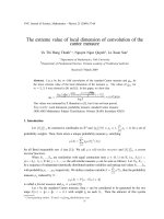
cancers. Representative images of positive and negative
SENP1 immunostainings are given in Fig. 1. Strong
SENP1 immunostaining was significantly linked to advanced pathological tumor stage (p < 0.0001), high Gleason grade (p < 0.0001), presence of lymph node metastases
(p = 0.0019) and high preoperative PSA-levels (p = 0.0037)
when all tumors were jointly analyzed (Table 2). SENP1
immunostaining showed no significant association with
positive resection margin status (p = 0.3216).
Association with TMPRSS2:ERG fusion status and ERG
protein expression
To evaluate whether SENP1 expression is associated
with ERG status in prostate cancers, we used data from
previous studies (expanded from [20, 21]). Data on
Fig. 1 Representative pictures of SENP1 immunostaining in prostate cancer with a) negative, b) weak, c) moderate, and d) strong staining
Burdelski et al. BMC Cancer (2015) 15:538
Page 5 of 13
Table 2 Association between SENP1 immunostaining results and prostate cancer phenotype in all cancers
Parameter
All cancers
SENP1
p value
n evaluable
Negative
(%)
Weak
(%)
Moderate
(%)
Strong
(%)
9,516
65.5
12.3
14.9
7.3
<0.0001
Tumor stage
pT2
6,143
68.2
11.1
14.1
6.6
pT3a
2,137
61.8
13.7
15.9
8.7
pT3b-4
1,203
58.1
15.8
17.6
8.5
<0.0001
Gleason grade
≤3 + 3
2,135
72.5
9.2
11.6
6.7
3+4
5,451
65.3
11.7
15.5
7.5
4+3
1,445
58.4
16.2
17.6
7.8
≥4 + 4
442
58.1
20.8
14.9
6.1
N0
5,472
62.3
12.6
16.6
8.5
N+
526
56.8
18.4
17.5
7.2
0.0019
Lymph node metastasis
0.0037
Preop. PSA level (ng/ml)
<4
1160
64.2
12.2
15.9
7.6
4-10
5702
66.8
11.2
15.0
7.0
10-20
1892
63.4
14.6
14.5
7.5
>20
666
62.3
14.9
14.3
8.6
negative
7,549
65.9
12.1
14.9
7.1
positive
1,797
63.8
13.0
15.2
8.0
0.3216
Surgical margin
TMPRSS2:ERG fusion status obtained by FISH were
available from 5,677 and by immunohistochemistry from
8,459 tumors with evaluable SENP1 immunostaining.
Data on both ERG FISH and IHC were available from
5,468 cancers, and an identical result (ERG IHC positive
and break by FISH or ERG IHC negative and missing
break by FISH) was found in 5,231 of 5,468 (95.7 %) cancers. SENP1 immunostaining was slightly more frequent
in TMPRSS2:ERG rearranged and ERG positive prostate
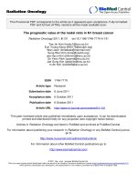
cancers than in ERG negative tumors. Positive SENP1
immunostaining was seen in 41.7 % (ERG IHC) and
40.9 % (ERG FISH) of ERG positive cancers but in only
28.6 % and 30 % of cancers without ERG staining and
ERG rearrangement, respectively (p < 0.0001 each; Fig. 2).
SENP1 immunostaining was similarly linked to unfavorable tumor features in subsets of both ERG negative and
ERG positive cancers (Additional file 1: Table S1 and
Additional file 2: Table S2).
Association to other key genomic deletions
Earlier studies had provided evidence for recurrent
chromosomal deletions delineating further molecular
subgroups amongst ERG positive and ERG negative
prostate cancers. In particular, deletions of PTEN and
3p13 define subgroups in ERG positive and deletions of
5q21 and 6q15 define subgroups in ERG negative cancers [22, 23, 25]. To examine, whether SENP1 expression might be particularly associated with one of these
genomic deletions, SENP1 data were compared to preexisting findings on PTEN (10q23), 3p13 (FOXP1), 6q15
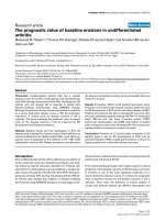
(MAP3K7) and 5q21 (CHD1) deletions. Elevated SENP1
expression levels were strongly linked to deletions of
PTEN both in ERG positive and ERG negative cancers
(p < 0.0001 each, Fig. 3). However, SENP1 was largely
unrelated to all other deletions irrespective of whether
all cancers or subgroups of ERG positive or ERG negative cancers were analyzed.
Association to tumor cell proliferation (Ki67LI)
Strong SENP1 staining was significantly linked to accelerated cell proliferation as measured by Ki67LI in all
cancers (p < 0.0001). This association held also true with
high significance in most subgroups of cancers with
identical Gleason grade (≤3 + 3; 3 + 4; 4 + 3; ≥4 + 4), and
also in the subset of ERG positive tumors lacking PTEN
deletions (p = 0.0315). All comparisons with the Ki67LI
are summarized in Table 3.
Burdelski et al. BMC Cancer (2015) 15:538
Page 6 of 13
Fig. 2 Association between SENP1 immunostaining results and the ERG-status determined by IHC and FISH analysis. Rearranged indicates breakage of
the ERG gene according to FISH analysis
Association with PSA recurrence
Follow-up data were available from 8,920 patients with interpretable SENP1 immunostaining on the TMA. Since
there was no significant prognostic impact of the level of
positive SENP1 staining (data not shown), all cancers with
weak, moderate, and strong SENP1 staining were combined into one group (“positive”) for follow-up analysis.
Tumors with positive SENP1 immunostaining showed a
significantly shortened PSA recurrence-free interval if all
cancers were jointly analyzed (p < 0.0001, Fig. 4a), as well
as in subsets of ERG-IHC-positive (p < 0.0001, Fig. 4b) or
ERG-IHC-negative cancers p < 0.0001, Fig. 4c). Because of
the strong link between SENP1 expression and PTEN deletion, we extended the analyses to tumor subgroups
stratified according to the SENP1/ PTEN status. These
analyses revealed that the prognostic impact of SENP1 expression was limited to cancers lacking PTEN deletions in
ERG positive (p < 0.0001 Fig. 4d), but not in ERG negative
tumors (p = 0.1251, Fig. 4e). SENP1 had no prognostic
relevance in cancers harboring PTEN deletions, neither in
ERG positive (p = 0.7745, Fig. 4d), nor in ERG negative
cancers (p = 0.7267, Fig. 4e).
Multivariate analysis
Four different types of multivariate analyses were performed evaluating the clinical relevance of SENP1 expression in different scenarios (Table 4). Scenario 1 evaluated
all postoperatively available parameters including pathological tumor stage, pathological lymph node status (pN),
surgical margin status, preoperative PSA value and pathological Gleason grade obtained after the morphological
evaluation of the entire resected prostate. In scenario 2, all
postoperatively available parameters with exception of
nodal status were included. The rational for this approach
was that the indication and extent of lymph node dissection is not standardized in the surgical therapy of prostate
cancer and that excluding pN in multivariate analysis can
markedly increase case numbers. Two additional scenarios
had the purpose to model the preoperative situation as
much as possible. Scenario 3 included SENP1 expression,
preoperative PSA, clinical tumor stage (cT stage) and
Gleason grade obtained on the prostatectomy specimen.
Since postoperative determination of a tumors Gleason
grade is “better” than the preoperatively determined Gleason grade (subjected to sampling errors and consequently
under-grading in more than one third of cases [27]), another multivariate analysis was added. In scenario 4, the
preoperative Gleason grade obtained on the original biopsy was combined with preoperative PSA, cT stage and
SENP1 expression. SENP1 largely did not provide independent prognostic information if all tumors or the subgroups of ERG positive and ERG negative cancers were
interrogated. A further subset analysis of ERG positive/
PTEN undeleted cancers revealed independent prognostic
impact, however, in 3 of 4 tested scenarios (Table 4 a-d).
Discussion
Immunohistochemically detectable SENP1 expression
was found in about 35 % of prostate cancers in our
study. This frequency is lower than what has been observed in two earlier IHC studies, reporting positive
SENP1 staining in 76.5 % of 115 [16] and high SENP1
expression in 47 % of 117 [17] analyzed prostate cancers
from Asian patients. These earlier studies also analyzed
tissue microarrays. Although both previous studies utilized a slightly larger core diameter (1 mm) than in our
study (0.6 mm), it seems unlikely that the lower fraction
of SENP1 positive cancers in our study was caused by
sampling bias due to this small difference in core diameter. Rather, different antibodies, immunohistochemistry
protocols, and scoring criteria might have contributed to
the slightly variable results between these studies. Given
the paramount impact of IHC protocols on the positivity
rates in TMA studies [18] we would not view our data
Burdelski et al. BMC Cancer (2015) 15:538
Page 7 of 13
Fig. 3 Association between positive SENP1 immunostaining results and deletions of PTEN, 5q21 (CHD1), 6q15 (MAP3K7), and 3p13 (FOXP1) in all
cancers as well as the subsets of ERG-negative and ERG-positive cancers according to ERG-IHC analysis
Burdelski et al. BMC Cancer (2015) 15:538
Page 8 of 13
Table 3 Associations between SENP1 immunohistochemistry results and cell proliferation as measured by Ki67 immunohistochemistry in
all cancers and subsets of cancers defined by Gleason grade, and the ERG/PTEN status. Ki67LIav = average Ki67 labeling index. * P-value
for SENP1 negative vs. positive (combined groups of weak, moderate, strong)
SENP1 IHC
All cancers
Gleason
≤3 + 3
3+4
4+3
≥4 + 4
ERG-positive cancers without PTEN deletion
Number
P
Ki67LI av
negative
3,880
2.58
±0.04
weak
679
3.05
±0.10
<0.0001
moderate
838
3.31
±0.09
* < 0.0001
strong
419
3.21
±0.13
negative
980
2.07
±0.07
weak
112
2.30
±0.19
<0.0001
moderate
137
2.55
±0.18
* < 0.0001
strong
75
2.65
±0.24
negative
2,238
2.51
±0.05
weak
396
3.02
±0.12
<0.0001
moderate
520
3.14
±0.10
* < 0.0001
strong
252
3.24
±0.15
negative
504
3.34
±0.16
weak
119
3.69
±0.32
0.4329
moderate
137
3.85
±0.30
*0.1209
strong
71
3.56
±0.42
negative
133
4.74
±0.39
weak
51
3.41
±0.63
0.0516
moderate
41
5.90
±0.70
*0.5643
strong
18
3.78
±1.06
negative
814
2.92
±0.09
weak
151
3.44
±0.21
0.0315
moderate
196
2.99
±0.19
*0.0293
strong
80
3.59
±0.29
as strong evidence in favor of possible ethnical differences in SENP1 expression in prostate cancers.
Our analysis revealed weak cytoplasmic SENP1 staining in secretory cells of normal prostate epithelium,
while more intense cytoplasmic and nuclear staining was
rare and occurred in only about 10 % of normal tissues.
Finding a markedly higher fraction of cytoplasmic/nuclear SENP1 staining in cancer as compared to normal
prostate suggests that SENP1 becomes upregulated in a
fraction of tumors. Comparable to our observation, Li
et al. [16] reported a gradual increase of SENP1 positivity from normal prostate (4.2 %) to prostatic intraepithelial neoplasia (PIN, 57.9 %) and cancer (76.5 %). SENP1
expression was significantly linked to adverse tumor features including advanced stage, high Gleason grade, and
presence of lymph node metastases, preoperative PSA
levels, and early biochemical recurrence in our analysis.
These findings are in line with earlier studies in prostate
cancer reporting significant associations with advanced
and high-grade cancers as well as poor prognosis in
Asian patients [16, 17]. Similar results have also been
observed in analyses of other solid cancer types, including cancers of the colon [11], bladder [12], head & neck
[13], and lung [14], where SENP1 overexpression was
consistently linked to advanced and high-grade cancers
and in some studies also with adverse clinical outcome
[11, 13]. A relevant tumor biological role of SENP1 is
also supported by our observation that SENP1 expression was linked to increased cell proliferation. Known
biological functions of SENP1 are consistent with a role
in cancer development and progression. SENP1 activity
affects the homeostasis of post-transcriptional SUMO
modification of various target proteins required for normal cell physiology. While both loss of SUMO conjugation as well as excessive SUMOylation results in
embryonic lethality [28, 29], more subtle changes of the
SUMOylation machinery lead to deregulation of multiple cellular pathways including those with relevance for
cell proliferation and differentiation [10]. Genes and
pathways known to be targeted by SENP1 include histone deacetylases [7], c-Jun- and ERK-dependent transcription [30, 31], cyclin D1 activity [32], Pi3K/AKT
Burdelski et al. BMC Cancer (2015) 15:538
Page 9 of 13
Fig. 4 Association between SENP1 expression and biochemical recurrence in a) all cancers, b) ERG-IHC positive cancers, c) ERG-IHC negative
cancers. Combined effect of SENP1 and PTEN deletion in d) all cancers, e) ERG-IHC positive cancers and f) ERG-IHC negative cancers
signaling pathway [33, 34], and HIF1α-dependent angiogenesis [29].
The high number of tumors in our TMA enabled us
to profoundly evaluate SENP1 in the context of key genomic alterations of prostate cancer. Gene fusions involving the androgen-regulated serine protease TMPRSS2
and ERG, a member of the ETS family of transcription
factors, occur in about 50 % of prostate cancers and result in strong AR-driven ERG protein overexpression
[35, 36] and massive transcriptional changes [37–40].
The increased SENP1 expression levels in ERG positive
cancers detected by two independent approaches (i.e.
ERG-IHC and -FISH) in our study apparently reflects
the AR dependency of both SENP1 and ERG, since
Burdelski et al. BMC Cancer (2015) 15:538
Page 10 of 13
Table 4 Multivariate analysis including SENP1 expression in a) all cancers, b) ERG-negative, c) ERG-positive cancers and d) ERG-positive
cancers lacking PTEN deletion
a)
Scenario
n analyzable
p -value
Preoperative
PSA-Level
pT
Stage
cT
Stage
Gleason grade
prostatectomy
Gleason grade
biopsy
N-Stage
R-Status
SENP1
Expression
1
5,273
<0.0001
<0.0001
-
<0.0001
-
0.0001
0.0008
0.9255
2
8,392
<0.0001
<0.0001
-
<0.0001
-
-
<0.0001
0.9136
3
8,268
<0.0001
-
<0.0001
<0.0001
-
-
-
0.6842
4
8,155
<0.0001
-
<0.0001
-
<0.0001
-
-
0.0227
Preoperative
PSA-Level
pT Stage
cT Stage
Gleason-grade
prostatectomy
Gleason grade
biopsy
N-Stage
R-Status
SENP1
Expression
b)
Scenario
n analyzable
p -value
1
2,681
0.0004
<0.0001
-
<0.0001
-
0.0006
0.1375
0.1487
2
4,179
<0.0001
<0.0001
-
<0.0001
-
-
0.0022
0.1962
3
4,145
<0.0001
-
<0.0001
<0.0001
-
-
-
0.3180
4
4,091
<0.0001
-
<0.0001
-
<0.0001
-
-
0.3539
n analyzable
p -value
preoperative
PSA-Level
pT Stage
cT Stage
Gleason-grade
prostatectomy
Gleason grade
biopsy
N-Stage
R-Status
SENP1
Expression
c)
Scenario
1
2,090
0.0003
<0.0001
-
<0.0001
-
0.0073
0.0065
0.4108
2
3,279
<0.0001
<0.0001
-
<0.0001
-
-
<0.0001
0.4351
3
3,203
<0.0001
-
<0.0001
<0.0001
-
-
-
0.3231
4
3,156
<0.0001
-
<0.0001
-
<0.0001
-
-
0.0143
preoperative
PSA-Level
pT Stage
cT Stage
Gleason-grade
prostatectomy
Gleason grade
biopsy
N-Stage
R-Status
SENP1
Expression
-
<0.0001
-
0.0007
0.0958
0.1174
d)
Scenario
n analyzable
p -value
1
872
0.0015
<0.0001
2
1,495
0.0012
<0.0001
-
<0.0001
-
-
0.0017
0.0157
3
1,463
<0.0001
-
0.0197
<0.0001
-
-
-
0.0057
4
1,444
<0.0001
-
0.0354
-
<0.0001
-
-
0.0005
SENP1 functions both as a transcriptional target as well
as an inducer of AR expression in a positive feedback
loop [32, 41].
Further subgroup analyses targeted highly recurrent
chromosomal deletions that are tightly linked to the
ERG status and that may delineate important molecular
subgroups within ERG positive and ERG negative cancers. For example, 3p13 and PTEN deletions are linked
to ERG positivity and deletions at 5q21 and 6q15 to
ERG negativity and all these deletions have high prognostic impact within these subgroups [23–25, 42–44].
This analysis revealed that SENP1 expression was not
only linked to a positive ERG status but to an even
stronger extent to PTEN deletions. The classical function of PTEN involves control of the PI3K/AKT signaling pathway by antagonizing PI3K activity [45]. A
functional relationship of PTEN and SENP1 is conceivable because SENP1 induced SUMOylation is known to
occur and to have biological impact in the PTEN/PI3K/
AKT signaling pathway [33, 34]. Comparison of large
enough molecularly defined subgroups with clinical data
is one approach to further interrogate functional interrelationships “in vivo”. The complete lack of a difference
in clinical outcome between PTEN deleted cancers with
and without SENP1 expression argues against a clinically
relevant cooperative effect of reduced PTEN function
and SENP1 activation. The very strong association between SENP1 overexpression and PTEN would, however,
be consistent with models suggesting a role of SENP1
activation for development of PTEN deletions. This
could be driven by the effect of SENP1 on histone modification and its impact on the epigenetic machinery.
Burdelski et al. BMC Cancer (2015) 15:538
Both histone configuration and epigenetic events are
thought to predispose to the development of specific
genomic alterations including deletions [46–48]. In such
a scenario, the additional PTEN deletion would result in
such a strong disruption of cancer cell physiology that
SENP1 expression no longer has a critical additional effect on tumor aggressiveness.
The overall prognostic impact of SENP1 expression
was – although statistically highly significant - rather
small in absolute numbers. Several models for multivariate analyses were used in this study in order to - as
much as possible - model the application of prognostic
features in pre- and postoperative scenarios. Unfortunately, in the real world, prognostic molecular features
can hardly be analyzed on preoperative biopsies because
these are typically distributed among many different
pathology laboratories, and even if they were available
for analyses such precious collection of tissues would be
used up after only a few studies. The application of
multivariate models revealed that SENP1 largely lacked
independent prognostic value if all tumors and the classical molecular subgroups of ERG positive and ERG
negative cancers were analyzed. However, our subgroup
analyses demonstrated that the significant impact of
SENP1 expression on outcome was entirely driven by
the subgroup of ERG positive PTEN non-deleted cancers. Accordingly, independent prognostic relevance was
seen for SENP1 expression in this particular subgroup.
In earlier studies, we have identified other molecular
markers that seemed to exert their prognostic impact
only in specific molecularly defined subgroups such as
in ERG positive and PTEN deleted cancers [49, 50], ERG
negative cancers lacking PTEN deletion [51], ERG positive cancers [25], ERG negative cancers [52], cancers
lacking PTEN deletion [53, 54], or in all cancers irrespective of ERG and PTEN status [55].
The frequent finding of subtype specific prognostic
features challenges the concept of molecular classifiers
that apply to all prostate cancers. For example, several
multiparametric prognostic tests were recently suggested
in prostate cancer [56–59] and several tests are now
commercially available to patients [60, 61]. It might be
interesting to see, how these tests perform in molecularly defined prostate cancer subgroups.
With SENP1 being one of the most important deSUMOylating enzymes, it has been hypothesized that
targeting SENP1 with inhibitory drugs may restore the
balance of the SUMO modification system [10], and several experimental SENP1 specific inhibitors have been
successfully designed as to yet [8, 62–64]. Such inhibitors may even cooperate with other treatment modalities
that are commonly used in prostate cancer. Recently,
Wang et al. used RNAi for depletion of SENP1 in lung
cancer cell lines and found that inhibition of SENP1
Page 11 of 13
markedly enhanced the radiosensitivity of lung carcinoma by promoting irradiation-induced cell cycle arrest,
γ-H2AX expression and apoptosis [14]. Although clinical
studies are so far lacking, these first attempts emphasize
the potential druggability of SENP1 in human cancers.
Given that prostate cancer is characterized by AR-driven
SENP1 expression, it is possible that drugs targeting
SENP1, possibly in combination with anti-androgenic
therapy, will also be effective in prostate cancer.
Conclusions
Overall, our study demonstrates that SENP1 overexpression is frequent in ERG positive prostate cancer and
linked to PTEN deletions. Moreover, SENP1 overexpression has strong prognostic value in the subset of ERGpositive prostate cancers lacking PTEN deletions.
Additional files
Additional file 1: Table S1. Association between SENP1
immunostaining results and prostate cancer phenotype in ERG–fusion
negative tumors. (DOC 63 kb)
Additional file 2: Table S2. Association between SENP1
immunostaining results and prostate cancer phenotype in ERG–fusion
positive tumors. (DOC 63 kb)
Abbreviations
SUMO: Small ubiquitin-like modifiers; SENP1: SUMO1/Sentrin specific
peptidase 1; ERG: v-ets avian erythroblastosis virus E26 oncogene related;
PSA: Prostate-specific antigen; FISH: Fluorescence In Situ Hybridization;
PTEN: Phosphatase and tensin homolog; TMA: Tissue micro array;
CHD1: Chromodomain helicase DNA binding protein 1; MAP3K7:
Mitogen-activated protein kinase kinase kinase 7; FOXP1: Forkhead box P1;
Ki67: Marker of proliferation Ki-67; IHC: Immunohistochemistry;
PI3K: Phosphatidylinositol-4,5-bisphosphate 3-kinase; AKT: v-akt murine
thymoma viral oncogene homolog 1; HIF1α: Hypoxia-inducible factor
1-alpha; ETS family: (erythroblast transformation- specific) family of
transcription factors; AR: Androgen receptor; RNAi: RNA interference.
Competing interests
The authors declare that they have no competing interests.
Authors’ contributions
CB, DM, C H-M, GS, RS and TK conceived and designed the study, analyzed
the data and drafted the manuscript. CB and DM performed most of the key
immunohistochemical analyses. GS and RS were involved in the original
conception of the study. MCT, MK, NM, C H-M and SM provided data. CB,
DM, MCT, SM, CK, CW, GS, SS and TK participated in tissue processing,
pathological diagnosis and immunohistochemical analysis. CK, MG, HH, GS,
RS and TS provided materials, clinical follow-up data and technical assistance.
All authors have read and approved the manuscript.
Acknowledgements
We thank Sophia Krech and Stefan Kraft for their support, discussion, and
thoughtful feedback. This work was supported by the Institute of Pathology,
University Medical Center Hamburg-Eppendorf, Germany.
Author details
1
General, Visceral and Thoracic Surgery Department and Clinic, University
Medical Center Hamburg-Eppendorf, Hamburg, Germany. 2Institute of
Pathology, University Medical Center Hamburg-Eppendorf, Hamburg,
Germany. 3Martini-Clinic, Prostate Cancer Center, University Medical Center
Hamburg- Eppendorf, Martinistr. 25, 20246 Hamburg, Germany. 4Department
Burdelski et al. BMC Cancer (2015) 15:538
of Urology, Section for translational Prostate Cancer Research, University
Medical Center Hamburg-Eppendorf, Hamburg, Germany.
Received: 26 November 2014 Accepted: 14 July 2015
References
1. Siegel R, Naishadham D, Jemal A. Cancer statistics, 2013. CA Cancer J Clin.
2013;63(1):11–30.
2. Hay RT. SUMO: a history of modification. Mol Cell. 2005;18(1):1–12.
3. Geiss-Friedlander R, Melchior F. Concepts in sumoylation: a decade on. Nat
Rev Mol Cell Biol. 2007;8(12):947–56.
4. Guo D, Li M, Zhang Y, Yang P, Eckenrode S, Hopkins D, et al. A functional
variant of SUMO4, a new I kappa B alpha modifier, is associated with type 1
diabetes. Nat Genet. 2004;36(8):837–41.
5. Gong L, Millas S, Maul GG, Yeh ET. Differential regulation of sentrinized
proteins by a novel sentrin-specific protease. J Biol Chem.
2000;275(5):3355–9.
6. Bailey D, O’Hare P. Characterization of the localization and proteolytic
activity of the SUMO-specific protease, SENP1. J Biol Chem.
2004;279(1):692–703.
7. Cheng J, Wang D, Wang Z, Yeh ET. SENP1 enhances androgen
receptor-dependent transcription through desumoylation of histone
deacetylase 1. Mol Cell Biol. 2004;24(13):6021–8.
8. Huang W, He T, Chai C, Yang Y, Zheng Y, Zhou P, et al. Triptolide inhibits
the proliferation of prostate cancer cells and down-regulates SUMO-specific
protease 1 expression. PLoS One. 2012;7(5):e37693.
9. Kaikkonen S, Jaaskelainen T, Karvonen U, Rytinki MM, Makkonen H, Gioeli D,
et al. SUMO-specific protease 1 (SENP1) reverses the hormone-augmented
SUMOylation of androgen receptor and modulates gene responses in
prostate cancer cells. Mol Endocrinol. 2009;23(3):292–307.
10. Bawa-Khalfe T, Yeh ET. SUMO Losing Balance: SUMO Proteases Disrupt
SUMO Homeostasis to Facilitate Cancer Development and Progression.
Genes Cancer. 2010;1(7):748–52.
11. Xu Y, Li J, Zuo Y, Deng J, Wang LS, Chen GQ. SUMO-specific protease 1
regulates the in vitro and in vivo growth of colon cancer cells with the
upregulated expression of CDK inhibitors. Cancer Lett. 2011;309(1):78–84.
12. Brems-Eskildsen AS, Zieger K, Toldbod H, Holcomb C, Higuchi R, Mansilla F,
et al. Prediction and diagnosis of bladder cancer recurrence based on
urinary content of hTERT, SENP1, PPP1CA, and MCM5 transcripts. BMC
Cancer. 2010;10:646.
13. Ni RS, Shen X, Qian X, Yu C, Wu H, Gao X. Detection of differentially
expressed genes and association with clinicopathological features in
laryngeal squamous cell carcinoma. Oncol Lett. 2012;4(6):1354–60.
14. Wang RT, Zhi XY, Zhang Y, Zhang J. Inhibition of SENP1 induces
radiosensitization in lung cancer cells. Exp Ther Med. 2013;6(4):1054–8.
15. Park HC, Seong J, An JH, Kim J, Kim UJ, Lee BW. Alteration of cancer
pain-related signals by radiation: proteomic analysis in an animal model
with cancer bone invasion. Int J Radiat Oncol Biol Phys. 2005;61(5):1523–34.
16. Ponce DM, Sauter C, Devlin S, Lubin M, Gonzales AM, Kernan NA, et al. A
Novel Reduced-Intensity Conditioning Regimen Induces a High Incidence of
Sustained Donor-Derived Neutrophil and Platelet Engraftment after DoubleUnit Cord Blood Transplantation. Biol Blood Marrow Transplant. 2013.
17. Wang Q, Xia N, Li T, Xu Y, Zou Y, Zuo Y, et al. SUMO-specific protease 1
promotes prostate cancer progression and metastasis. Oncogene.
2013;32(19):2493–8.
18. Schlomm T, Iwers L, Kirstein P, Jessen B, Kollermann J, Minner S, et al.
Clinical significance of p53 alterations in surgically treated prostate cancers.
Mod Pathol. 2008;21(11):1371–8.
19. Kononen J, Bubendorf L, Kallioniemi A, Barlund M, Schraml P, Leighton S,
et al. Tissue microarrays for high-throughput molecular profiling of tumor
specimens. Nat Med. 1998;4(7):844–7.
20. Weischenfeldt J, Simon R, Feuerbach L, Schlangen K, Weichenhan D, Minner
S, et al. Integrative genomic analyses reveal an androgen-driven somatic
alteration landscape in early-onset prostate cancer. Cancer Cell.
2013;23(2):159–70.
21. Minner S, Enodien M, Sirma H, Luebke AM, Krohn A, Mayer PS, et al. ERG
status is unrelated to PSA recurrence in radically operated prostate cancer
in the absence of antihormonal therapy. Clin Cancer Res.
2011;17(18):5878–88.
Page 12 of 13
22. Burkhardt L, Fuchs S, Krohn A, Masser S, Mader M, Kluth M, et al. CHD1 is a
5q21 tumor suppressor required for ERG rearrangement in prostate cancer.
Cancer Res. 2013;73(9):2795–805.
23. Kluth M, Hesse J, Heinl A, Krohn A, Steurer S, Sirma H, et al. Genomic
deletion of MAP3K7 at 6q12-22 is associated with early PSA recurrence in
prostate cancer and absence of TMPRSS2:ERG fusions. Mod Pathol.
2013;26(7):975–83.
24. Krohn A, Diedler T, Burkhardt L, Mayer PS, De Silva C, Meyer-Kornblum M,
et al. Genomic deletion of PTEN is associated with tumor progression and
early PSA recurrence in ERG fusion-positive and fusion-negative prostate
cancer. Am J Pathol. 2012;181(2):401–12.
25. Krohn A, Seidel A, Burkhardt L, Bachmann F, Mader M, Grupp K, et al.
Recurrent deletion of 3p13 targets multiple tumour suppressor genes and
defines a distinct subgroup of aggressive ERG fusion-positive prostate
cancers. J Pathol. 2013;231(1):130–41.
26. Minner S, Jessen B, Stiedenroth L, Burandt E, Kollermann J, Mirlacher M, et al.
Low level HER2 overexpression is associated with rapid tumor cell proliferation
and poor prognosis in prostate cancer. Clin Cancer Res. 2010;16(5):1553–60.
27. Epstein JI, Feng Z, Trock BJ, Pierorazio PM. Upgrading and downgrading of
prostate cancer from biopsy to radical prostatectomy: incidence and predictive
factors using the modified Gleason grading system and factoring in tertiary
grades. Eur Urol. 2012;61(5):1019–24.
28. Nacerddine K, Lehembre F, Bhaumik M, Artus J, Cohen-Tannoudji M, Babinet C,
et al. The SUMO pathway is essential for nuclear integrity and chromosome
segregation in mice. Dev Cell. 2005;9(6):769–79.
29. Cheng J, Kang X, Zhang S, Yeh ET. SUMO-specific protease 1 is essential for
stabilization of HIF1alpha during hypoxia. Cell. 2007;131(3):584–95.
30. Cheng J, Perkins ND, Yeh ET. Differential regulation of c-Jun-dependent
transcription by SUMO-specific proteases. J Biol Chem.
2005;280(15):14492–8.
31. Kubota Y, O’Grady P, Saito H, Takekawa M. Oncogenic Ras abrogates MEK
SUMOylation that suppresses the ERK pathway and cell transformation. Nat
Cell Biol. 2011;13(3):282–91.
32. Jiang Z, Fan Q, Zhang Z, Zou Y, Cai R, Wang Q, et al. SENP1 deficiency
promotes ER stress-induced apoptosis by increasing XBP1 SUMOylation. Cell
Cycle. 2012;11(6):1118–22.
33. Huang J, Yan J, Zhang J, Zhu S, Wang Y, Shi T, et al. SUMO1 modification of
PTEN regulates tumorigenesis by controlling its association with the plasma
membrane. Nat Commun. 2012;3:911.
34. Choe CU, Nabuurs C, Stockebrand MC, Neu A, Nunes P, Morellini F, et al.
L-arginine:glycine amidinotransferase deficiency protects from metabolic
syndrome. Hum Mol Genet. 2013;22(1):110–23.
35. Tomlins SA, Rhodes DR, Perner S, Dhanasekaran SM, Mehra R, Sun XW, et al.
Recurrent fusion of TMPRSS2 and ETS transcription factor genes in prostate
cancer. Science. 2005;310(5748):644–8.
36. Tomlins SA, Mehra R, Rhodes DR, Smith LR, Roulston D, Helgeson BE, et al.
TMPRSS2:ETV4 gene fusions define a third molecular subtype of prostate
cancer. Cancer Res. 2006;66(7):3396–400.
37. Jhavar S, Reid A, Clark J, Kote-Jarai Z, Christmas T, Thompson A, et al. Detection
of TMPRSS2-ERG translocations in human prostate cancer by expression
profiling using GeneChip Human Exon 1.0 ST arrays. J Mol Diagn.
2008;10(1):50–7.
38. Gupta S, Iljin K, Sara H, Mpindi JP, Mirtti T, Vainio P, et al. FZD4 as a mediator of
ERG oncogene-induced WNT signaling and epithelial-to-mesenchymal
transition in human prostate cancer cells. Cancer Res. 2010;70(17):6735–45.
39. Brase JC, Johannes M, Mannsperger H, Falth M, Metzger J, Kacprzyk LA, et al.
TMPRSS2-ERG -specific transcriptional modulation is associated with prostate
cancer biomarkers and TGF-beta signaling. BMC Cancer. 2011;11:507.
40. Iljin K, Wolf M, Edgren H, Gupta S, Kilpinen S, Skotheim RI, et al. TMPRSS2
fusions with oncogenic ETS factors in prostate cancer involve unbalanced
genomic rearrangements and are associated with HDAC1 and epigenetic
reprogramming. Cancer Res. 2006;66(21):10242–6.
41. Bawa-Khalfe T, Cheng J, Wang Z, Yeh ET. Induction of the SUMO-specific
protease 1 transcription by the androgen receptor in prostate cancer cells.
J Biol Chem. 2007;282(52):37341–9.
42. Lapointe J, Li C, Giacomini CP, Salari K, Huang S, Wang P, et al. Genomic
profiling reveals alternative genetic pathways of prostate tumorigenesis.
Cancer Res. 2007;67(18):8504–10.
43. Taylor BS, Schultz N, Hieronymus H, Gopalan A, Xiao Y, Carver BS, et al.
Integrative genomic profiling of human prostate cancer. Cancer Cell.
2010;18(1):11–22.
Burdelski et al. BMC Cancer (2015) 15:538
44. Berger MF, Lawrence MS, Demichelis F, Drier Y, Cibulskis K, Sivachenko AY,
et al. The genomic complexity of primary human prostate cancer. Nature.
2011;470(7333):214–20.
45. Worby CA, Dixon JE. Pten. Annu Rev Biochem. 2014;83:641–69.
46. Dovey OM, Foster CT, Conte N, Edwards SA, Edwards JM, Singh R, et al.
Histone deacetylase 1 and 2 are essential for normal T-cell development
and genomic stability in mice. Blood. 2013;121(8):1335–44.
47. Li X, Liu L, Yang S, Song N, Zhou X, Gao J, et al. Histone demethylase
KDM5B is a key regulator of genome stability. Proc Natl Acad Sci U S A.
2014;111(19):7096–101.
48. Lahue RS, Frizzell A. Histone deacetylase complexes as caretakers of
genome stability. Epigenetics. 2012;7(8):806–10.
49. Grupp K, Ospina-Klinck D, Tsourlakis MC, Koop C, Wilczak W, Adam M, et al.
NY-ESO-1 expression is tightly linked to TMPRSS2-ERG fusion in prostate
cancer. Prostate. 2014;74(10):1012–22.
50. Muller J, Ehlers A, Burkhardt L, Sirma H, Steuber T, Graefen M, et al. Loss of
pSer2448-mTOR expression is linked to adverse prognosis and tumor
progression in ERG-fusion-positive cancers. Int J Cancer.
2013;132(6):1333–40.
51. Grupp K, Boumesli R, Tsourlakis MC, Koop C, Wilczak W, Adam M, et al. The
prognostic impact of high Nijmegen breakage syndrome (NBS1) gene
expression in ERG-negative prostate cancers lacking PTEN deletion is driven
by KPNA2 expression. Int J Cancer. 2014;135(6):1399–407.
52. Stumm L, Burkhardt L, Steurer S, Simon R, Adam M, Becker A, et al. Strong
expression of the neuronal transcription factor FOXP2 is linked to an
increased risk of early PSA recurrence in ERG fusion-negative cancers. J Clin
Pathol. 2013;66(7):563–8.
53. Grupp K, Habermann M, Sirma H, Simon R, Steurer S, Hube-Magg C, et al.
High nuclear karyopherin alpha 2 expression is a strong and independent
predictor of biochemical recurrence in prostate cancer patients treated by
radical prostatectomy. Mod Pathol. 2014;27(1):96–106.
54. Grupp K, Sanader S, Sirma H, Simon R, Koop C, Prien K, et al. High
lysophosphatidylcholine acyltransferase 1 expression independently predicts
high risk for biochemical recurrence in prostate cancers. Mol Oncol.
2013;7(6):1001–11.
55. Tsourlakis MC, Weigand P, Grupp K, Kluth M, Steurer S, Schlomm T, et al.
betaIII-tubulin overexpression is an independent predictor of prostate
cancer progression tightly linked to ERG fusion status and PTEN deletion.
Am J Pathol. 2014;184(3):609–17.
56. Erho N, Crisan A, Vergara IA, Mitra AP, Ghadessi M, Buerki C, et al. Discovery
and validation of a prostate cancer genomic classifier that predicts early
metastasis following radical prostatectomy. PLoS One. 2013;8(6):e66855.
57. Bismar TA, Alshalalfa M, Petersen LF, Teng LH, Gerke T, Bakkar A, et al.
Interrogation of ERG gene rearrangements in prostate cancer identifies a
prognostic 10-gene signature with relevant implication to patients’ clinical
outcome. BJU Int. 2014;113(2):309–19.
58. Rizzi F, Belloni L, Crafa P, Lazzaretti M, Remondini D, Ferretti S, et al. A novel
gene signature for molecular diagnosis of human prostate cancer by
RT-qPCR. PLoS One. 2008;3(10):e3617.
59. Nakagawa T, Kollmeyer TM, Morlan BW, Anderson SK, Bergstralh EJ, Davis BJ,
et al. A tissue biomarker panel predicting systemic progression after PSA
recurrence post-definitive prostate cancer therapy. PLoS One.
2008;3(5):e2318.
60. Cooperberg MR, Simko JP, Cowan JE, Reid JE, Djalilvand A, Bhatnagar S,
et al. Validation of a cell-cycle progression gene panel to improve risk
stratification in a contemporary prostatectomy cohort. J Clin Oncol.
2013;31(11):1428–34.
61. Knezevic D, Goddard AD, Natraj N, Cherbavaz DB, Clark-Langone KM, Snable
J, et al. Analytical validation of the Oncotype DX prostate cancer assay - a
clinical RT-PCR assay optimized for prostate needle biopsies. BMC
Genomics. 2013;14:690.
62. Chen Y, Wen D, Huang Z, Huang M, Luo Y, Liu B, et al. 2-(4-Chlorophenyl)
-2-oxoethyl 4-benzamidobenzoate derivatives, a novel class of SENP1
inhibitors: Virtual screening, synthesis and biological evaluation. Bioorg Med
Chem Lett. 2012;22(22):6867–70.
63. Madu IG, Namanja AT, Su Y, Wong S, Li YJ, Chen Y. Identification and
characterization of a new chemotype of noncovalent SENP inhibitors. ACS
Chem Biol. 2013;8(7):1435–41.
64. Qiao Z, Wang W, Wang L, Wen D, Zhao Y, Wang Q, et al. Design, synthesis,
and biological evaluation of benzodiazepine-based SUMO-specific protease
1 inhibitors. Bioorg Med Chem Lett. 2011;21(21):6389–92.
Page 13 of 13
Submit your next manuscript to BioMed Central
and take full advantage of:
• Convenient online submission
• Thorough peer review
• No space constraints or color figure charges
• Immediate publication on acceptance
• Inclusion in PubMed, CAS, Scopus and Google Scholar
• Research which is freely available for redistribution
Submit your manuscript at
www.biomedcentral.com/submit