Isolation and identification of bacteria from swollen head syndrome (SHS) affected chicken flock
Bạn đang xem bản rút gọn của tài liệu. Xem và tải ngay bản đầy đủ của tài liệu tại đây (557.49 KB, 8 trang )
Int.J.Curr.Microbiol.App.Sci (2020) 9(8): 359-366
International Journal of Current Microbiology and Applied Sciences
ISSN: 2319-7706 Volume 9 Number 8 (2020)
Journal homepage:
Original Research Article
/>
Isolation and Identification of Bacteria from Swollen Head Syndrome (SHS)
Affected Chicken Flock
M. Ruthra*, A. Arulmozhi, A.Balasubramaniam and G. A. Balasubramaniam
Department of Veterinary Pathology, Veterinary College and Research Institute,
TANUVAS, Namakkal- 637002, Tamil Nadu, India
*Corresponding author
ABSTRACT
Keywords
Swollen Head
Syndrome (SHS),
bacterial culture,
PCR, Chicken
Article Info
Accepted:
10 July 2020
Available Online:
10 August 2020
The purpose of this study was designed to isolate and identify the bacterial
organisms from the chicken flocks which are affected by Swollen Head
Syndrome (SHS) in and around Namakkal, Tamil Nadu. Clinical materials like
infraorbital sinus swabs, chonal cleft swabs, tracheal swabs and lungs were
collected for the bacterial culture. Suspected bacterial cultures were used for
biochemical tests and PCR. Out of 48 positive flocks, 9 got positive for
Avibacterium paragallinarum¸ 8 for Mycoplasma gallisepticum and remaining
flocks are positive for Escherichia coli in combination with Avian
metapneumovirus, Staphlylococcus spp., Pseudomonas spp., and Pasteurella
spp., This suggests that SHS can be associated with most of the bacteria other
than pneumovirus.
gallisepticum, Pasteurella spp., Bordetella
spp. and Ornithobacterium rhinotracheale
etc. that lead to a respiratory syndrome called
swollen head syndrome (Jones et al., 1988).
The present study was conducted to rule out
the concurrent bacterial infections in the SHS
affected birds.
Introduction
Swollen Head Syndrome (SHS) is an acute
highly contagious upper respiratory tract
infection primarily of chicken and turkeys
caused by avian metapneumovirus (aMPV)
(OIE manual, 2009). The damage to upper
respiratory organs like sinus, turbinates and
trachea may lead to clinical signs such as
nasal discharge, coughing, sneezing and more
complicated respiratory problems. This stress
on the cilia and upper respiratory tract can
facilitate the multiplication of E. coli and
other
bacterial
infections
such
as
Staphylococcus
spp.,
Mycoplasma
Materials and Methods
Clinical materials
From 48 positive flocks, choanal cleft swabs,
infraorbital sinus swabs, trachea and lung
samples were collected from the birds with
359
Int.J.Curr.Microbiol.App.Sci (2020) 9(8): 359-366
the respiratory signs like coughing,
respiratory rales and swollen infraorbital
sinus.
concurrent bacterial infections. E. coli alone
was isolated from 19 affected flocks. E.coli
with Staphylococcus spp. was isolated from 6
flocks and Pseudomonas spp. with E.coli and
Staphylococcus spp. in 3 flocks, E.coli with
Avibacterium paragallinarum (AP) in 9
flocks, E.coli with Mycoplasma gallisepticum
(MG) in 8 flocks and Pasteurella spp. was
isolated from 2 flocks.
Culture media
The media like nutrient agar, mannitol salt
agar, Brain Heart Infusion (BHI) agar,
chocolate agar, MacConkey agar, Eosin
Methylene Blue agar (EMB) and Triple Sugar
Iron (TSI) broth were used to isolate and
identify the bacteria associated with SHS.
Detection of secondary bacterial agents by
culture
Infraorbital sinus and heart blood swabs were
collected from all the 54 flocks and were
subjected to bacteriological isolation using
selective culture media.
PCR
Avibacterium paragallinarum primers
Sequence of primers used for amplification of
16S rRNA gene that produced a 500 bp
fragment (Nouri et al., 2014) was as follows:
Escherichia coli
Escherichia coli organisms were identified
based on round, smooth, glistening and
lactose fermenting pink colour colonies in
MacConkey’s agar and green metallic sheen
colonies in Eosin Methylene Blue (EMB)
agar (Fig. 1). The confirmatory test was done
with triple sugar iron (TSI) slant. The slant
and butt was turned into yellow colour
without H2S production (Fig. 2). In the
present study, E. coli was the major organism
responsible for SHS in most of the flocks. E.
coli affected the chicken as individual or
combined infection with aMPV and other
bacteria. These findings are in accordance
with Barnes et al., (2003) and Paul (1998)
who recovered E. coli from the purulent
lesions in skull bone of SHS affected chicken.
Forward primer:
5’-TGA GGG TAG
TCT TGC ACG CGA AT-3’
Reverse primer:
5’-CAA
GAT CGT CTC TCT ACT-3’
GGT
ATC
Mycoplasma gallisepticum primers
Sequence of primers used for amplification of
16S rRNA that produced a 530 bp fragment
(Kiss et al., 1997) was as follows:
Forward primer:
5’- AAC ACC AGA
GGC GAA GGC GAG G-3’
Reverse primer:
5’-ACG GAT
CAA CTG TTT GTA TTG G-3’
TTG
Avibacterium paragallinarum
Results and Discussion
In chocolate agar, transparent minute colonies
of Avibacterium paragallinarum were
observed and their size ranged from pinpoint
to 1 mm diameter within 24 h and 0.5 - 1.5
mm in 48 h (Fig. 3). Smears prepared from
these colonies revealed pleomorphic Gram-
Clinical signs suggestive of swollen head
syndrome (SHS) were investigated in 54
flocks of broiler and layer chicken situated in
and around Namakkal district. Out of these,
48 flocks were positive for viral and
360
Int.J.Curr.Microbiol.App.Sci (2020) 9(8): 359-366
negative coccobacilli (Fig. 4). The isolates
were found to be negative for catalase, urease,
indole production and positive for oxidase.
These findings are in agreement with the early
report by Quinn et al., (1994). The fastidious
nature of Avibacterium paragallinarum and
subsequent requirement for special media has
made the isolation and identification of this
organism difficult (Chen et al., 1998).
Staphylococcus spp.
Pasteurella spp.
These results are comparable with the
findings of Quinn et al., (2002). Paul (1998)
and Nakamura et al., (1997) recovered
Staphylococcus spp. and Proteus spp. from
the nasal cavities of chicken affected with
SHS.
Round,
milky
white
colonies
of
Staphylococcus spp. were produced in
nutrient and BHI agar. Whereas, minute,
yellow colour colonies were observed (Fig. 7)
in Mannitol Salt Agar (MSA). Smears
prepared from these colonies revealed clusters
or bunches of Gram-positive cocci.
Fine, dew drop like colonies of Pasteurella
spp. were noticed in Brain Heart Infusion
(BHI) agar (Fig. 5). Smear prepared from
single colony revealed Gram-negative rods
which were non-motile and non-spore
forming. Arrangement was either single or
paired and occasionally as chains or
filamentous appearance. Bipolar organisms
were observed in lung impression smears by
Giemsa staining (Fig. 6) denoted the
emergence of Pasteurella spp. as and when
birds at stake due to any kind of stress,
infection with other pathogens as observed in
this study. Similar findings were reported by
Rhoades and Rimler (1991).
Pseudomonas spp.,
Green colour discolouration of MacConkey’s
and nutrient agar (Fig. 8) was noticed due to
production and release of pyocyanin by
Pseudomonas spp. These observations are in
accordance with Quinn et al., (2002).
Presence of Pseudomonas spp. organisms
indicates
that
there
was
severe
immunosuppression among ailing birds from
which samples were drawn.
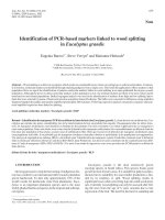
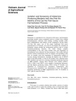
Fig.1 E.coli: Green metallic sheen appearance of colonies in Eosin Methylene Blue (EMB) agar
361
Int.J.Curr.Microbiol.App.Sci (2020) 9(8): 359-366
Fig.2 E.coli: Yellow slant, yellow butt and no H2S production in Tripe Sugar Iron (TSI) slant
Fig.3 A. paragallinarum: Transparent minute colonies in chocolate agar
Fig.4 A. paragallinarum: Gram-negative pleomorphic coccobacilli
in Gram staining Gram’s x 1000
362
Int.J.Curr.Microbiol.App.Sci (2020) 9(8): 359-366
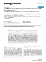
Fig.5 Pasteurella spp: Fine dew drops like colonies in Brain Heart Infusion (BHI) agar
Fig.6 Pasteurella spp: Bipolar organisms (arrows) in lung impression smear by Giemsa staining
Giemsa x1000
Fig.7 Staphylococcus spp: Minute yellow coloured colonies in mannitol salt agar
363
Int.J.Curr.Microbiol.App.Sci (2020) 9(8): 359-366
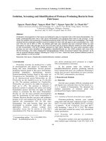
Fig.8 Pseudomonas spp.: Green colour discolouration of nutrient agar due to pyocyanin
production
Fig.9 Avibacterium paragallinarum: 500 bp PCR product of 16S rRNA gene
on 1.5% agarose gel
Fig.10 Mycoplasma gallisepticum: 530 bp PCR product of 16S rRNA gene on 1.5% agarose gel
364
Int.J.Curr.Microbiol.App.Sci (2020) 9(8): 359-366
Jones, R.C., Williams R.A., Baxter-Jones, C.,
Savage, C.E and Wilding, G.P. 1988.
Experimental infection of laying
turkeys with rhinotracheitis virus:
distribution of virus in the tissues and
serological response. Avian Pathol., 17:
841 - 850.
Kiss, I., Matiz, K., Kaszaryitzky, E., Chavez,
Y and Johansson, K.F. 1997. Detection
and
identification
of
avian
mycoplasmas by polymerase chain
reaction and restriction fragment length
polymorphism assay. Vet. Microbiol.,
58: 23 - 30.
Nakamura, K., Mase, M., Tanimura, N.,
Yamaguchi, S., Nakazawa, M and
Yuasa, N. 1997. Swollen head
syndrome in broiler chickens in Japan:
its pathology, microbiology and
biochemistry. Avian Pathol., 26: 139 154.
Nouri, A., Banani, M., Goudrzi, H.,.
Pourbakhsh, S.A and Mirzaei, S.G.
2014. Retrospective detection of
Avibacterium paragallinarum serovar
B in egg yolk materials by PCR.
Archives of Razi Institute. 69: 179 183.
OIE Terrestrial Manual. 2009. Turkey
rhinotracheitis (avian metapneumovirus infections). Chapter 2.3.15, pp 1 13.
Paul
McMullin.
1998.
Diagnosis,
management and control of avian
pneumovirus infection in broiler parent
chickens. Poultry Health Services.
Poultry Health Centre, Main Site Lane,
Dalton, Thirsk, North Yorkshire, YO7
3JA U.K.
Quinn, P.J., Carter, M.E., Markey, B and
Carter, G.R. 1994. Haemophilus
paragallinarum. In Clinical Veterinary
Microbiology. Mosby Year Book
Europe Limited. Wolfe. Pp. 277.
Quinn, P.J., Markey, B.K., Carter, M.E.,
Donnelly, W.J and Leonard, F.C. 2002.
Detection of secondary bacterial agents by
PCR
Avibacterium paragallinarum
Isolation and identification of Avibacterium
paragallinarum by PCR reduces the
complexity of the diagnostic task (Chen et al.,
1998). Avibacterium paragallinarum nucleic
acid was detected in 9 out of 48 flocks. DNA
was extracted by Genomic DNA purification
kit from the isolated colonies and screened for
16S rRNA gene which produced band at 500
bp in gel electrophoresis (Fig. 9). These
results are supported by Badouei et al., (2014)
who used a primer pair complementary to
specific gene designated for the detection of
AP. In this study, all culture positive samples
were also positive by PCR for Avibacterium
paragallinarum. This denotes that PCR could
be relied upon for detection of AP, thus
saving time and labour.
Mycoplasma gallisepticum
DNA was extracted by Genomic DNA
purification kit from triturated tissue samples
of trachea, lung and liver. The samples were
subjected to PCR to screen 16S rRNA gene of
MG at 530 bp (Fig. 10) and showed positivity
for 8 flocks. The obtained results are
concurred with earlier reports by Kiss et al.,
(1997) and Ramadass et al., (2006).
References
Barnes, H.J., Vaillancourt, J.P. and Gross,
W.B. 2003. Colibacillosis, In: Saif
Y.M. et al., (eds.): Diseases of Poultry,
11th Edn. Iowa State University Press,
Ames, Iowa, USA. pp. 631 - 652.
Chen, X., Chen, Q., Zhang, P., Feng, W and
Blackall, P.J. 1998. Evaluation of PCR
test for the detection of Haemophilus
paragallinarum in china. Avian
Pathol., 27: 296-300.
365
Int.J.Curr.Microbiol.App.Sci (2020) 9(8): 359-366
Veterinary Microbiology and Microbial
disease. Blackwell science Ltd.
Ramadass, P., Ananth, R., Senthilkumar,
T.M.A.,
Venkatesh,
G
and
Ramaswamy, V. 2006. Isolation and
characterization
of
Mycoplasma
gallisepticum
and
Mycoplasma
synoviae from poultry. Indian J. Anim.
Sci., 76 (10): 796 - 798.
Rhoades, K.R and Rimler, R.B. 1991.
Pasteurellosis, In: B.W. Calnek, H.J.
Barnes, C.W. Beard, W.M. Reid and
H.W. Yoder Jr (Eds), In Diseases of
Poultry, 9th edn. Iowa state university
press, Ames, Iowa. 145 - 171.
How to cite this article:
Ruthra, M., A. Arulmozhi, A.Balasubramaniam and Balasubramaniam, G. A. 2020. Isolation
and Identification of Bacteria from Swollen Head Syndrome (SHS) Affected Chicken Flock.
Int.J.Curr.Microbiol.App.Sci. 9(08): 359-366. doi: />
366