Discovery and anti-cancer evaluation of two novel non-ATP-competitive FGFR1 inhibitors in non-small-cell lung cancer
Bạn đang xem bản rút gọn của tài liệu. Xem và tải ngay bản đầy đủ của tài liệu tại đây (1.76 MB, 9 trang )
Wu et al. BMC Cancer (2015) 15:276
DOI 10.1186/s12885-015-1307-9
RESEARCH ARTICLE
Open Access
Discovery and anti-cancer evaluation of two
novel non-ATP-competitive FGFR1 inhibitors in
non-small-cell lung cancer
Jianzhang Wu1†, Tao Wei1†, Qinqin Tang1, Bixia Weng1, Wulan Li2, Xin Jiang1, Ting Ding3, Xiaokun Li1,
Guang Liang1, Yuepiao Cai1* and Jiansong Ji1,4*
Abstract
Background: Fibroblast growth factor receptor 1 (FGFR1) is correlated closely with the occurrence and development
of lung cancer. FGFR1 kinase inhibitors have exhibited significant therapeutic effects against non-small-cell lung cancer.
Recently, non-ATP competitive FGFR1 inhibitors have attracted extensive attention due to their low side effects.
Methods: Caliper Mobility Shift Assay was used for FGFR1 inhibition test and kinase inhibitory mode study. Hoechst
staining and Annexin V/PI staining were used to evaluate the cell apoptosis induction. Western blot were then
performed to confirm the intracellular FGFR1 inhibition and apoptotic protein expression. Finally, the anti-tumor effect
and mechanism of Af23 and Ad23 was evaluated in vivo.
Results: In this study, we designed, synthesized and discovered two novel non-ATP competitive FGFR1 inhibitors, Af23
and Ad23, using NDGA as a leading compound. They had IC50 values of 0.6 μM and 1.4 μM against FGFR1 kinase,
respectively. The kinase inhibitory assay carried at different ATP concentrations showed that the FGFR1 inhibition mode
of both Ad23 and Af23 was non-ATP-competitive. Further, Af23 and Ad23 significantly suppressed FGFR1 phosphorylation
and cell proliferation in non-small-cell lung cancer (NSLCLC) H460 cells and induced cell apoptosis. Af23 and Ad23 also
showed significant anti-tumor activity in the H460 xenograft mouse model, accompanied with the inhibition of FGFR1,
ERK, and AKT phosphorylation without exhibiting toxicity.
Conclusions: These results indicate that Ad23 and Af23 are potential agents for the treatment of non-small-cell lung
cancer. This work also provides a structural lead for the design of new non-ATP-competitive FGFR1 inhibitors.
Keywords: Fibroblast growth factor receptor 1, Non-small-cell lung cancer, Non-ATP competitive FGFR1 inhibitors,
NDGA, Anti-cancer
Background
Lung cancer is the most common cause of death from cancer worldwide, and non-small-cell lung cancer (NSCLC)
accounts for 85% of the total incidence [1]. Recently, molecular targeted chemotherapy with RTK inhibitors has
been used in clinics for the treatment of NSCLC because
of the importance of a series of receptor tyrosine kinases
(RTKs) in the development of NSCLC [1,2]. Smallmolecule inhibitors of epidermal growth factor receptor
* Correspondence: ;
†
Equal contributors
1
Chemical Biology Research Center, College of Pharmaceutical Sciences,
Wenzhou Medical University, Wenzhou, Zhejiang 325035, China
Full list of author information is available at the end of the article
(EGFR), such as gefitinib and erlotinib, have been approved by the U.S. Food and Drug Administration (FDA)
for the treatment of NSCLC [3,4]. Unfortunately, NSCLC
treatment remains challenging because of some problems,
including adverse effects and drug resistance associated
with the ATP-competitive kinase inhibition mode of the
majority of RTK inhibitors [5-7]. The ATP-binding pocket
is highly conserved among members of the kinase family,
and it is difficult to find selective agents [7,8]. Moreover,
the ATP-competitive inhibitors must compete with high
intracellular ATP levels leading to a discrepancy between
IC50s measured by biochemical versus cellular assays.
The non-ATP-competitive inhibitors, called type II or
type III inhibitors, offer the possibility of overcoming
© 2015 Wu et al.; licensee BioMed Central. This is an Open Access article distributed under the terms of the Creative
Commons Attribution License ( which permits unrestricted use, distribution, and
reproduction in any medium, provided the original work is properly credited. The Creative Commons Public Domain
Dedication waiver ( applies to the data made available in this article,
unless otherwise stated.
Wu et al. BMC Cancer (2015) 15:276
these problems [7,9]. Thus, the development of RTK inhibitors that do not compete with ATP is an urgent need
for the treatment of NSCLC.
Fibroblast growth factor receptors (FGFRs) belong to
the family of RTK superfamily, and FGFRs, especially
FGFR1, was reported to be highly related to the development and progress of NSCLC [1,10-17]. Amplification
and activating mutations of FGFR1 have been observed in
NSCLC [13,14,17], while silencing the expression of
FGFR1 by siRNA or pharmacological inhibition of FGFR1
by small molecules suppresses the development of NSCLC
[11,12,16,17]. These observations make FGFRs increasingly a attractive target for the therapeutic intervention
in cancer. Two FGFR inhibitors, namely AZD4547 and
BGJ398, have entered phase II clinical trials, while more
small-molecule inhibitors, such as SU5402, PD173074,
TKI-258, and SU6668, have failed in clinical trials due to
various complications associated with the side effects
caused by the ATP-competitive inhibition mode [8]. To
date, only five non-ATP-competitive FGFR inhibitors
have been identified, including nordihydroguaiaretic acid
(NDGA) [18], NF449 [19], ARQ069 [20], and recently reported A114 and A117 [21]. Despite advances in the field,
identifying highly-selective, small-molecule inhibitors
that target an inactive conformation or a new domain of
FGFR continues to be a significant challenge.
Page 2 of 9
In this report, we characterize two nordihydroguaiaretic
acid (NDGA) analogs, i.e., Ad23 and Af23, as two kinase
inhibitors that effectively target FGFR1 (Figure 1A). Furthermore, we provide data that indicates these kinase inhibitors have a distinct ATP-independent mode of action.
Furthermore, these two compounds have shown excellent
anti-cancer activity against NSCLC H460 cells both
in vitro and in vivo. These data further confirm that
bisaryl-1,4-dien-3-one structures could be used as nonATP-competitive FGFR1 inhibitors for the treatment of
NSCLC.
Methods
Cell lines, compounds, and reagents
NSCLC cell line, H460, was purchased from ATCC
(Manassas, VA). FGFR1-overexpressing HEK 293 cell
lines were kindly gifted by the Institute of Materia Medical, Xi’an Jiaotong University. Various compounds were
designed and synthesized in our laboratory, including
Af23 (3,5-bis(3,4-dihydroxybenzylidene)-tetrahydropyran4-one) and Ad23 (3,5-bis(3,4-dihydroxybenzylidene)-tetrahydrothiopyran-4-one) (Figure 1A). Their purity was
detected by HPLC (>98.0%) before they were used in
biological experiments. The compounds were dissolved
in DMSO solution for in vitro assay. Their watersoluble formulations for in vivo studies were prepared
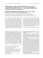
Figure 1 NDGA analogs Af23 and Ad23 inhibited FGFR1 activities in a non-ATP competitive manner. (A) The profile of design and FGFR1 kinase
inhibition assay of NDGA analogs. FGFR1 kinase inhibition rates of the compounds were evaluated by caliper mobility shift assay, and IC50 values
were calculated using conversion rates. The data were shown as a mean of 3–5 independent tests. (B) Af23 and Ad23 inhibit FGFR1 through a
mechanism that is independent of the concentration of ATP. Selective ATP-competitive kinase assay of compounds Ad23 (B), Af23 (C), and NDGA
(D) with FGFR1 was carried out through caliper mobility shift assay. The conversion data were fitted with Graphpad for global fitting, using “mixed
model inhibition”.
Wu et al. BMC Cancer (2015) 15:276
using the pharmaceutical method described previously
[22]. All the antibodies were purchased from Santa
Cruz Biotechnology (Santa Cruz, CA). Hoechst staining
kit was purchased from Beyotime Biotech (Nantong,
China). Recombinant FGFR1 proteins were obtained
from Carna Biosciences, Inc. (Kobe, Japan).
Cell-free FGFR1 kinase assays
Using the method described previously in Ref. [21], the
FGFR1 kinase inhibition assay of NDGA and its analogs
were performed by Caliper Mobility Shift Assay with
ATP concentration at its Km value (262 μM). Staurosporine was used as a positive control. For the determination of IC50, the compounds were tested in duplicate at
10 concentrations, ranging from 5 nM to 100 μM. Then,
conversion data were collected on Caliper EZ reader
(Hopkinton, MA). The IC50 values were obtained by
GraphPad Prism 5 (GraphPad, San Diego, CA).
Page 3 of 9
reactive bands were visualized by using an ECL kit (BioRad, Hercules, CA).
Hoechest staining
After the H460 cells were incubated with the compounds for 72 h, cells were stained with Hoechst 33342
dye according to the protocol provided with the kit
(Beyotime Biotech, Nantong, China). The cells were imaged under fluorescent microscope (Nikon, Tokyo, Japan),
and the pictures were taken at 200× objective.
Analysis of cell apoptosis
H460 cells were placed in 60-mm plates for 12 h and
then treated with NDGA, Ad23, or Af23 at the indicated
concentrations for 24 h. Then, the cells were harvested
and stained with annexin V and propidium iodide (PI) in
the presence of 100 mg/mL of RNAse and 0.1% Triton
X-100 for 30 min at 37°C. Flow-cytometric analysis was
performed using FACS calibur (BD Sciences, CA).
ATP competitive inhibition assay
This experiment was performed to test the relationship
between the compounds and ATP in which the concentration of the substrate was constant, while the concentrations of ATP were set at 4192, 2096, 1048, 524, 262,
131, 66, and 33 μM. The global competitive inhibition fit
for the compounds was performed based on percent
conversion = (Vmax*X)/{km*[(1 + I/Ki)n] + X}, where X
is the ATP concentration, and n is the Hill coefficient.
Specific details of this method were presented in a previous report [21].
MTT assay
The anti-proliferative activities of compounds were detected by MTT assay. H460 cells were seeded in a 96well plate with RPMI-1640 medium that contained 0.1%
FBS for 24 h. Then, the cells were treated with the compounds at the indicated concentrations (0.74, 2.22, 6.67,
20, and 60 μM) for 72 h. The proliferation of the H460
cells was detected through MTT assay, and the IC50
values were calculated by GraphPad software.
Western blot analysis
Cells or homogenated tumor tissues were lysated. Protein
concentrations in all the samples were determined by
using the Bradford protein assay kit (Bio-Rad, Hercules,
CA). The lysates were separated by SDS-PAGE electrophoresis, and electro-transferred to a 0.22-μm polyvinyldene difluoride membrane. After blocking with TBS that
contained 5% non-fat milk for 1.5 h at room temperature, the membranes were incubated with different
primary antibodies overnight at 4°C. Following the
TBST wash, immuno-reactive bands were detected by
incubating with respective secondary antibody conjugated with horseradish peroxidase for 1 h. Immuno-
In vivo anti-tumor study
All animal experiments complied with the Wenzhou
Medical College Policy on the Care and Use of Laboratory Animals (Wenzhou Medical College Animal Policy
and Welfare Committee, Document ID: 201100103).
Five to six-week-old athymic nu/nu female BALB/cA
mice (18–22 g) were purchased from the Animal Center
of the China Pharmaceutical University (Nanjing, China).
The animals were housed at a constant room temperature
with a 12:12-hr (light: dark) cycle and fed a standard rodent diet and water. H460 cells were harvested and mixed
with Matrigel at proportions of 1:1. Then, the cells were
injected subcutaneously into the right flank (2 × 10⁶ cells
in 200 μL of PBS) of 7-week-old, BALB/cA nude mice.
Two days after the H460 cells were injected, the mice
were injected intraperitoneally (i.p.) with a water-soluble
preparation of either compound Ad23 or compound Af23
in PBS at a dosage of 5 mg/kg/day for 28 days, whereas
the control mice were injected with the liposome vehicle
in PBS (n = 10 in each group). The volume of the tumors
were determined by measuring their length (l) and width
(w) and calculated using the formula; V = 0.52 × l × w2.
The weight of the tumors were recorded on the day the
mice were killed.
Immunohistochemistry analysis
On day 30 after tumor induction, the mice were killed in
a CO2 chamber, and the tumor tissues were dissected
and weighed. Some of the tissues were lysed for protein
isolation and then processed for the determination of
signaling pathway proteins using Western blot method.
A part of harvested tumor tissues were fixed in 10% formalin at room temperature overnight, processed, and
embedded in paraffin. The paraffin-embedded tissues
Wu et al. BMC Cancer (2015) 15:276
Page 4 of 9
were sectioned (5-μm thick) followed by staining with
primary antibodies. The signal was detected by biotinylated secondary antibody and developed with 3,3-diaminobenzidine (DAB).
ATP-competitive FGFR1 inhibitors, i.e., Ad23 and Af23,
from the leading NDGA.
Statistical analysis
The inhibitory effects of these two compounds on FGFR1
activation were determined in FGFR1-overexpressing 293
cells and human NSCLC H460 cells. As shown in
Figures 2A and B, pre-treatment with Ad23 or Af23
dose-dependently reduced the bFGF-induced phosphorylation of FGFR1 in both the cell lines. Also, both Ad23
and Af23 inhibited the phosphorylation of FRS2, a proliferative substrate of FGFR1, in a dose-dependent manner
in H460 cells (Figure 2C). Consistent with the cell-free
results, Ad23 and Af23 had greater activity than NDGA,
and Ad23 showed stronger inhibition than Af23 against
cellular FGFR1 phosphorylation.
All in vitro experiments were repeated at least three
times. Data were presented as means ± SD or mean ±
SEM. The statistical significance of differences between
groups was obtained by the student’s t test or ANOVA
multiple comparisons in GraphPad Prism 5 (License
Number: GPW5-415777-RAG-2191, GraphPad Software
Inc., San Diego, CA). P values less than 0.05 (p < 0.05)
were considered to be significant.
Results
Af23 and Ad23 inhibits FGFR1 kinase via a non-ATP
dependent manner
The leading compound NDGA is a natural product isolated from creosote bush. It exhibits multiple pharmacological effects, such as anti-oxidation, anti-inflammation,
and anti-tumor [18,23]. Recently, Meyer et al. found that
NDGA could inhibit the autophosphorylation of FGFR3
kinase both in vitro and in vivo [18]. In our previous
cell-free assay, we found that the IC50 values of NDGA
against FGFR1 and FGFR3 were 24.5 and 72.4 μM, respectively, indicating that NDGA exhibits better inhibitory activity against FGFR1 than FGFR3 (Figure 1A).
Therefore, using NDGA as a leading compound, we designed and synthesized a series of structural analogs
(Figure 1A). Next, we tested the inhibitory activity of
synthetic NDGA analogs against FGFR1 kinase by mobility shift assay.
The inhibitory potency of 72 bisaryl-1,4-dien-3-one
compounds against FGFR1 kinase was evaluated by
in vitro kinase assays. Out of the 72 compounds, Ad23
and Af23 were found to exhibit much stronger inhibition
against FGFR1 kinase activity than NDGA and other analogs (IC50: Ad23,0.6 μM; Af23,1.4 μM) (Figure 1A).
Thus these two were chosen for further studies. Subsequently, the kinase inhibition modes of both Ad23 and
Af23 were studied. As shown in Figure 1B, the velocity
of FGFR1 substrate phosphorylation without inhibitors
increases as the ATP concentration increased, and it was
reached to the peak at an ATP concentration of
2000 μM. At concentrations greater than the IC50 value
(1.4 μM for Ad23; 1.88 μM for Af23; 100 μM for
NDGA), the kinase activity was decreased by more than
90%, and further increases in the concentration of ATP,
even up to 4190 μM, had no effect on the inhibitory potency of the compounds (Figure 1B). These results
showed that the inhibition of FGFR1 kinase activity by
Ad23, Af23, and NDGA was not dependent on the concentration of ATP. Thus, we obtained two novel non-
Ad23 and Af23 inhibits the cellular FGFR1
phosphorylation
Ad23 and Af23 inhibited the proliferation and induced
the apoptosis of H460 cell line
Next, the growth inhibition of H460 cells by NDGA,
Ad23, and Af23 was studied by MTT assay. Our data
showed that Af23, Ad23, and NDGA exhibited marked
inhibitory effects against H460 cells (Figure 3A). The inhibitory potencies (IC50 values) of Af23 and Ad23 were
much greater when compared to NDGA. Further, Western
blot analysis showed that the cleavage of caspase-3 and
caspase-9 increased after treatment with Ad23 or Af23, indicating that these compounds could induce apoptosis in
H460 cells after a 12-h treatment (Figure 3B). Further,
Hoechst staining was performed 12 h after treatment with
the compounds (Figure 3C). A concentration-dependent
increase in the number of cells with nuclear condensation
and fragmentation was observed in both the groups. Next,
we assessed the effects of Ad23 and Af23 on the induction
of apoptosis in H460 cells by flow cytometric analysis.
Figure 3D shows that both Ad23 and Af23 dosedependently increased the H460 apoptosis after 24-h treatment. At a concentrations of 20 μM, both Ad23-treated
group (Annexin V+/PI+, 44.8 ± 7.07%) and Af23-treated
group (48.2 ± 11.12%) induced a greater rate of cell apoptosis than NDGA (27.2 ± 5.27%). We have also tested the
growth inhibition of Ad23 and Af23 against human liver
cells HL7702 and human fetal lung cell line MRC-5 which
expresses low level of FGFR1. The results showed that all
the IC50 values of Ad23, Af23, and NDGA against HL7702
or MRC-5 cells were greater than 30 μM (Figure 3A).
Ad23 and Af23 significantly suppressed the H460 tumor
growth in xenograft mouse model
In order to further assess the anti-tumor activities of the
NDGA analogs, we tested the efficacies of Af23 and
Ad23 in the H460 xenograft mouse model. Treatment
with Af23 or Ad23 for 28 days resulted in significant
Wu et al. BMC Cancer (2015) 15:276
Page 5 of 9
Figure 2 Compounds Ad23 and Af23 inhibited intracellular FGFR1/
FRS2 phosphorylation. FGFR1 over-expression 293 cells (A) or H460
cells (B and C) were pretreated with compounds at indicated
concentrations or vehicle (0.1% DMSO), respectively. Then, cells were
stimulated with bFGF (30 ng/mL) for 10 min, and the phosphorylation
levels of FGFR1 (A and B) and FRS2 (C) in cell lysates was measured by
western blot analysis. The figures were representative of 3 separate
experiments. The column figures show the normalized optical density
as a percentage of total protein control. Bars represent the mean ±
SEM of 3 independent experiments. Statistical significance relative to
bFGF alone group was expressed, *P <0.05, **P < 0.01, ***P < 0.001.
reduction in tumor volume (Figure 4A). The weight of
the tumors were also reduced markedly in the Af23treated and Ad23-treated groups, with the inhibition rate
of 67.4 and 75.8%, respectively (Figure 4B). To evaluate
whether the inhibition of tumor growth by Ad23 or
Af23 was associated with the inhibition of FGFR1 activity in vivo, we analyzed the expression of p-FGFR1 in
the tumor tissues. As shown in Figure 4C, Ad23 and Af23
exhibited a significant increased inhibitory effect on
FGFR1 phosphorylation in H460 tumor xenografts than
the vehicle control tumors. Also, no obvious toxicity was
observed in the Af23-treated group or the Ad23-treated
group, evidenced by no obvious loss of weight among the
two groups during the period of treatment (Figure 4D).
Prior studies have revealed that phosphorylation of
FGFR1 can lead to the activation of downstream signaling cascades including ERK and AKT, which plays an
important role in the proliferation and survival of cancer
cells. Thus, the phosphorylation of ERK and AKT in
tumor samples was tested by immunohistochemical assays (Figure 4E). Similarly, these compounds inhibited
the FGFR1-downstream ERK and AKT phosphorylation.
Finally, the immunochemical data also showed that the
expression of Bcl-2, Cyclin D1 and COX-2 was reduced
to a far greater extent in the Ad23-treated group and the
Af23-treated group than compared to the vehicle-treated
group (Figure 4E), indicating that these two FGFR1 inhibitors also induced tumor-cell apoptosis in H460
xenografts.
Discussion
FGFR1 is highly related to the development of lung cancer [10,12-15,17,24]. Ren et al. identified four NSCLC
cell lines and two, newly-established primary lungcancer cultures that showed high FGFR1 expression
levels [24]. They also found that treatment with ponatinib, an FGFR1 inhibitor, could inhibit cell growth in
NSCLC cell lines [24]. Terai et al. revealed that the activation of the FGF2-FGFR1 autocrine pathway may be a
novel mechanism of acquired resistance to the EGFR inhibitor, gefitinib, in NSCLC [12]. There has been increasing evidence indicating that FGFR1 inhibitors could
Wu et al. BMC Cancer (2015) 15:276
Page 6 of 9
Figure 3 Af23 and Ad23 inhibited proliferation and induced apoptosis in H460 cells. (A) H460 cells were treated with Af23, Ad23 or NDGA at
different concentration (0.74, 2.22, 6.67, 20, and 60 μM) for 72 h. The viability of H460 cells were detected by MTT assay, and the IC50 values of
compounds were fitted with GraphPad. (B) Effects of Af23 and Ad23 on caspase activation in H460 cells. H460 cells were harvested and lysated
after incubated with Af23 (10 μM), Ad23 (10 μM), or NDGA (10 μM) for 12 h. The levels of cleaved caspase-3 and cleaved caspase-9 were
determined by western blot analysis. (C) Morphological changes and hoechst staining were observed in H460 cells cultured with and without
Af23, Ad23, or NDGA at indicated concentrations for 12 h (200×). The figures were representative of more than three separate experiments.
D. Af23 and Ad23 induced cell apoptosis in H460 cells. H460 cells were treated with Af23, Ad23, or NDGA at indicated concentrations for 24 h,
and then stained with Annexin V and PI, followed by detection using flow cytometry. The representative pictures are shown.
be a promising candidate for the treatment of NSCLC in
clinics. Although a variety of ATP-competitive FGFR1
inhibitors with therapeutic prospects for lung cancer
have been identified, most of them failed in pre-clinical
or clinical studies because of their low efficacy or high
toxicity. At present only two selective FGFR1 inhibitors,
i.e., AZD4547 and BGJ398, are being studied in clinical
trials (phase II) [8]. The ATP-competitive FGFR1 inhibitors functions by targeting the ATP-binding pocket of
FGFR1, which may lead to a decrease in their efficiency
when high physiological or intracellular concentrations
of ATP exist [8]. In addition, since the ATP-binding site
is highly conserved in RTKs, most inhibitors exhibit limited selectivity within RTKs, which induces side effects
during treatment, such as nausea, weakness, and elevated blood pressure [25]. For instance, PD173074 and
SU5402 failed to enter phase II clinical trials due to their
high toxicities [25]. Therefore, the exploration of nonATP-competitive FGFR1 inhibitors has attracted extensive attention in recent years.
NDGA was reported previously to inhibit FGFR3 kinase [18]. In this study, we found that NDGA exhibited
better inhibitory activity against FGFR1 than FGFR3.
Using NDGA as a leading compound, we designed and
synthesized several NDGA analogs with the basic skeleton of bisaryl-1,4-dien-3-one (Figure 1A). After screening for kinase inhibition, we obtained two FGFR1
inhibitors (Ad23 and Af23) that had inhibitory activities
better than compared to NDGA. Interestingly, NDGA
and its analogs retained their potency for the inhibition
of kinase activity when the concentration of ATP increased, suggesting that the inhibitory effects of these
compounds were independent of the concentration of
ATP (Figure 1B). So far, ARQ069 is the only published
molecule that inhibits FGFR1/2 in a manner that does not
compete with ATP. ARQ069 exhibits FGFR1/2 kinase inhibition with IC50 values of 0.84 and 1.23 μM, respectively
[20]. Although the IC50 values of Ad23, Af23, and
ARQ069 are at micromolar concentration levels, the observation that the inhibition of FGFR1 autophosphorylation
Wu et al. BMC Cancer (2015) 15:276
Page 7 of 9
Figure 4 Anti-tumor effects of compounds Af23 and Ad23 in H460 xenograft models. Xenografts were established in nude mice. Two days after
the mice were treated with liposome vehicle (once daily, i.p.), Af23 (once daily, i.p., 5 mg/kg/day) or Ad23 (once daily, i.p., 5 mg/kg/day) for
28 days (n = 10 in each group). (A) Tumor volume (mm3) and (B) Tumor weight (g) were recorded (n = 10). Points, mean of 10 mice; bars, SD.
**P < 0.01. (C) pFGFR1 expressions in tumor tissues were detected by Western Blot with FGFR1 as internal control (3 mice in each group were
used); (D) Body weight of each group was recorded (n = 10 in each group); (E) The levels of p-ERK, p-AKT, Bcl-2, CyclinD1, and COX-2 in tumor
tissues were detected by immunohistochemical staining (6 mice in each group were used). Representative pictures are shown.
(Figure 2) and the inhibition of the direct FGFR1 downstream substrate (Figure 1A) occur at different dose levels
was consistently detectable. A literature search on kinase
inhibitors yielded several reports describing that nonATP-competitive kinase inhibitors may not display identical IC50 values with that of classic ATP-competitive
inhibitors for the inhibition of both kinase autophosphorylation and downstream signaling pathways [20,24]. Our
novel NDGA analogs (Ad23 and Af23) also showed
micromole-grade FGFR1 inhibitory effects in a manner
that was independent of ATP.
Figure 2 further revealed the anti-FGFR1 ability of
Ad23 and Af23 in the cellular levels using FGFR1overexpressed 293 cells and NSCLC H460 cells. These
two compounds also dose-dependently inhibited FRS2
phosphorylation in H460 cells. These data led us to
investigate the anti-cancer efficacy of Ad23 and Af23
further. We evaluated the anti-proliferative effects of
Ad23 and Af23 in vitro. As shown in Figure 3, Ad23 and
Af23, as well as NDGA, inhibited cell growth and induced cell apoptosis in NSCLC H460 cells; they also had
low toxicities against normal MRC-5 cells and normal
human liver cells (HL-7702 cells). Further, we showed
the potent anti-tumor ability of these two inhibitors in
the H460 xenograft mouse model. Previously, studies on
ARQ069, a non-ATP-competitive inhibitor, did not report the in vivo anti-tumor activity. In the present study,
the data revealed that the compounds Ad23 and Af23
suppressed the phosphorylation of FGFR1 in H460
tumor tissues, thereby inhibited the downstream phosphorylation of ERK and AKT, and reduced the expression of BCL-2, COX-2, and Cyclin D1 (Figure 4). At the
Wu et al. BMC Cancer (2015) 15:276
same time, we observed that Ad23 and Af23 exhibited
high safety in vivo (Figure 4D). As already known, the
ATP-competitive inhibitory mode leads to the biggest
problems (toxicity and side effects) of current FGFR1 inhibitors. Our findings for Ad23 and Af23 indicated that
the non-ATP-competitive FGFR1 inhibition might be a
new cancer therapeutic alternative with much lower toxicity in vivo. However, continued research is needed to
examine the underlying FGFR1-binding mechanism and
preclinical evaluation of these two compounds. In spite
of the predicted “DFG-OUT” docking model, it is unclear how Ad23/Af23 exactly binds to the FGFR1 kinase
domain or other domains, which needs to be demonstrated by X-ray diffraction-based structural biology
study. Also, it is important to test the RTK-inhibitory selectivity of these two compounds at both molecular and
cellular levels in the future.
Conclusion
Given the critical importance of FGFR1 in the pathogenesis and development of NSCLC, this study identified
two novel, non-ATP-competitive inhibitors of FGFR1
kinase, i.e., Ad23 and Af23, both of which exhibited
good anti-tumor activity in vitro and in vivo. These two
compounds were shown to have the potential to be developed as novel agents for the treatment of NSCLC.
The results of the present study indicate that Ad23 and
Af23deserve further studies both in the pre-clinical
evaluation and in the field of medicinal chemistry for developing structurally different and more effective bisaryl1,4-dien-3-one-containing FGFR1 inhibitors.
Competing interests
The authors declare that they have no competing interests.
Authors’ contributions
YC and JW analyzed data and wrote the paper; JW, TW, QT, BW, XJ, and WL
performed experiments; GL, XL, and JJ designed the research project and
experiments, JJ and YC analyzed data and revised the paper; TD and GL
revised the paper. All authors read and approved the final manuscript.
Acknowledgements
The work was supported by National Natural Science Foundation of China
(81173083, 81272462, and 81473242), Natural Science Funding of Zhejiang
Province (LY13H160022, LY12H16003, and LY12H30005), Key Science Funding
of Zhejiang Department of Health (WKJ2013-2-021), and Zhejiang Key Group
Project in Scientific Innovation (2010R50042).
Author details
1
Chemical Biology Research Center, College of Pharmaceutical Sciences,
Wenzhou Medical University, Wenzhou, Zhejiang 325035, China. 2College of
Information Science and Computer Engineering, Wenzhou Medical
Universtiy, Wenzhou, Zhejiang 325035, China. 3Department of Pharmacy, the
Sixth Affiliated Hospital of Wenzhou Medical University, Lishui, Zhejiang
323000, China. 4Department of Interventional Radiology, The Fifth Affiliated
Hospital of Wenzhou Medical University, Lishui, Zhejiang 323000, China.
Received: 4 June 2014 Accepted: 31 March 2015
Page 8 of 9
References
1. Kono SA, Marshall ME, Ware KE, Heasley LE. The fibroblast growth factor
receptor signaling pathway as a mediator of intrinsic resistance to EGFRspecific tyrosine kinase inhibitors in non-small cell lung cancer. Drug Resist
Updat. 2009;12(4–5):95–102.
2. Saijo N. Targeted therapies: Tyrosine-kinase inhibitors–new standard for
NSCLC therapy. Nat Rev Clin Oncol. 2010;7(11):618–9.
3. Sordella R, Bell DW, Haber DA, Settleman J. Gefitinib-sensitizing EGFR
mutations in lung cancer activate anti-apoptotic pathways. Science.
2004;305(5687):1163–7.
4. Horn L, Sandler A. Epidermal growth factor receptor inhibitors and
antiangiogenic agents for the treatment of non-small cell lung cancer. Clin
Cancer Res. 2009;15(16):5040–8.
5. Zhou W, Ercan D, Chen L, Yun CH, Li D, Capelletti M, et al. Novel mutantselective EGFR kinase inhibitors against EGFR T790M. Nature.
2009;462(7276):1070–4.
6. Hammerman PS, Janne PA, Johnson BE. Resistance to Epidermal Growth
Factor Receptor Tyrosine Kinase Inhibitors in Non-Small Cell Lung Cancer.
Clin Cancer Res. 2009;15(24):7502–9.
7. Zhang J, Yang PL, Gray NS. Targeting cancer with small molecule kinase
inhibitors. Nat Rev Cancer. 2009;9(1):28–39.
8. Liang G, Liu Z, Wu J, Cai Y, Li X. Anticancer molecules targeting fibroblast
growth factor receptors. Trends Pharmacol Sci. 2012;33(10):531–41.
9. Fang Z, Grutter C, Rauh D. Strategies for the selective regulation of kinases
with allosteric modulators: exploiting exclusive structural features. ACS
Chem Biol. 2013;8(1):58–70.
10. Kohler LH, Mireskandari M, Knosel T, Altendorf-Hofmann A, Kunze A,
Schmidt A, et al. FGFR1 expression and gene copy numbers in human lung
cancer. Virchows Arch. 2012;461(1):49–57.
11. Dutt A, Ramos AH, Hammerman PS, Mermel C, Cho J, Sharifnia T, et al.
Inhibitor-sensitive FGFR1 amplification in human non-small cell lung cancer.
PLoS One. 2011;6(6):e20351.
12. Terai H, Soejima K, Yasuda H, Nakayama S, Hamamoto J, Arai D, et al.
Activation of the FGF2-FGFR1 autocrine pathway: a novel mechanism
of acquired resistance to gefitinib in NSCLC. Mol Cancer Res.
2013;11(7):759–67.
13. Tran TN, Selinger CI, Kohonen-Corish MR, McCaughan BC, Kennedy CW,
O’Toole SA, et al. Fibroblast growth factor receptor 1 (FGFR1) copy number
is an independent prognostic factor in non-small cell lung cancer. Lung
Cancer. 2013;81(3):462–7.
14. Preusser M, Berghoff AS, Berger W, Ilhan-Mutlu A, Dinhof C, Widhalm G,
et al. High rate of FGFR1 amplifications in brain metastases of squamous
and non-squamous lung cancer. Lung Cancer. 2014;83(1):83–9.
15. Heist RS, Mino-Kenudson M, Sequist LV, Tammireddy S, Morrissey L,
Christiani DC, et al. FGFR1 amplification in squamous cell carcinoma of the
lung. J Thorac Oncol. 2012;7(12):1775–80.
16. Yang J, Zhao H, Xin Y, Fan L. MicroRNA-198 inhibits proliferation and
induces apoptosis of lung cancer cells via targeting FGFR1. J Cell Biochem.
2013. doi:10.1002/jcb.24742.
17. Zhang J, Zhang L, Su X, Li M, Xie L, Malchers F, et al. Translating the
therapeutic potential of AZD4547 in FGFR1-amplified non-small cell lung
cancer through the use of patient-derived tumor xenograft models. Clin
Cancer Res. 2012;18(24):6658–67.
18. Meyer AN, McAndrew CW, Donoghue DJ. Nordihydroguaiaretic acid inhibits
an activated fibroblast growth factor receptor 3 mutant and blocks
downstream signaling in multiple myeloma cells. Cancer Res.
2008;68(18):7362–70.
19. Krejci P, Murakami S, Prochazkova J, Trantirek L, Chlebova K, Ouyang Z, et al.
NF449 is a novel inhibitor of fibroblast growth factor receptor 3 (FGFR3)
signaling active in chondrocytes and multiple myeloma cells. J Biol Chem.
2010;285(27):20644–53.
20. Eathiraj S, Palma R, Hirschi M, Volckova E, Nakuci E, Castro J, et al. A novel
mode of protein kinase inhibition exploiting hydrophobic motifs of
autoinhibited kinases: discovery of ATP-independent inhibitors of fibroblast
growth factor receptor. J Biol Chem. 2011;286(23):20677–87.
21. Wang Y, Cai Y, Ji J, Liu Z, Zhao C, Zhao Y, et al. Discovery and identification
of new non-ATP competitive FGFR1 inhibitors with therapeutic potential on
non-small-cell lung cancer. Cancer Lett. 2014;344:82–9.
22. Wu J, Li J, Cai Y, Pan Y, Ye F, Zhang Y, et al. Evaluation and discovery of
novel synthetic chalcone derivatives as anti-inflammatory agents. J Med
Chem. 2011;54(23):8110–23.
Wu et al. BMC Cancer (2015) 15:276
Page 9 of 9
23. Lu JM, Nurko J, Weakley SM, Jiang J, Kougias P, Lin PH, et al. Molecular
mechanisms and clinical applications of nordihydroguaiaretic acid (NDGA)
and its derivatives: an update. Med Sci Monit. 2010;16(5):RA93–100.
24. Ren M, Hong M, Liu G, Wang H, Patel V, Biddinger P, et al. Novel FGFR
inhibitor ponatinib suppresses the growth of non-small cell lung cancer
cells overexpressing FGFR1. Oncol Rep. 2013;29(6):2181–90.
25. Liang G, Chen G, Wei X, Zhao Y, Li X. Small molecule inhibition of fibroblast
growth factor receptors in cancer. Cyto Growth Factor Rev. 2013;24(5):467–75.
Submit your next manuscript to BioMed Central
and take full advantage of:
• Convenient online submission
• Thorough peer review
• No space constraints or color figure charges
• Immediate publication on acceptance
• Inclusion in PubMed, CAS, Scopus and Google Scholar
• Research which is freely available for redistribution
Submit your manuscript at
www.biomedcentral.com/submit