FGF9 from cancer-associated fibroblasts is a possible mediator of invasion and anti-apoptosis of gastric cancer cells
Bạn đang xem bản rút gọn của tài liệu. Xem và tải ngay bản đầy đủ của tài liệu tại đây (992.46 KB, 9 trang )
Sun et al. BMC Cancer (2015) 15:333
DOI 10.1186/s12885-015-1353-3
RESEARCH ARTICLE
Open Access
FGF9 from cancer-associated fibroblasts is a possible
mediator of invasion and anti-apoptosis of gastric
cancer cells
Chao Sun1,2†, Hirokazu Fukui1*†, Ken Hara1, Xinxing Zhang1,3, Yoshitaka Kitayama1, Hirotsugu Eda1, Toshihiko Tomita1,
Tadayuki Oshima1, Shojiro Kikuchi4, Jiro Watari1, Mitsuru Sasako4 and Hiroto Miwa1
Abstract
Background: Cancer-associated fibroblasts (CAFs), which reside around tumor cells, are suggested to play a pivotal
role in tumor progression. Here we performed microarray analyses to compare gene expression profiles between CAFs
and non-cancerous gastric fibroblasts (NGFs) from a patient with gastric cancer and found that fibroblast growth factor
9 (FGF9) was a novel growth factor overexpressed in CAFs. We then examined the biological effects of FGF9 during
progression of gastric cancer.
Methods: Expression of FGF9 in CAFs and NGFs, and their secreted products, were examined by Western blotting.
The effects of FGF9 on AGS and MKN28 gastric cancer cells in terms of proliferation, invasion and anti-apoptosis were
assessed by WST-1 assay, invasion chamber assay and FACS, respectively. Furthermore, the intracellular signaling by
which FGF9 exerts its biological roles was examined in vitro.
Results: FGF9 was strongly expressed in CAFs in comparison with NGFs, being compatible with microarray data
indicating that FGF9 was a novel growth factor overexpressed in CAFs. Treatment with FGF9 promoted invasion and
anti-apoptosis through activation of the ERK and Akt signaling pathways in AGS and MKN28 cells, whereas these effects
were attenuated by treatment with anti-FGF9 neutralizing antibody. In addition, FGF9 treatment significantly enhanced
the expression of matrix metalloproteinase 7 (MMP7) in both cell lines.
Conclusions: FGF9 is a possible mediator secreted by CAFs that promotes the anti-apoptosis and invasive capability of
gastric cancer cells.
Keywords: FGF, Cancer-associated fibroblast, Invasion, Anti-apoptosis, ERK, Akt, Gastric cancer
Background
The formation of cancerous lesions is intimately associated with their unique microenvironment. Progression is
closely correlated with the capability of cancer cells to recruit and activate the surrounding stromal cells, and subsequently exploit them [1]. Cancer-surrounding stromal
cells, such as fibroblasts, endothelial cells, and immune
cells, can orchestrate tumorigenesis and metastasis
through cell-to-cell interaction and/or production of soluble growth factors/cytokines/chemokines [1-3]. In this
* Correspondence:
†
Equal contributors
1
Division of Gastroenterology, Department of Internal Medicine, Hyogo
College of Medicine, l-1, Mukogawa, Nishinomiya 663-8501, Japan
Full list of author information is available at the end of the article
context, fibroblasts in cancerous lesions are known as
cancer-associated fibroblasts (CAFs) and have received
much attention with regard to their role in tumor progression [4,5]. Although CAFs are known to be largely different from normal fibroblasts in non-neoplastic tissues in
terms of their gene profile, the mechanism by which CAFs
promote tumor progression is unclear. Therefore, we
compared the gene expression profiles of CAFs, focusing
especially on growth factors/cytokines/chemokines, with
those of non-cancerous gastric fibroblasts (NGFs) using
microarray assay, and subsequently isolated fibroblast
growth factor 9 (FGF9) as a novel gene that was overexpressed in CAFs in gastric cancer.
FGF9, a secretory protein of the FGF family, is reportedly expressed in stromal cells including fibroblasts [6-8].
© 2015 Sun et al.; licensee BioMed Central. This is an Open Access article distributed under the terms of the Creative
Commons Attribution License ( which permits unrestricted use, distribution, and
reproduction in any medium, provided the original work is properly credited. The Creative Commons Public Domain
Dedication waiver ( applies to the data made available in this article,
unless otherwise stated.
Sun et al. BMC Cancer (2015) 15:333
In general, FGF signaling occurs via FGF receptors
(FGFRs) to regulate a variety of cell biological behavior, including proliferation, differentiation, survival and motility
[9], and expression of FGF9, FGFR2c, FGFR3b and
FGFR3c has been detected in gastric and colon cancers
[10]. Thus, like other FGF family proteins, FGF9 may
play a pivotal role in the interaction between cancer
cells and their surrounding stromal cells, and it is noteworthy that FGF9 is strongly expressed in CAFs in gastric cancer. In the present study, we screened for
differences in gene expression between CAFs and NGFs
from a patient with gastric cancer. We examined the effect of FGF9 on proliferation, invasion and antiapoptosis of gastric cancer cells, and moreover clarified
the intracellular signaling by which FGF9 exerts its biological effects on gastric cancer cells.
Methods
Reagents and cell culture
Human recombinant FGF9 and anti-human FGF9 neutralizing antibody were purchased from R&D Systems
(Minneapolis, MN, USA). Anti-extracellular signal-regulated
protein kinase (ERK), anti-phospho-specific ERK (p-ERK),
anti-Akt, anti-phospho-specific Akt (p-Akt; Ser473), and
anti-β-actin antibodies were purchased from Cell Signaling
Technology (Beverly, MA, USA).
The gastric cancer cell lines AGS was cultured in
Ham’s F-12 medium (Sigma, Aurora, Ohio, USA) with
10% fetal bovine serum (FBS; Biowest, Nuaillé, France)
in a humidified incubator at 37°C with an atmosphere of
5% CO2. Similarly, MKN28 was cultured in RPMI 1640
medium with 10% fetal bovine serum, and other five gastric cancer cell lines MKN1, MKN 45, MKN74, GCIY, and
KATOIII were maintained as previously described [11].
Page 2 of 9
studies were done with the approval of the Review Board
of Hyogo College of Medicine, and informed consent was
obtained from the patient.
Microarray analysis
Using Trizol reagent (Invitrogen, Carlsbad, CA, USA),
total RNA was extracted from three sets of CAFs and
NGFs cultured. cDNA labeling, hybridizations, scanning
and data analysis were performed by Hokkaido System
Science Co., Ltd (Sapporo, Japan). Briefly, cyanine-3
(Cy3)-labeled cRNA was prepared from total RNA (0.05
μg) using a Low Input Quick Amp Labeling Kit (Agilent)
in accordance with the manufacturer’s instructions,
followed by RNAeasy column purification (QIAGEN,
Valencia, CA). Dye incorporation and cRNA yield were
checked with a NanoDrop ND-1000 Spectrophotometer.
Cy3-labeled cRNA (0.60 μg) was fragmented at 60°C for
30 min in a reaction volume of 25 μl containing 1x Agilent
fragmentation buffer and 2x Agilent blocking agent in accordance with the manufacturer’s instructions. On completion of the fragmentation reaction, 25 μl of 2x Agilent
hybridization buffer was added to the fragmentation mixture and hybridized to Agilent SurePrint G3 Human Gene
Expression Microarray (8x60K ver.2.0) for 17 h at 65°C in
a rotating Agilent hybridization oven. After hybridization,
the microarrays were washed for 1 min at room
temperature with GE Wash Buffer 1 (Agilent) and for 1
min at 37°C with GE Wash buffer 2 (Agilent), then dried
immediately by brief centrifugation. Slides were scanned
immediately after washing on an Agilent DNA Microarray
Scanner (G2565CA) using one color scan setting for
8x60K array slides (Scan Area 61×21.6 mm, Scan resolution 3μm, Dye channel for Green PMT set to 100%).
The scanned images were analyzed and normalized with
Feature Extraction Software 10.7.3.1 (Agilent).
Isolation and culture of human gastric fibroblasts
Human gastric cancer (poorly-differentiated adenocarcinoma) specimens were obtained from a patient who
underwent gastrectomy at Hyogo College of Medicine
Hospital in 2012. Cancer-associated fibroblasts (CAFs)
were prepared from the cancerous portion in the stomach.
Non-cancerous gastric fibroblasts (NGFs) were prepared
from non-cancerous portion with atrophic gastritis at least
50 mm far from tumor in the stomach. The tissue specimens were trimmed of fat and necrotic tissue, minced
with scalpels and washed in PBS containing antibioticantimycotic reagent (Anti-Anti®, GIBCO). The tissue
pieces were transferred to a 12-well microplate (IWAKI,
Tokyo, Japan) at one fragment/well. The cells were cultured in DMEM medium (GIBCO, Grand Island, NY,
USA) with 10% heat-inactivated FBS at 37°C in an atmosphere of 5% CO2. The fibroblasts that initially grew in
a monolayer were collected, transferred to another dish
and used for experiments within the 10th passage. These
RNA extraction and reverse transcription-polymerase
chain reaction (RT-PCR)
Total RNA was extracted from gastric cancer cell lines using
Trizol reagent (Invitrogen). Four microgram of total RNA
was reverse-transcribed by using oligo dT (Applied Biosystems, Branchburg, NJ, USA) and 200 U of Superscript™ II
reverse transcriptase (Invitrogen) in a total volume of 20μl.
For the following PCR, pairs of oligonucleotide primers
for human FGFRs were prepared as previously described
[12]. Human FGFR2c: 5′-TGGTCGGAGGAGACGTAGA
G-3′ (Forward) and 5′-AAAGTTACATTCCGAATATAGA
GAACC-3′ (Reverse); human FGFR3b: 5′-GGAGTTCCA
CTGCAAGGTGT-3′ (Forward) and 5′ -GTGAACGCTCA
GCCAAAAG-3′ (Reverse); human FGFR3c: 5′-GGAGTTC
CACTGCAAGGTGT-3′ (Forward) and 5′-AAGCGGGAG
ATCTTGTGC-3′ (Reverse); human GAPDH: 5′-GGCTGC
TTTTAACTCTGGTA-3′ (Forward) and 5′-ATGCCAGT
GAGCTTCCCGT-3′ (Reverse). One microliter of RT
Sun et al. BMC Cancer (2015) 15:333
product (cDNA) was amplified by PCR in a 50-μl reaction
volume containing 20 pmol of the above sets of primers,
1.25 U of Ampli-Taq DNA polymerase (Applied Biosystems,
Foster City, Calif., USA), and the final PCR buffer: 20 mM
Tris–HCl (pH 8.4), 50 mM KCl, 2.5 mM MgCl2, 10 mM
dithiothreitol, and 1 mM dNTP. The PCR amplification
was performed as follows: for FGFRs, at 95°C for 5 min
once; 40 cycles at 95°C for 30 s, at 57°C for 30 s, and at
72°C for 1 min; then at 72°C for 7 min; for GAPDH, at
95°C for 7 min once; 40 cycles at 95°C for 30 s, at 55°C
for 1 min, and at 72°C for 30 sec; then at 72°C for 7 min.
Real-time RT-PCR
Real-time RT-PCR was performed using 7900H Fast
Real-Time PCR System (Applied Biosystem) as previously described [13]. The following sets of primers for
human matrix metalloproteinase 2 (MMP2), MMP3,
MMP7, MMP9, and GAPDH were prepared (Additional
file 1: Table S1). Real-time RT-PCR assays were carried
out with 200 ng RNA equivalent cDNA, SYBR Green
Master Mix (Applied Biosystems), and 500 nmol/l gene
specific primers. The PCR cycling conditions were 50°C
for 15 s, and 60°C for 60 s. The intensity of the fluorescent dye was determined, and each of mRNA expression
levels was normalized to GAPDH mRNA expression
levels.
Page 3 of 9
using a microscope in five different visual fields (magnification, x200).
Apoptosis assay
AGS (2 × 10⁵) and MKN28 (2.5 × 10⁵) cells were seeded
in six-well plates in routine medium for 24 h. The cells
were then deprived of serum and treated with or without
recombinant FGF9 (1–10 ng/ml) for 48h. To inhibit the
effects of FGF9, anti-FGF9 antibody (1 μg/ml) was also
added to the culture medium. After treatment, both floating and attached cells were harvested, washed with PBS
and stained with AnnexinV-FITC and propidium iodide
(PI) using a MEBCYTO Apoptosis Kit (MBL, Nagoya,
Japan). Stained cells were analyzed on a FACScalibur flow
cytometer (Becton Dickinson, Franklin Lakes, NJ, USA),
and the data obtained were analyzed using CELLQUEST
software (Becton Dickinson, Mountain View, CA, USA).
Western blot analysis
Western blot analyses were performed as described previously [14]. Briefly, after treatment with or without reagent,
cells were lysed in protein extraction buffer, and protein
extract (30 μg) was fractioned by sodium dodecyl sulfate
polyacrylamide gel electrophoresis and transferred to a
nitrocellulose blotting membrane. The membrane was
Cell proliferation assay
AGS (4 × 10³) and MKN28 cells (1 × 10⁴) were seeded in
complete medium in 96-well microplates. The medium
was then replaced with one containing recombinant
FGF9 (0—10 ng/ml). WST-1 solution was added after 72
h incubation, and the plates were incubated at 37°C for
1 h. The plates were analyzed using an ELISA plate
reader at 450 nm with the reference wavelength set at
600 nm.
Cell invasion assay
Cell invasion assay was performed using BioCoat Matrigel
invasion chambers (BD Biosciences, Bedford, MA, USA)
according to the manufacturer’s protocol. Briefly, AGS
cells (1 × 10⁵) or MKN28 cells (3 × 10⁵) were seeded in
the insert of the Matrigel-coated invasion chamber (24
wells, 8-μm pore size) filled with serum-free medium containing different concentrations of FGF9 (0–10 ng/ml).
Then, the cells were incubated with medium containing
10% FBS in the lower chamber at 37°C in 5% CO2. To inhibit the effects of FGF9, anti-FGF9 antibody (1 μg/ml)
was also added to the upper chamber. After incubation for
27 h, non-invading cells were removed using a cotton
swab and the cells that had invaded into the lower surface
of the membrane were fixed with ethanol. The invading
cells were then stained with hematoxylin and counted
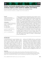
Figure 1 Expression of FGF9 and its receptors in CAFs and gastric cancer
cells. (A) Morphology of gastric CAFs and NGFs. (B) Production of FGF9 in
gastric CAFs, NGFs and their conditioned medium (CM). (C) Expression of
FGF9 in CAFs of the gastric cancer lesion. Arrows indicating CAFs.
(D) Expression of FGF receptors responsible for FGF9 in gastric
cancer cell lines.
Sun et al. BMC Cancer (2015) 15:333
Page 4 of 9
incubated with a primary antibody and then with a
peroxidase-conjugated secondary antibody. Proteins were
detected using an enhanced chemiluminescence system
(Amersham Biosciences, Buckinghamshire, UK).
Immunohistochemistry
A total of 20 gastric cancers tissues were obtained from
specimens resected surgically at Hyogo College of Medicine.
The tissue specimen were fixed in 10% formalin solution
and embedded in paraffin. This study was approved by the
Review Board of Hyogo College of Medicine, and informed consent was obtained from all patients. The characteristics of gastric cancer patients were showed in
Additional file 2: Table S2.
Immunohistochemical staining for FGF9 was performed
with an LSAB+ kit using anti-FGF9 antibody (1:40; R&D
Systems, Minneapolis, MN, USA) as described previously
[15]. Finally, the sections were incubated in 3,3′-diaminobenzide tetrahydrochloride with 0.05% H2O2 for 3 min,
and then counterstained with Mayer’s haematoxylin. To
evaluate the immunoreactivity of FGF9, at least five
different visual fields were observed at the invasive front
of gastric cancer lesions. A specimen was considered positive when FGF9-positive fibroblastic nests were observed
in the visual fields examined.
Statistics analysis
All values were expressed as the mean ± SD. The data
were analyzed using unpaired two-tailed t-test. P values
of less than 0.05 were considered to indicate statistical
significance.
Results
Microarray analyses of CAFs in gastric cancer tissues
We isolated CAFs and NGFs (Figure 1A) and compared
the gene expression profile of CAFs with that of NGFs
using microarray assay. Ten representative genes that
were upregulated in CAFs are listed in Table 1. Among
these genes, we targeted FGF9 as the most highly
expressed gene to examine the role of this CAFproduced growth factor on gastric cancer cells, and in
Table 1 Representative genes differentially expressed in CAFs from NGFs
Accession No.
Symbol
Gene name
Fold
change
NM_014333
CADM1
Cell adhesion molecule 1, transcript variant 1
273.6
CB178477
XLOC_l2_007424
gb|is39c09.y1 HR85 islet Homo sapiens cDNA clone
IMAGE:6554705 5′, mRNA sequence
254.8
NM_001113207
TSTD1
Thiosulfate sulfurtransferase (rhodanese)-like domain
containing 1, transcript variant 1
237.5
NM_000867
HTR2B
5-hydroxytryptamine (serotonin) receptor 2B
171.5
NM_001008539
SLC7A2
Solute carrier family 7 (cationic amino acid transporter,
y + system), member 2, transcript variant 2
142.5
NM_002010
FGF9
Fibroblast growth factor 9 (glia-activating factor)
141.1
NM_005559
LAMA1
Laminin, alpha 1
119.0
Up-regulated
NM_001040058
SPP1
Secreted phosphoprotein 1, transcript variant 1
116.1
A_24_P247454
A_24_P247454
Unknown
112.6
NM_014398
LAMP3
Lysosomal-associated membrane protein 3
111.3
Down-regulated
NM_001141919
XG
Xg blood group (XG), transcript variant 2,
0.0035
NM_175569
XG
Xg blood group (XG), transcript variant 1
0.0044
NM_000609
CXCL12
Chemokine (C-X-C motif) ligand 12 (CXCL12), transcript variant 2
0.0071
NM_014817
TRIL
TLR4 interactor with leucine-rich repeats
0.0099
NM_002839
PTPRD
Protein tyrosine phosphatase, receptor type D, transcript variant 1
0.0138
NR_021485
EGFEM1P
EGF-like and EMI domain containing 1, pseudogene, non-coding RNA
0.0141
NM_198285
WDR86
WD repeat domain 86
0.0145
NM_001164000
MECOM
MDS1 and EVI1 complex locus (MECOM), transcript variant 6
0.0166
NM_004335
BST2
Bone marrow stromal cell antigen 2
0.0168
ENST00000484765
XLOC_002912
Hypothetical LOC100507661 (LOC100507661), miscRNA
0.0172
Fold change values were evaluated as a ratio of normalized CAFs/normalized NGFs.
Sun et al. BMC Cancer (2015) 15:333
fact before starting in vitro studies we confirmed that
CAF cells produced much larger amount of FGF9 protein than NGF cells (Figure 1B). Moreover, we confirmed
that FGF9 is strongly expressed in the fibroblasts in the
stroma of the gastric cancer lesion from which CAF was
isolated (Figure 1C).
Expression of FGFR2c and FGFR3b/c in gastric cancer cell
lines
FGF9 has been reported to show high affinity for the
FGFR2c isoform and FGFR3b/c isoforms [12]. Therefore, we examined the expression of these FGFRs in
various gastric cancer lines using RT-PCR. As shown in
Figure 1D, expression of FGFR2c was detected in all
seven gastric cancer cell lines, whereas expression of
Page 5 of 9
FGFR3b/c was detected in six of the seven, with the exception of MKN74. These findings suggested that gastric cancer cells have the capacity to respond to FGF9
stimulation.
FGF9 activates the ERK and AKT signaling pathways in
gastric cancer cells
We investigated the effect of FGF9 stimulation on possible pathways including ERK and Akt in gastric cancer
cell lines [16]. Expression of both p-Akt and p-ERK was
dose-dependently enhanced by FGF9 stimulation in AGS
and MKN28 cells (Figure 2A). The enhancement was
evident from 15 min after FGF9 (10 ng/mL) treatment
in both cell lines (Figure 2B). Moreover, we examined
Figure 2 Effect of FGF9 treatment on intracellular signaling in gastric cancer cells. (A) Phosphorylation of Akt and ERK in gastric cancer cells treated
with FGF9. AGS (4 × 10⁵) and MKN28 (4 × 10⁵) were cultured in six-well plates and treated with various concentrations of FGF9 for 30 min. Extracted
protein was analyzed by Western blotting, as described in Materials and Methods. (B) Time course change in Akt and ERK phosphorylation in gastric
cancer cells treated with FGF9. AGS and MKN28 cells were similarly treated with FGF9 (10 ng/ml) for the indicated times. (C) Effect of anti-FGF9
neutralizing antibody on FGF9-induced Akt and ERK phosphorylation in gastric cancer cells. AGS and MKN28 cells were pretreated with anti-FGF9 antibody
(Ab; 1 μg/ml) for 45 min and then stimulated with FGF9 (10 ng/ml) for 30 min.
Sun et al. BMC Cancer (2015) 15:333
the effect of anti-FGF9 neutralizing antibody on gastric
cancer cells and found that the increased expression of
p-Akt and p-ERK elicited by FGF9 stimulation was attenuated by concomitant administration of anti-FGF9
neutralizing antibody (Figure 2C).
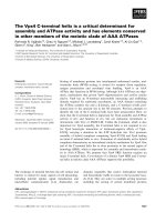
Effect of FGF9 on cell proliferation, invasion and
anti-apoptosis in gastric cancer cells
Since FGF9 is known to have a mitogenic effect on some
cell types [17], we first tested the effect of FGF9 on the
growth kinetics of gastric cancer cells. However, we
found no effect of FGF9 on cell proliferation in the AGS
and MKN28 cell lines (Figure 3A).
To further identify the possible role of FGF9 in tumor
progression, we examined whether exogenous FGF9 confers an anti-apoptotic effect on gastric cancer cells. FACS
analyses revealed that the number of annexin V-positive
AGS cells was significantly smaller in the FGF9-treated
Page 6 of 9
group than in the control group, and similar findings
were obtained in MKN28 cells (Figure 3B and C). Furthermore, this effect of FGF9 was abolished by concomitant administration of anti-FGF9 neutralizing
antibody in both cell lines (Figure 3D).
Moreover, we next examined the effect of FGF9 on the
invasive ability of gastric cancer cells. When AGS cells
were stimulated with FGF9 (1–10 ng/mL), the number
of invasive cells was significantly increased (Figure 4A).
Similarly, the invasive ability of MKN28 cells was significantly enhanced dose-dependently by FGF9 stimulation
(Figures 4A). We then examined whether this pro-invasive
effect of FGF9 could be abolished by adding a neutralizing
antibody. In both cell lines, after concomitant administration of FGF9 neutralizing antibody (1 μg/ml), the number
of invasive cells was significantly decreased in comparison
with cells treated with FGF9 alone (10 ng/mL) (Figure 4B).
In addition, we examined whether FGF9 induced the expression of MMPs, which play a pivotal role in invasion of
Figure 3 Effect of FGF9 on growth and anti-apoptosis of gastric cancer cells. (A) Effect of FGF9 on growth of gastric cancer cells. (B-D) Effect of
FGF9 on anti-apoptosis capability of gastric cancer cells. (B) Representative graphs of FACS analysis using Annexin V-FITC staining. AGS cells were
treated with FGF9 (10 ng/ml) and evaluated as described in Materials and Methods. (C) Changes in the number of apoptotic AGS and MKN28 cells
treated with FGF9. (D) Effect of anti-FGF9 neutralizing antibody (Neu Ab; 1 μg/ml) on FGF9 (10 ng/ml)-induced anti-apoptosis in AGS and MKN28 cells.
All the results are expressed as the mean ± SD of four samples. Significantly lower than control: *P <0.05, **P <0.01. Significantly greater than
the FGF9-treated group: #P <0.05, ##P <0.01.
Sun et al. BMC Cancer (2015) 15:333
Page 7 of 9
Figure 4 Effect of FGF9 on invasion and MMPs expression of gastric cancer cells. (A) Effect of FGF9 on gastric cancer cell invasion. Change in
number of invasive AGS and MKN28 cells treated with FGF9 were examined. Photographs showing representative images of invasive gastric
cancer cells in the control and FGF9-treated groups. (B) Effect of anti-FGF9 neutralizing antibody (Neu Ab; 1 μg/ml) on FGF9 (10 ng/ml)-induced
invasion of AGS and MKN28 cells. (C) Effect of FGF9 on expression of MMPs in gastric cancer cells. All the results are expressed as the mean ± SD
of four samples. Significantly greater than control: *P <0.05, **P <0.01. Significantly lower than the FGF9-treated group: #P <0.05, ##P <0.01.
various cancers. As shown in Figure 4C, FGF9 stimulation
commonly enhanced the expression of MMP7 in both
AGS and MKN28 cells.
Clinicopathological significance of FGF9 expression in
gastric cancer-associated fibroblasts
Of the 20 gastric cancer tissue samples examined, sixteen
(80%) was positive for FGF9 expression. Regarding the
clinicopathological features, none of the parameters ─
age, gender, tumor location, histological type, or tumor
stage ─ had a significant relationship to FGF9 expression
(Additional file 2: Table S2).
Discussion
It is believed that tumor development and progression
depend on cross-talk between cancer cells and their surrounding stromal cells. As a major stromal population,
“activated” fibroblasts, referred to as CAFs, have been
suggested to promote tumorigenesis using various molecular signals, and in the present study we isolated
FGF9 as a novel gene that was overexpressed in CAFs in
gastric cancer. Accumulating evidence, including our
present data, have shown that CAFs differ from NGFs in
terms of not only morphology but also gene expression
profiles [18,19], although the underlying mechanisms responsible for these differences are still unclear. On the
basis of recent evidence, it is tempting to propose that
tumor cells initiate a switch from NGFs to CAFs
through some form of signaling [20] or that CAFs originate from cancer cells through epithelial-mesenchymal
transition [21,22]. Interestingly, it is widely accepted that
the FGF family is crucial for epithelial-mesenchymal
transition, not only during development but also in carcinogenesis [9,23]. In the present study, we showed that
Sun et al. BMC Cancer (2015) 15:333
CAFs are a possible source of FGF9, and that furthermore gastric cancer cells have receptors that are responsive to FGF9, suggesting that FGF9 may be a potential
mediator between CAFs and gastric cancer cells.
What is the possible role of FGF9 in the pathogenesis
of gastric cancer? Since FGF9 serves as mitogen for
prostate or ovarian cancer [17,24], we first investigated
the proliferation of gastric cancer cells in the presence of
FGF9 in vitro. Subsequently, FGF9 failed to promote the
proliferation of both AGS and MKN28 gastric cancer
cell lines; however, we cannot exclude the possibility that
FGF9 may serve as mitogen for other types of gastric
cancer cells because FGF9 promotes the proliferation of
other gastric cancer cells such as AZ521 or SGC-7901
[25,26]. On the other hand, we clarified in the present
study that FGF9 has an anti-apoptotic effect on gastric
cancer cells, in accord with the findings of several previous studies [26,27]. In support of this finding, Akt and
ERK pathway signaling, crucial for anti-apoptosis, was
commonly activated in both gastric cancer cell lines.
Furthermore, FGF9-induced Akt and/or ERK phosphorylation and the resulting cell invasion was abolished by
blocking FGF9 stimulation, suggesting that FGF9 at least
acts as an anti-apoptotic factor.
We also examined whether FGF9 promotes the invasive ability of gastric cancer cells. In vitro studies demonstrated that FGF9 treatment increased the number of
invading gastric cancer cells, whereas this increased invasive ability was suppressed by adding FGF9 neutralizing antibody. Although it is unclear how FGF9 promotes
the invasion of gastric cancer cells, degradation of the
extracellular matrix is an important part of this process
[28]. In this context, it is accepted that MMPs play a
central role in such cell behavior and promote the invasive capacity of various malignancies [29]. Therefore, we
screened for MMPs that are linked to FGF9 stimulation
and found that MMP7 was potentially involved. Interestingly, recent studies have emphasized the importance of
MMP7 in gastric cancer progression, since MMP7 has
potential to not only degrade the extracellular matrix
but also confer an anti-apoptotic effect on cancer cells
[30-32]. Accordingly, in a future study, we intend to
focus on the FGF/MMP7 axis in the context of gastric
cancer cell invasion.
Conclusion
In summary, we have shown that gastric CAFs differ
from their corresponding NGFs in terms of their gene
expression profiles, and propose that FGF9 is a potential
molecule that mediates cross-talk between CAFs and
gastric cancer cells. Moreover, we have clarified that
FGF promotes the anti-apoptosis and invasive capability
of gastric cancer cells. Taken together, our data suggest
that FGF9 is a possible mediator secreted from cancer-
Page 8 of 9
associated fibroblasts that promotes the anti-apoptosis
and invasive capability of gastric cancer cells.
Additional files
Additional file 1: Table S1. Primers for real-time RT-PCR analysis.
Additional file 2: Table S2. Relationship between clinicopathological
features and stromal FGF9 expression in patients with gastric cancer.
Abbreviations
FGF: Fibroblast growth factor; MMP: Matrix metalloproteinase;
ERK: Extracellular signal-regulated protein kinase; CAF: Cancer-associated
fibroblast.
Competing interests
The authors declare that they have no competing interests.
Authors’ contributions
CS and HF designed and performed all the experiments, analyzed data, and
wrote the manuscript. KH, XZ and SK performed the experiments. YK, HE, TT,
TO, and JW collected samples. MS and HM supervised the study. All authors
read and approved the final manuscript.
Acknowledgements
This work was supported in part by Grants-in-aid for Scientific Research from
the Ministry of Education, Culture, Sports, Science and Technology, Japan
(23591949 to M.S, 25460466 to S.K and 26460953 to H.F). This work was also
supported in part by Grant-in-Aid for Researchers, Hyogo College of Medicine
and Supported Program for the Strategic Research Foundation at Private
Universities (to M.S).
The authors thank Noriko Kamiya (Division of Gastroenterology, Department
of Internal Medicine) and Yuko Yasui (Department of Surgery) for their technical
assistance and also appreciate Nobuyuki Adachi and Ryota Shinozaki (HCM
Joint-Use Research facilities) for technical contributions.
Author details
1
Division of Gastroenterology, Department of Internal Medicine, Hyogo
College of Medicine, l-1, Mukogawa, Nishinomiya 663-8501, Japan.
2
Department of Digestive Diseases, Tianjin Medical University General
Hospital, Tianjin, China. 3Department of Geriatric Digestive Internal Medicine,
Sichuan Academy of Medical Science & Sichuan People’s Hospital, Chengdu,
China. 4Department of Surgery, Hyogo College of Medicine, Nishinomiya,
Japan.
Received: 10 September 2014 Accepted: 23 April 2015
References
1. Xing F, Saidou J, Watabe K. Cancer associated fibroblasts (CAFs) in tumor
microenvironment. Front Biosci. 2010;15:166–79.
2. Silzle T, Randolph GJ, Kreutz M, Kunz-Schughart LA. The fibroblast: sentinel
cell and local immune modulator in tumor tissue. Int J Cancer.
2004;108:173–80.
3. Mishra P, Banerjee D, Ben-Baruch A. Chemokines at the crossroads of
tumor-fibroblast interactions that promote malignancy. J Leukoc Biol.
2011;89:31–9.
4. Marsh T, Pietras K, McAllister SS. Fibroblasts as architects of cancer
pathogenesis. Biochim Biophys Acta. 1832;2013:1070–8.
5. Bhowmick NA, Neilson EG, Moses HL. Stromal fibroblasts in cancer initiation
and progression. Nature. 2004;432:332–7.
6. Giri D, Ropiquet F, Ittmann M. FGF9 is an autocrine and paracrine prostatic
growth factor expressed by prostatic stromal cells. J Cell Physiol.
1999;180:53–60.
7. Coffey E, Newman DR, Sannes PL. Expression of fibroblast growth factor 9 in
normal human lung and idiopathic pulmonary fibrosis. J Histochem
Cytochem. 2013;61:671–9.
8. Tsai SJ, Wu MH, Chen HM, Chuang PC, Wing LY. Fibroblast growth factor-9
is an endometrial stromal growth factor. Endocrinology. 2002;143:2715–21.
Sun et al. BMC Cancer (2015) 15:333
9.
10.
11.
12.
13.
14.
15.
16.
17.
18.
19.
20.
21.
22.
23.
24.
25.
26.
27.
28.
29.
30.
31.
32.
Turner N, Grose R. Fibroblast growth factor signalling: from development to
cancer. Nat Rev Cancer. 2010;10:116–29.
Jang JH, Shin KH, Park JG. Mutations in fibroblast growth factor receptor 2
and fibroblast growth factor receptor 3 genes associated with human gastric
and colorectal cancers. Cancer Res. 2001;61:3541–3.
Fukui H, Fujii S, Takeda J, Kayahara T, Sekikawa A, Nanakin A, et al. Expression of
Reg Iα protein in human gastric cancers. Digestion. 2004;69:177–84.
Wang CK, Chang H, Chen PH, Chang JT, Kuo YC, Ko JL, et al. Aryl hydrocarbon
receptor activation and overexpression upregulated fibroblast growth factor-9
in human lung adenocarcinomas. Int J Cancer. 2009;125:807–15.
Fukui H, Sekikawa A, Tanaka H, Fujimori Y, Katake Y, Fujii S, et al. DMBT1 is a
novel gene induced by IL-22 in ulcerative colitis. Inflamm Bowels Dis.
2011;17:1177–88.
Sekikawa A, Fukui H, Fujii S, Ichikawa K, Tomita S, Imura J, et al. REG Iα protein
mediates an anti-apoptotic effect of STAT3 signaling in gastric cancer cells.
Carcinogenesis. 2008;29:76–83.
Fukui H, Zhang X, Sun C, Hara K, Kikuchi S, Yamasaki T, et al. IL-22 produced
by cancer-associated fibroblasts promotes gastric cancer cell invasion via
STAT3 and ERK signaling. Br J Cancer. 2014;111:763–71.
Lai MS, Cheng YS, Chen PR, Tsai SJ, Huang BM. Fibroblast growth factor 9
activates akt and MAPK pathways to stimulate steroidogenesis in mouse
leydig cells. PLoS One. 2014;9:e90243.
Hendrix ND, Wu R, Kuick R, Schwartz DR, Fearon ER, Cho KR. Fibroblast growth
factor 9 has oncogenic activity and is a downstream target of Wnt signaling in
ovarian endometrioid adenocarcinomas. Cancer Res. 2006;66:1354–62.
Chen SF, Nieh S, Jao SW, Wu MZ, Liu CL, Chang YC, et al. The paracrine effect
of cancer-associated fibroblast-induced interleukin-33 regulates the invasiveness
of head and neck squamous cell carcinoma. J Pathol. 2013;231:180–9.
Peng Q, Zhao L, Hou Y, Sun Y, Wang L, Luo H, et al. Biological characteristics
and genetic heterogeneity between carcinoma-associated fibroblasts and their
paired normal fibroblasts in human breast cancer. PLoS One. 2013;8:e60321.
Cirri P, Chiarugi P. Cancer associated fibroblasts: the dark side of the coin.
Am J Cancer Res. 2011;1:482–97.
Petersen OW, Nielsen HL, Gudjonsson T, Villadsen R, Rank F, Niebuhr E, et al.
Epithelial to mesenchymal transition in human breast cancer can provide a
nonmalignant stroma. Am J Pathol. 2003;162:391–402.
Rasanen K, Vaheri A. Activation of fibroblasts in cancer stroma. Exp Cell Res.
2010;316:2713–22.
Chaffer CL, Dopheide B, Savagner P, Thompson EW, Williams ED. Aberrant
fibroblast growth factor receptor signaling in bladder and other cancers.
Differentiation. 2007;75:831–42.
Teishima J, Shoji K, Hayashi T, Miyamoto K, Ohara S, Matsubara A. Relationship
between the localization of fibroblast growth factor 9 in prostate cancer cells
and postoperative recurrence. Prostate Cancer Prostatic Dis. 2012;15:8–14.
Matsumoto-Yoshitomi S, Habashita J, Nomura C, Kuroshima K, Kurokawa T.
Autocrine transformation by fibroblast growth factor 9 (FGF-9) and its
possible participation in human oncogenesis. Int J Cancer. 1997;71:442–50.
Deng M, Tang HL, Lu XH, Liu MY, Lu XM, Gu YX, et al. miR-26a suppresses
tumor growth and metastasis by targeting FGF9 in gastric cancer. PLoS
One. 2013;8:e72662.
Lum M, Turbic A, Mitrovic B, Turnley AM. Fibroblast growth factor-9 inhibits
astrocyte differentiation of adult mouse neural progenitor cells. J Neurosci
Res. 2009;87:2201–10.
Kessenbrock K, Plaks V, Werb Z. Matrix metalloproteinases: Regulators of the
tumor microenvironment. Cell 2010, 141: 52–67.
Birkedal-Hansen H, Moore WG, Bodden MK, Windsor LJ, Birkedal-Hansen B,
DeCarlo A, et al. Matrix metalloproteinases: a review. Crit Rev Oral Biol Med.
1993;4:197–250.
Sakamoto N, Naito Y, Oue N, Sentani K, Uraoka N, Zarni Oo H, et al. MicroRNA-148a
is downregulated in gastric cancer, targets MMP7, and indicates tumor invasiveness
and poor prognosis. Cancer Sci. 2014;105:236–43.
Koskensalo S, Mrena J, Wiksten JP, Nordling S, Kokkola A, Hagström J, et al.
MMP-7 overexpression is an independent prognostic marker in gastric cancer.
Tumour Biol. 2010;31:149–55.
Wang WS, Chen PM, Wang HS, Liang WY, Su Y. Matrix metalloproteinase-7
increases resistance to Fas-mediated apoptosis and is a poor prognostic factor
of patients with colorectal carcinoma. Carcinogenesis. 2006;27:1113–20.
Page 9 of 9
Submit your next manuscript to BioMed Central
and take full advantage of:
• Convenient online submission
• Thorough peer review
• No space constraints or color figure charges
• Immediate publication on acceptance
• Inclusion in PubMed, CAS, Scopus and Google Scholar
• Research which is freely available for redistribution
Submit your manuscript at
www.biomedcentral.com/submit