Radiographers supporting radiologists in the interpretation of screening mammography: A viable strategy to meet the shortage in the number of radiologists
Bạn đang xem bản rút gọn của tài liệu. Xem và tải ngay bản đầy đủ của tài liệu tại đây (625.48 KB, 12 trang )
Torres-Mejía et al. BMC Cancer (2015) 15:410
DOI 10.1186/s12885-015-1399-2
RESEARCH ARTICLE
Open Access
Radiographers supporting radiologists in the
interpretation of screening mammography: a
viable strategy to meet the shortage in the
number of radiologists
Gabriela Torres-Mejía1*, Robert A. Smith2, María de la Luz Carranza-Flores3, Andy Bogart4,
Louis Martínez-Matsushita1, Diana L. Miglioretti4,5, Karla Kerlikowske6,7, Carolina Ortega-Olvera1,
Ernesto Montemayor-Varela1, Angélica Angeles-Llerenas1, Sergio Bautista-Arredondo8,
Gilberto Sánchez-González8, Olga G. Martínez-Montañez9, Santos R. Uscanga-Sánchez10,
Eduardo Lazcano-Ponce1 and Mauricio Hernández-Ávila1
Abstract
Background: An alternative approach to the traditional model of radiologists interpreting screening mammography is
necessary due to the shortage of radiologists to interpret screening mammograms in many countries.
Methods: We evaluated the performance of 15 Mexican radiographers, also known as radiologic technologists, in the
interpretation of screening mammography after a 6 months training period in a screening setting. Fifteen
radiographers received 6 months standardized training with radiologists in the interpretation of screening
mammography using the Breast Imaging Reporting and Data System (BI-RADS) system. A challenging test set of 110
cases developed by the Breast Cancer Surveillance Consortium was used to evaluate their performance. We estimated
sensitivity, specificity, false positive rates, likelihood ratio of a positive test (LR+) and the area under the subject-specific
Receiver Operating Characteristic (ROC) curve (AUC) for diagnostic accuracy. A mathematical model simulating the
consequences in costs and performance of two hypothetical scenarios compared to the status quo in which a
radiologist reads all screening mammograms was also performed.
Results: Radiographer’s sensitivity was comparable to the sensitivity scores achieved by U.S. radiologists who took the
test but their false-positive rate was higher. Median sensitivity was 73.3 % (Interquartile range, IQR: 46.7–86.7 %) and the
median false positive rate was 49.5 % (IQR: 34.7–57.9 %). The median LR+ was 1.4 (IQR: 1.3-1.7 %) and the median AUC
was 0.6 (IQR: 0.6–0.7). A scenario in which a radiographer reads all mammograms first, and a radiologist reads only
those that were difficult for the radiographer, was more cost-effective than a scenario in which either the radiographer
or radiologist reads all mammograms.
Conclusions: Given the comparable sensitivity achieved by Mexican radiographers and U.S. radiologists on a test set,
screening mammography interpretation by radiographers appears to be a possible adjunct to radiologists in countries
with shortages of radiologists. Further studies are required to assess the effectiveness of different training programs in
order to obtain acceptable screening accuracy, as well as the best approaches for the use of non-physician readers to
interpret screening mammography.
Keywords: Radiographers, Film readers, Screening mammography, BI-RADS system
* Correspondence:
1
Centro de Investigación en Salud Poblacional, Instituto Nacional de Salud
Pública, Avenida Universidad No. 655, Colonia Santa María Ahuacatitlán,
Cuernavaca 62100, Morelos, Mexico
Full list of author information is available at the end of the article
© 2015 Torres-Mejía et al.; licensee BioMed Central. This is an Open Access article distributed under the terms of the Creative
Commons Attribution License ( which permits unrestricted use, distribution, and
reproduction in any medium, provided the original work is properly credited. The Creative Commons Public Domain
Dedication waiver ( applies to the data made available in this article,
unless otherwise stated.
Torres-Mejía et al. BMC Cancer (2015) 15:410
Background
In Mexico, since 2006, breast cancer is the leading cause
of death from cancer in women [1] with epidemiological
and demographic transitions contributing to an increasing trend in rates. Approximately 90 % of breast cancer
cases are diagnosed at an advanced stage, which in part
is due to the lack of an organized screening program,
limited access to mammography and shortage of radiologists to interpret screening mammography, particularly
in rural areas [2]. These conditions may contribute to
delays in diagnosis and less successful treatment [2–5].
The Mexican Official Norm for breast cancer (NOM041-SSA2-2011) recommends mammography every two
years for healthy women ages 40–69 years and provides
the guidelines for the breast cancer national program to
achieve greater coverage and quality [6]. However, the
results of the recent Mexican National Health Survey,
showed that only 17.2 % and 29.4 % of women between
40 and 49 years and between 50 and 69 years, had a
mammogram within the previous 2 years, respectively
[7]. Only 291 radiologists participate in the screening
mammography program in Mexico, among whom 260
focus exclusively on breast imaging. With these current
infrastructure and human resources, it will clearly not be
possible to increase the coverage up to 70 % as suggested
by the World Health Organization (WHO) [8], given
that there are close to 14 million women eligible for
screening [2, 3, 9].
Since the mid-1980s, there has been a growing literature suggesting that shortages of radiologists could be
overcome, and costs reduced if radiographers (also
known as radiologic technologists) [10, 11], or mid-level
practitioners (physician assistants or nurse practitioners)
could interpret mammograms and serve as first readers,
determining the presence or absence of abnormal images
in cases that warrant further evaluation by a radiologist.
[12] In 1995 in the United Kingdom (UK), there was
both a shortage and falling recruitment rates of radiologists, and therefore the possibility of radiographers as
readers was explored in order to maintain double reading
[13]. Since then, several studies have demonstrated radiographers’ ability to identify abnormalities on screening
mammograms [14] albeit with higher false positive rates,
but similar sensitivity compared with radiologists [15–19].
Furthermore, trained radiographers have been shown to
perform well as pre-readers in a clinical setting [20].
To our knowledge there are no studies examining the
potential for radiographers or physician extenders to contribute to mammography interpretation in Latin America.
Given Mexico’s public health policy goals for breast cancer
screening and the shortage of radiologists to interpret
mammograms, we designed a study to evaluate the performance of 15 radiographers in the interpretation of
mammograms after a 6 months training period in a
Page 2 of 12
screening setting. Additionally, using the results of the
study, we modeled the effects on costs and performance
of two hypothetical screening scenarios; both were compared with the status quo in which the standard practice
is a single reading by a radiologist. We conducted this
study to determine the feasibility of having radiographers
as first readers, in order to consider a strategy for integrating non-radiologist readers into mammography screening
in countries that, face the problem of shortages of radiologists with specialization and interest in mammography.
Methods
Study population
Fifteen radiographers from 12 states in Mexico were invited to participate in the study. Inclusion criteria were:
1) having a formal role in the mammography facility operating under the auspices of the Secretariat of Health,
2) having completed radiographers training and obtained
a degree, 3) at least 6 months experience in x-ray and
breast imaging, and 4) permission from the institution
where he or she worked in order to engage in the study.
Screening setting
Health-care coverage in Mexico is delivered by a range
of different institutions. Although there are isolated efforts on the implementation of an organized populationbased screening program, the breast cancer screening
program in Mexico is based on an opportunistic model
[21]. For this study, the training was held in Mexico
City in a Digital Diagnostic Center, which forms part of
a network of 20 digital mammography machines in
health facilities distributed across Mexico City with a
goal of performing and interpreting 90, 000 mammograms annually [22]. All participants received a grant
for maintenance and transportation. The setting offered
a class-room, a reading area with a work station and
trainees.
Training program
The training program was designed by a group of radiologists and epidemiologists. It had a total duration of
6 months and consisted of clinical lectures, training inservice by three radiologists who guided the radiographers. The participants read a progressive number of
digital mammographic studies using the Breast Imaging
Reporting and Data System (BI-RADS) system. During
the first month, each student interpreted at least 10
screening mammograms a day; as they improved their
performance, the number of mammograms assigned
daily increased to approximately 40 mammograms per
day. Each day, the radiographers, in teams of two,
reviewed 10–30 mammograms with the advice of a radiologist, and on a weekly basis they received feedback, in
class, using a subsample of images. Prior to the final test
Torres-Mejía et al. BMC Cancer (2015) 15:410
set evaluation, the median number of mammograms
interpreted by the radiographers during the six-month
training program was 777. The classroom component
consisted of 122 training hours on breast anatomy and
mammographic features of normal, benign and malignant breast conditions based on updated literature on
the standards for training specialized health professionals dealing with breast cancer [23]. They also received an introduction to breast cancer epidemiology
and ethics.
Evaluation
At the end of training (6 months), a formal evaluation
was performed using a self-administered test set of
mammograms developed by the Breast Cancer Surveillance Consortium (BCSC) [24] for the evaluation of U.S.
radiologists in a prospective study of skills assessment
and training. The evaluation was carried out in the computer lab of the National Institute of Public Health using
software developed by the American College of Radiology for the BCSC.
The test set consisted of 110 screening examinations
of which 15 cases were biopsy confirmed cancers, 14
were non-cancers that 3 U.S. expert radiologists judged
should be recalled, and 81 were non-cancers that the experts judged had no findings to justify recall. Of the 15
confirmed breast cancer cases, the 3 U.S. expert radiologists who helped assemble the test set judged 3 to be obvious, 7 intermediate, and 5 subtle, from which 100 %,
85.7 %, and 80 % were recalled in the U.S. clinical practices, respectively. U.S. experts’ consensus was that all
cancers should have been recalled. Among the 14 noncancer cases which were recalled by the expert panel, 12
(85.7 %) were recalled in clinical practice (personal communication from Andy Bogart, BCSC Statistical Coordinating Center). In the main analysis, the biopsy confirmed
cancer cases were treated as the true positives for evaluation purposes. Each of the 110 cases had 4 images (mediolateral oblique and cephalocaudal views for each breast).
Participants also had access to screening mammograms
performed within 2 years before the test films were taken
in order to have a baseline comparison.
Interpretive performance was measured in terms of
sensitivity, specificity, false positives, likelihood ratio of a
positive test (LR+) and the area under the subjectspecific Receiver Operating Characteristic (ROC) curve
(AUC). The assessments performed by the 3 U.S. expert
radiologists were defined as the gold standard. The three
breast imaging experts were selected based on their expertise in breast imaging. Two of the three breast imaging experts were males and all were over 55 years. All
worked in an academic setting and had over 30 years’
experience interpreting mammograms. Their annual volume of mammography interpretation ranged from 2500
Page 3 of 12
to 7000 per year. All experts were fellows of the Society
of Breast Imaging (SBI), fellows of the American College
of Radiology, past presidents of National Breast Societies, and gold medalists of the SBI. For each case the
radiographer was asked to identify the BI-RADS interpretation category corresponding to the observed images: BIRADS 0 (needs further evaluation), 4 (suspicious) or 5
(highly suggestive of malignancy) or BI-RADS 1 (negative)
or 2 (benign finding). BI-RADS 3 category was not an interpretation category, because it should not be used in
screening [25]. For evaluation purposes, sensitivity was estimated as the percent of cancer cases that were recalled
by radiographers out of the total number of cancer films
in the test set (n = 15), and false negatives as its complement. Specificity was calculated as the percent of noncancer films which were correctly not recalled by the
radiographers of the total of non-cancer films in the test
set (n = 95) and false positives as its complement.
For recalled mammograms, the most significant finding type was described as either a mass, calcifications,
asymmetry, or architectural distortion. After the evaluation, the results were sent via the Internet to a server in
the BCSC Statistical Coordinating Center, Group Health
Cooperative, Seattle WA, U.S. to be analyzed by one of
the BCSC members. The database was sent back via secure file transfer protocol to Mexico for further analysis.
Subsequently, specific results were sent back to each
participant with confidentiality maintained.
Statistical analysis
An exploratory analysis was performed to describe the
socio-demographic, academic and experience characteristics of the participants and of the variables related to
diagnostic interpretive performance, measured by sensitivity, specificity, false positive rate, the AUC and the
likelihood ratio of a positive test (LR+). We report the
median and the inter-quartile range (IQR) of performance measures across radiographers. For continuous
variables we used measures of central tendency and dispersion. For categorical variables we used measures of
frequency.
To evaluate performance, sensitivity, specificity and
false positive (FP) rate were calculated. Overall observer
performance was measured by calculating the AUC and
the LR+ for each participant. The AUC was estimated
as the median of the sensitivity and specificity as described by Cantor and Kattan [26]. The LR+ for each
radiographer was calculated by dividing the sensitivity
by 1-specificity. This measure combines sensitivity and
specificity into a single index that measures how many
times it is more likely that a patient recalled by experts
has an abnormal mammogram compared to women
whom the experts did not recall [27].
Torres-Mejía et al. BMC Cancer (2015) 15:410
Page 4 of 12
Mathematical modeling
We designed a mathematical model to assess the costs
and outcomes of screening mammography interpretation
by the radiographers in this study in terms of true positives, false negatives and false positives. The model compares three hypothetical scenarios: (A) the status quo in
which a radiologist reads all mammograms; (B) a radiographer reads all mammograms; and (C) a radiographer
reads all mammograms first, recommends obvious abnormal findings for diagnostic evaluation, and refers to a
radiologist for a second reading any images that appear
abnormal, but for which the need for recall is uncertain
(i.e., radiographer’s percentage of “FP, but not obvious
breast cancer” images). Both scenarios (B and C) were
compared with the status quo, where a radiologist reads
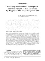
all mammograms (A) (Fig. 1).
We compared the results in terms of diagnostic accuracy and costs. The parameters of the model were: salary
per month for a full time radiographer and a radiologist
[28], mammogram and diagnosis costs [29], prevalence
of breast cancer in a screening setting [30], average
number of mammograms read per month (the number
of mammograms per month was assumed to be the
same for both radiologists and radiographers) [31], and
test set sensitivity and specificity obtained from this
study for radiographers and a previous clinical setting
for radiologists [31] (Table 1). All model input parameters were introduced as triangular probability distributions, whose minimum, mean and maximum values are
Status quo
indicated in Table 1. The triangular distribution allowed
us to assign probability distributions to the costs and effectiveness parameters rather than simple point estimates. This is a common approach when the actual
distribution of the parameter is unknown, but three parameters (minimum, maximum and some modal value)
are known or can be guessed.
Results of both scenarios were calculated using second-order Monte Carlo simulations. The costs and
effectiveness parameters were drawn from the triangular probabilistic distributions described above. This
method implies that during the simulation these parameters are sampled randomly from the triangular distributions, and each sample drawn represents one
radiologist or radiographer performance and costs. In
total 1000 radiologists and 1000 radiographers were
simulated and the results of all simulations were averaged. The results therefore, explicitly consider the uncertainty in the input parameters of the model. Instead
of performing simple point-estimate sensitivity analysis,
the results are reported with confidence intervals. This
approach is referred to as probabilistic-sensitivity, or
multiway sensitivity analysis [32]. The labor costs used
in the model were in terms of one month full time
equivalent unit of either radiologist or radiographer.
Ethics
The study was approved by the Institutional Review
Board at the National Institute of Public Health. All
Two hypothetical scenarios
TN FN
FP
FN
TN
Fig. 1 Decision tree model to assess the costs and effects of screening mammography interpretation by the radiographers in this study in terms
of true positives, false negatives and false positives. The model compares three hypothetical scenarios: (A) the status quo in which one radiologist
reads all mammograms; (B) a radiographer reads all mammograms; and (C) a radiographer reads all mammograms first, sends obvious abnormal
findings for diagnostic evaluation and leaves to the radiologist, for a second reading, only those images which he/she considers difficult to
interpret. Both scenarios (B and C) were compared with the status quo, where a radiologist reads all mammograms (A)
Torres-Mejía et al. BMC Cancer (2015) 15:410
Page 5 of 12
Table 1 Parameters of the mathematical model, Mexico 2012
Parameter
Mean/Median value
95 % CI
Reference
Radiologists' salary per montha
663
600-720
[24]
Radiographers' salary per month
407
360-440
Mammogram's costa
24
22-26
Confirmation (diagnosis) cost
802
720-880
Number of invasive cancers detected per 1000 screens
5
0.45-0.55
[26]
[27]
a
a
Average number of mammographies read per month
93
58-200
Radiologists' sensitivity
0.729
0.604-0.833
Radiologists' specificity
0.844
0.781-0.906
Radiographers' sensitivity
0.733
0.467-0.867
Radiographers' specificity
0.505
0.421-0.653
a
[25]
Present study
Salaries and costs are indicated in USD
participants gave their consent to participate in the study.
The study was in agreement with the regulations established by the Breast Cancer Surveillance Consortium
Assessing and Improving Mammography (BCSC-AIM)
Collaborative Research Agreement supported by the
American Cancer Society and the U.S. National Cancer
Institute.
Results
Performance of radiographers
Eighty percent of the radiographers who participated in
the study were women (80 %) and the median age was
38 years (IQR: 28–47 years) (Table 2). The median duration of educational training to become a radiographer
was 2.5 years (IQR: 2–3 years) and the median time of
experience performing mammography studies was 8 years
(IQR: 2–18). Prior to the educational training, they had no
previous experience in interpreting mammograms. Most
radiographers worked for second level of attention general
hospitals (42.9 %) and very few reported having previously
attended breast disease courses (Table 2).
The median sensitivity was 73.3 % (IQR: 46.7-86.7 %)
whereas the average false positive rate was 49.5 % (IQR:
34.7–57.9 %). (Table 3, Fig. 2). The PPV was 18.3 %
(IQR: 16.9 %–21.3 %) and the NPV was 92 % (IQR:
88.7–94.3 %). The median likelihood ratio of a positive
test was 1.4 (IQR: 1.3–1.7 %) and the median AUC was 0.6
(IQR: 0.6–0.7). The median time to interpret a study per
radiographer was 115.9 s (IQR: 105.2–131.6 s) (Table 3).
In relation to the characteristics of the breast cancers,
sensitivity was highest for identification of masses and
architectural distortions (100 % for both) and lowest for
asymmetries (25 %) (Table 4). Radiographer’s sensitivity
decreased with increasing difficulty of the lesion, with
median sensitivity of 100 % for obvious lesions, 71.4 %
for intermediate lesions, and 60 % for subtle lesions
(Table 4).
Model outcomes
1. Radiographer vs radiologist
When comparing the monthly cost of mammography
screening interpretation by radiographer vs radiologist,
it was more efficient to employ a radiologist, despite the
differential in salaries. The total monthly cost of Scenario A, where the radiologist interprets all mammograms, was less expensive than the total monthly cost of
Scenario B, where the radiographer interprets the same
number of mammograms (US$17,019 vs US$44,165, respectively) (Table 5). These scenarios were comparable
in terms of percentage of true positives (0.36 % vs 0.34 %,
respectively), in terms of false-negative results (0.14 % vs
0.16 %, respectively) and in terms of total mammograms
interpreted per month. However, the results in terms of
false positive readings were very different (15.6 % vs
47.1 %, respectively) because the false-positives were
based on a clinical setting for the radiologists and a test
set for the radiographers where the test set is enriched
with abnormal examinations [31]. As a result of this difference, the cost per breast cancer detected was significantly higher in scenario B compared with scenario A
(US$139,263 vs US$51,403, respectively).
2. Radiographer and radiologist working together vs
radiologist only
Our model showed that the monthly cost and average
cost per breast cancer case detected for scenario C (radiologist & radiographer) was slightly lower (US$16,331
and US$51,347, respectively) than for Scenario A (radiologist only) (US$17,019 and US$51,403, respectively).
Discussion
Our study used a test set to evaluate the effect of a 6month screening mammography interpretation training
Torres-Mejía et al. BMC Cancer (2015) 15:410
Page 6 of 12
Table 2 Characteristics of radiographers (n = 15), Mexico 2012
Age (years)
Median
38
Interquartile range
(28–47)
Range
(24–54)
Female
12 (80 %)
Male
3 (20 %)
Sex
Years since graduation
Median
8
Interquartile range
(5, 19)
Range
(2, 26)
Technical education cumulative grade scorea
Median
8.5
Interquartile range
(8–9.7)
Range
(7.8–10)
Technical education length (years)
Median
2.5
Interquartile range
(2–3)
Range
(1–4)
Experience in performing mammography studies (years)b
Median
8
Interquartile range
(2–18)
Range
(0–25)
Number of mammograms per week performed before trainingc
Median
100
Interquartile range
(50–125)
Range
(10–200)
Health care level
First
4 (28.6 %)
Second
6 (42.9 %)
Third
4 (28.5 %)
Number of additional breast courses
Median
1
Interquartile range
(0–5)
Range
(0–10)
a
The cumulative grade score is on a scale of 1–10
b
Radiographers were not necessarily devoted exclusively to this activity
c
Radiographers in Mexico do not interpret, they only perform mammograms
program for radiographers. The median sensitivity was
73.3 %, but was achieved at the expense of a high percentage of false positives (49.5 %) on a test set enriched
with abnormal examinations with an expert recall rate of
26.4 % (29/110). Our results are consistent with other
studies evaluating performance on consecutive series of
patients or test set studies where sensitivity rates have
ranged from 73 % to 90 % [33]. Recall rates tend to be
higher in test set situations than one expects to find in
screening conditions [34]. In addition, this test set was
developed to be challenging, especially for the noncancer cases. For example, the clinical sensitivity obtained from U.S. radiologists on the same films read in
clinical practice, was 86.7 % (13/15) while the false
Torres-Mejía et al. BMC Cancer (2015) 15:410
Page 7 of 12
Table 3 Radiographers’ test set performance evaluation after
6 months of training, Mexico 2012
Table 4 Radiographers' sensitivity by lesion type and
difficulty after 6 months of training, Mexico 2012
Performance post-training
Median
Interquartile range
Performance post-training
Sensitivity ( %)a
73.3
46.7–86.7
Specificity ( %)a
50.5
42.1–65.3
Breast cancer lesions types
False positivies (1 – specificity) ( %)
Appropiate recalls ( %)b
c
Median
% Sensitivitya
Interquartile
range
49.5
34.7–57.9
Mass
100.0
66.7–100.0
78.6
78.6–92.9
Calcification
83.3
66.7–100.0
In appropiate recalls ( %)
36.8
25.3–44.2
Asymmetry
25.0
25.0–50.0
Positive predictive value ( %)
18.3
16.9–21.3
Architectural distortion
100.0
50.0–100.0
Negative predictive value ( %)
92.0
88.7–94.3
Difficulty of cancer lesion identified by radiographers
LR + d
1.4
1.3–1.7
Obvious
100.0
66.7–100.0
e
AUC
0.6
0.6–0.7
Intermediate
71.4
57.1–85.7
Time spent per interpretationf
115.9
105.2–131.6
Subtle
60.0
40.0–80.0
a
a
Percentage of histologically confirmed breast cancer lesions that were
recalled by radiographers, by type of lesion (mass = 3, calcifications = 6,
asymmetry = 4, architectural distortion = 2) and difficulty (obvious = 3,
intermediate = 7, subtle = 5)
The biopsy confirmed cancer cases were treated as true positives for
evaluation purposes
Percent non-cancer appropriate recalls
c
Percent non-appropriate recalls
d
LR+ Likelihood ratio of a positive test = (sensitivity)/(1-specificity)
e
AUC Area under the subject-specific receiver operator characteristic
(ROC) curve
f
Time in seconds
b
0
10
20
30
Sensitivity %
40 50 60
70
80
90 100
positives rate for the test set films was 38.9 % (37/95)
(personal communication: Andy Bogart BCSC Statistical Coordinating Center).
Among the advantages of our study was that training
took place in a center far from the radiographers’ work
environment, enabling them to dedicate themselves
exclusively to learning to interpret mammograms. The
test was conducted in a computer lab and we ensured
that radiographers did not communicate with each other
during the exam. The interpretations were sent directly
to an external evaluator (BCSC) so that neither the researchers nor the radiographers knew the results of the
assessment at the time of examination. In our study, we
also measured sensitivity by treating recalled biopsyproven benign cases as true positives when the abnormality was judged by expert radiologists to warrant
recall (median = 75.9; IQR: 65.5–86.2; data not shown).
This is a reasonable and clinically relevant approach for
both radiologists and radiographers, since some screening exams, although eventually determined to be benign,
must be recalled due to the suspicious nature of the
abnormality. Since non-radiologist readers may be expected to interpret exams with a lower threshold for
0
10
20
30
40
50
60
70
80
90
100
1-Specificity %
Fig. 2 Sensitivity vs. percentage of false positives (1-specificity) on the test set performance evaluation among 15 radiographers after 6 months of
training. Mexico 2012
Torres-Mejía et al. BMC Cancer (2015) 15:410
Page 8 of 12
Table 5 Model outcomes for different scenarios, Mexico 2012
Strategy
Total cost per month
(in US Dollars)
(A)
Radiologist only
Mean
95 % CI
(B)
Radiographers only
Mean
95 % CI
(C)
Radiographers and radiologist
Mean
95 % CI
Incremental (B-A)
Mean
95 % CI
Incremental (C-A)
Mean
95 % CI
17,019
16,376–17,651
44,165
43,556–45,067
16,331
15,755–16,921
27,146
25,376–28,916
−687
−2415–1040
% of truepositives
results
% of falsenegatives
results
% of falsepositives
results
Average cost per case found
(in US Dollars)
0.356
0.14
15.55
51,403
0.23–0.48
0.23–0.23
14.88–16.26
38,124–79,563
0.341
0.155
47.13
139,263
0.22–0.45
0.08–0.23
46.11–48.1
105,531–215,858
0.342
0.154
16.99
51,347
0.23–0.47
0.08–0.24
14.33–17.68
37,363–76,350
−0.015
0.015
31.58
87,860
−0.364–0.334
−0.219–0.249
29.14–34.02
15,356–160,365
−0.014
0.015
1.44
−56
−0.351–0.323
−0.219–0.249
−4.04–6.93
−3625–3512
Note: Average cost per case found = total cost per month/(-% of true-positives results/100*average number of mammographies read per month)The denominator
of percentages of true positives, false negatives and false positives is the whole sample. The exchanged rate used was 13 Mexican Pesos per USD
(January, 2014)
suspicion compared with radiologists, this approach may
be even more appropriate for non-radiologists [15, 35].
Finally, not knowing the number/proportion of cancers
in the test set, or the fraction of non-cancers for which
recall was expected, prevented the examination of cases
with a “counting down” approach to the identification of
abnormal exams.
Our investigation has some limitations. In many centers
in Mexico, mammography examinations are performed
using full-field digital mammography units and the radiographers were trained utilizing digital mammography images, while the test set was constructed with digitized
analog images, which may have affected the radiographers’
performance. Radiologists who trained the radiographers
may have varied in their ability to accurately interpret
screening mammograms, such as it has been previously
observed [31, 36, 37]. The participants in this study
achieved the stated performance levels with relatively low
levels of overall training and experience in the field, compared with some other settings in which radiographers
read mammograms. For example, in the U.K., radiographers are all initially trained to a bachelor’s degree standard, and to work in breast screening they must undertake
a master’s level course of approximately one year’s duration including the reading of 1500 to 2000 mammograms
with feedback [38]. In contrast, an average Mexican radiographer studies 2–3 years after junior high-school, and
rarely is exposed to curricula specifically related to breast
cancer screening (e.g., only 3 out of 27 schools analyzed),
which they typically will receive as on-the-job training
once they begin working. Further, in the present study the
radiographer read fewer mammograms (mean = 770; SD
174), and only received feedback in class on a subsample
of the homework.
The radiologists’ specificity used for the mathematical
model is not directly comparable with the radiographers’
specificity obtained in our study, given that the conditions from which the measures were derived were different. If the evaluation had been comparable, i.e., U.S. test
set, radiologists likely would have a higher false positive
rate, and would be more expensive (Scenario A, Radiologist would cost US $19, 061 total cost per month assuming a mean value of sensitivity = 73.3 and specificity = 53.7,
data not shown vs. US $17, 019 (Table 5)). An important
difference between scenarios A and C, the implications of
which are not quantified in our results, is that scenario C
could increase access to mammography screening by increasing the number of film readers, reducing the screening load of the radiologists, and providing greater time for
the radiologist to devote to evaluating difficult and abnormal screening exams. In a setting where there are too few
radiologists to achieve recommended screening goals, scenario C offers a potential solution. The model presented
in this study does not provide sufficient evidence for the
alternative scenarios, but provides a first estimate of how
these scenarios would compare to usual practice. Further,
we would expect that radiographers’ accuracy, both sensitivity and specificity, would improve with continuing
experience and training, thus steadily improving the costeffectiveness. Lastly, the accuracy of mammography interpretation by Mexican radiologists measured in an earlier
study was based on films interpreted with a film viewer rather than digitized images using a computer screen as was
used in our study, and no information is available about
the breast cancer lesion types and difficulty in that earlier
evaluation used for the Mexican radiologist’s evaluation
[31]. Finally, we had no baseline measure of performance
for the radiographers and no comparison group, so we are
Torres-Mejía et al. BMC Cancer (2015) 15:410
not able to directly measure the effect of the training program. However, radiographers in Mexico do not have a
formal role in the interpretation of mammograms, and
given that was their first experience, we believe our results
are a reasonable proxy of the effect of training.
Radiographers are good non-radiologist candidates for
the interpretation of mammograms because of their considerable experience with breast imaging, professional
dedication [18], and because they work under the supervision of a radiologist. Sumkin et al. showed that even without undergoing additional training, technologists classified
screening mammograms at a reasonable level of accuracy
[19]. In addition, when radiographers participate in the interpretation of mammograms it contributes to increased
realization of the importance of producing high quality
mammographic images [18], and their satisfaction at work
[35]. Besides radiographers, other health professionals
such as physician assistants, nurse practitioners, and general practitioners are worthy of consideration as candidates where there are shortages of radiologists, provided
that they have adequate initial training and supervision,
on-going training and evaluation, and can perform at preset target levels determined for the program. There also is
evidence that radiologists [39, 40], and other specialists
[41, 42], will accept other health professionals performing
services that traditionally only have been performed by
them if there has been formal training, and there are access problems, such as shortages of specialists in rural
areas [43, 44].
Compared with radiologists, radiographers or physician assistants have achieved similar sensitivity after initial training, although generally with higher false positive
rates in screening settings and on test sets [33]. A testset likely still underestimates clinical specificity performance because the participants would have known that a
recall decision in the test would not carry a cost (e.g.,
unnecessary procedures and psychological effects) for a
real woman [45]. Investigators have noted that it is realistic to anticipate that specificity would improve with
additional training and experience to the equivalent of
radiologists reading screening mammograms. [12, 16, 46,
47] Evidence from mature programs that have included
radiographers in the interpretation of mammograms, such
as the U.K. National Health Services Breast Screening
Program (NHSBSP), confirms that both sensitivity and
specificity are similar among radiographers and radiologists [48, 49]. Improvement in accuracy also has been observed in the learning curves of radiologists involved in
breast imaging [50].
Investigations focused on the ability of radiographers
and other non-radiologists to interpret mammograms
typically have taken place in settings where there was
not an acute shortage of radiologists [33, 51], although
consideration of the potential for non-radiologists to
Page 9 of 12
play a role in the interpretation of mammograms usually
has been motivated by affordability, anticipated personnel
shortages, and the pressure of a growing number of
women invited to screening due to demographic change
and program expansion. In the U.K., for example, increasing workloads led to interest in training radiographers to
reduce the time demands on radiologists while maintaining the programmatic commitment to double reading.
Presently radiographers contribute to a significant fraction
of screening interpretations in the NHSBSP, and the evidence indicates that there are no significant differences in
the interpretative accuracy of radiographers and radiologists [48, 49].
While pre-reading by radiographers has been proposed
as an alternative to the interpretation of mammograms
solely by radiologists in a screening setting [17] it has
not been supported by others [12] due, in part, to the
risk of missing lesions [33]. However, it has to be acknowledged that radiologists also do not achieve perfect
sensitivity in practice. Some false negatives are not visible in retrospect, and even the most skilled radiologist
does not detect all breast cancers. To consider the potential for radiographers as first readers, they must be
able to achieve similar, not necessarily superior, screening sensitivity in detecting cancers compared with radiologists, and the evidence consistently supports that
with adequate training they do achieve that benchmark.
Indeed, in some U.K. practices, radiographers are paired
for double reading, and radiologists only interpret nonconcordant exams [51].
While the ability to achieve the same sensitivity as radiologists is important, there are numerous options to
achieve that goal. After training, a radiographer could be
paired with a radiologist or experienced radiographer in
a program of double reading and periodic proficiency
testing with enriched tests sets until program leaders
were satisfied that the radiographer’s performance was
reliable. To assure confidence in their performance, periodic proficiency testing could be required for a period of
time after completion of training, and regular medical
audits afterwards. A program could follow the U.K.
model and have all exams double read by radiographers,
with discordant interpretations referred by a radiologist.
Alternatively, a program could accept lower specificity
as a way to reach the goal of high sensitivity. Radiographers also could be entirely or initially limited to reading
mammograms only from women without significant
breast density, leaving more difficult cases for radiologists. Each of these options reduces the amount of radiologist time in the interpretation of screening exams, for
which the large majority will be normal, while assuring
equivalent accuracy. A skills-mix model such as this
allows the physician to focus their time, which is
scarce, on supervision, refereeing discordant cases,
Torres-Mejía et al. BMC Cancer (2015) 15:410
and diagnostic evaluation of abnormal test results and
women who present with symptoms. Still, while scientific evidence and the U.K. experience leaves little
doubt that radiographers can perform effectively as interpreters of screening mammograms, the process of
their integration into a screening program requires adherence to high standards, and careful implementation
in order to assure the confidence of policy makers, radiologists, and the public.
Combining the expertise and skills of both a radiographer and a radiologist in the interpretation of screening mammograms could be an efficient alternative to the
traditional model where radiologists are responsible for
all screening and diagnostic mammography, especially in
a setting where there is a shortage of radiologists or
where growing imaging needs will eventually exceed
available specialty resources. Although the model does
not provide sufficient evidence for other alternative scenarios, our results suggest that taking advantage of the
high sensitivity of interpretations by radiographers and
high specificity of radiologists could result in an efficient
strategy for screening mammography. This would imply
a different use of radiologists’ time and a more rapid delivery of positive results to patients by letting the radiographers taking care of obvious interpretations, and
triage those that warrant evaluation by the radiologist.
Improved training of radiographers and practice could
improve these results so radiographers would not likely
be generating additional procedures beyond what the radiologists would generate if they were reading as single
readers.
While scientific evidence and the U.K. experience leaves
little doubt that radiographers can perform effectively as
interpreters of screening mammograms, the implementation of a mixed skills program faces numerous challenges.
Costs must be considered in the design of training programs, including the potential for enhanced salaries. There
also is the requirement for implementation of regulations
regarding the additional radiographer’s responsibility to
undertake mammographic image interpretation. In this
study, the mathematical model to assess the costs and outcomes of screening mammography interpretation, by radiologists and radiographers, was based on cases in which
abnormalities were detected. Going further, it would be
desirable to perform a cost-effectiveness study to estimate
the cost of breast cancer screening under different scenarios of personnel involved in interpretation with an emphasis on deaths averted from breast cancer or life years
saved, which is the ultimate goal of screening. Where
shortages of radiologists exist, there is a need to determine
whether there is an adequate pool of qualified radiographers, and whether recruiting them to be readers would
create personnel shortages of radiographers. There likely
would be a need to determine the training needs and
Page 10 of 12
costs, and compare the performance of non-radiographers
as interpreters. There also is the need to determine how
many non-radiologists are needed, and the volume of examinations they would be expected to interpret. Above all
else, the process of their integration into a screening program requires adherence to high standards, and careful
implementation in order to assure the confidence of policy
makers, radiologists, and the public.
Conclusions
Our findings and those of others have shown that well
trained radiographers could serve as first readers under
the supervision of a radiologist if there is dedication and
formal training. Mammography as part of an organized
screening program has been shown to reduce mortality
from breast cancer [52, 53]. In many middle and low resource countries the infrastructure and personnel are insufficient to provide mammograms to all eligible women
through an organized screening program; thus, it is necessary to find innovative options to solve this problem.
The existing evidence suggests that the use of nonradiologist readers could provide the opportunity to
offer mammography to a greater number of women. The
intention of this study was, in part, to present this as an
alternative means to interpret mammography, principally because in Mexico and in many other countries,
the number of radiologists is insufficient to meet the
current and growing need. With little realistic prospect
of increasing the numbers of radiologists prepared to
read a high volume of mammography, consideration of
non-radiologist readers must be examined seriously as
part of a set of measures.
Abbreviations
BI-RADS: Breast Imaging Reporting and Data System; LR+: Likelihood ratio of
a positive test; ROC: Receiver Operating Characteristic; AUC: Area under the
subject-specific ROC curve; U.S.: United States; IQR: Inter-quartile range;
NOM-041-SSA2-2011: The Mexican Official Norm for breast cancer;
WHO: World Health Organization; UK: United Kingdom; BCSC: The Breast
Cancer Surveillance Consortium; SBI: The Society of Breast Imaging; FP: False
positive; BCSC-AIM: Breast Cancer Surveillance Consortium Assessing and
Improving Mammography; NHSBSP: National Health Service Breast Screening
Program.
Competing interests
The authors declare that they have no competing interests.
Authors’ contributions
All authors made substantial contributions to conception and design,
analysis, and interpretation of data, and critical review of the manuscript.
GTM was involved in the study conception and design, conduction of the
study, collection and assembly of data, data analysis and interpretation,
manuscript writing and manuscript approval. RAS was involved in the study
conception and design, evaluation design, provision of images for
evaluation, data analysis and interpretation, manuscript writing and
manuscript approval. MLCF was involved in the training program design and
the training and evaluation of the radiographers. AB was involved in the
evaluation design, provision of images for evaluation, collection and
assembly of data, data analysis and interpretation, manuscript writing and
manuscript approval. LMM was involved in collection and assembly of data,
data analysis and interpretation and manuscript writing. DLM and KK were
Torres-Mejía et al. BMC Cancer (2015) 15:410
involved in the evaluation design, provision of images for evaluation, data
analysis and interpretation, manuscript writing and manuscript approval.
COO was involved in the study design, conduction of the study and the
training program design. EMV was involved in the conduction of the study
and the training and evaluation of the radiographers. AALl was involved in
the study design, conduction of the study, and manuscript writing. SBA was
involved in the designing of the mathematical decision tree model, analysis,
interpretation and manuscript writing. GSG was involved in the designing of
the mathematical decision tree model, analysis, interpretation and
manuscript writing. OGMM was involved in the study conception and design
and manuscript writing. SRUS was involved in the study design and
manuscript writing. ELP was involved in the study conception and design,
data interpretation, manuscript writing and manuscript approval. MHA was
involved in the study conception and design, data interpretation, manuscript
writing and manuscript approval. All authors read and approved the final
manuscript.
Page 11 of 12
5.
6.
7.
8.
9.
Acknowledgments
We thank the Radiographers for participating in the study, Dr. José D.
Contreras-Moreno and Dr. Francisco González-Alvarez for participating in the
training of the Radiographers; Dr. José G. Garnica-García for permitting using
the Digital Diagnostic Center “México España” for conducting the study;
Centro Nacional de Equidad de Género y Salud Reproductiva and Dirección
General de Programación y Presupuesto for funding the study; The Breast
Cancer Surveillance Consortium (BCSC) for permitting the use of a self-administered
test set of mammograms performed for the evaluation of American radiologists in a
prospective study of skills assessment and training, which was supported the
American Cancer Society and made possible by generous donation from the
Longaberger Company’s Horizon of Hope® Campaign (SIRSG-07-271, SIRSG-07-272,
SIRSG-07-273, SIRSG-07-274, SIRSG-07-275, SIRGS-06-281, SIRSG-09-270, SIRSG-09-271),
the Breast Cancer Stamp Fund, and the National Cancer Institute
(HHSN261201100031C); and the American College of Radiology for permitting the
use of their software.
10.
Author details
1
Centro de Investigación en Salud Poblacional, Instituto Nacional de Salud
Pública, Avenida Universidad No. 655, Colonia Santa María Ahuacatitlán,
Cuernavaca 62100, Morelos, Mexico. 2American Cancer Society, 250 Williams
St., Atlanta, GA 30303, USA. 3Centro de Diagnóstico Digital México-España,
Secretaria de Salud Pública del Distrito Federal, Mariano Escobedo No. 148
col. Anáhuac, Ciudad de México D. F. 11320, Mexico. 4Group Health Research
Institute, Group Health Cooperative, 1730 Minor Ave #1600, Seattle, WA
98101, USA. 5Division of Biostatistics, Department of Public Health Sciences,
School of Medicine, University of California, 1 Shields Ave, Davis, CA 95616,
USA. 6Department of Epidemiology and Biostatistics and the General Internal
Medicine Section, University of California, 4150 Clement St, San Francisco, CA
94121, USA. 7Department of Veterans Affairs, University of California, 4150
Clement St, San Francisco, CA 94121, USA. 8Dirección de Economía de la
Salud, Instituto Nacional de Salud Pública, Avenida Universidad No. 655,
Colonia Santa María Ahuacatitlán, CP. 62100 Cuernavaca, Morelos, Mexico.
9
Hospital de Oncología, Centro Médico Siglo XXI, Instituto Mexicano del
Seguro Social, Av. Cuauhtémoc 330, Cuauhtemoc Doctores, Ciudad de
México D.F. 06720, Mexico. 10Federación Mexicana de Colegios de
Ginecología y Obstetricia, Nueva York 38, Col. Nápoles, Benito Juárez, Ciudad
de México D.F. 03810, Mexico.
16.
11.
12.
13.
14.
15.
17.
18.
19.
20.
21.
22.
23.
Received: 15 December 2014 Accepted: 29 April 2015
24.
References
1. Palacio-Mejia LS, Lazcano-Ponce E, Allen-Leigh B, Hernandez-Avila M.
[Regional differences in breast and cervical cancer mortality in Mexico
between 1979–2006]. Salud Publica Mex. 2009;51 Suppl 2:s208–19.
2. Mohar A, Bargallo E, Ramirez MT, Lara F, Beltran-Ortega A. [Available resources
for the treatment of breast cancer in Mexico]. Salud Publica Mex. 2009;51 Suppl
2:s263–9.
3. Martinez-Montanez OG, Uribe-Zuniga P, Hernandez-Avila M. [Public policies
for the detection of breast cancer in Mexico]. Salud Publica Mex. 2009;51
Suppl 2:s350–60.
4. Unger-Saldana K, Infante-Castaneda CB. Breast cancer delay: a grounded
model of help-seeking behaviour. Soc Sci Med. 2011;72(7):1096–104.
25.
26.
27.
28.
29.
Bright K, Barghash M, Donach M, de la Barrera MG, Schneider RJ, Formenti
SC. The role of health system factors in delaying final diagnosis and
treatment of breast cancer in Mexico City, Mexico. Breast. 2011;20 Suppl
2:S54–9.
NORMA Oficial Mexicana NOM-041-SSA2-2011, Para la prevención, diagnóstico,
tratamiento, control y vigilancia epidemiológica del cáncer de mama. 2011.
/>Accessed 9 May 2015.
Torres-Mejía G, Ortega-Olvera C, Angeles-Llerenas A, Villalobos-Hernández A,
Lazcano-Ponce E, Salmerón-Castro J, et al. Patrones de utilización de
programas de prevención y diagnóstico temprano de cáncer en la mujer.
Salud Publica Mex. 2013;55(2):S241–8.
Breast cancer: prevention and control. 2015. />detection/breastcancer/en/index3.html. Accessed 9 May 2015.
Instituto Nacional de Estadística, Geografía e Informática (INEGI). Censo de
Población y Vivienda 2010. 2010. Accessed
9 May 2015.
What is a radiographer? 2015. Accessed
9 May 2015.
American Society of Radiologic Technologists. Who Are Radiologic
Technologists?. 2015. Accessed 9 May 2015.
Haiart dcaH JA. A comparison of interpretation of screening mammograms
by a radiographer, a doctor and a radiologist: results and implications.
Br J Clin Pract. 1991;45:43–5.
Field S. UK radiology workforce survey - breast imaging services. Roy Coll
Radiologists Newsletter. 1996;45:10–2.
Duijm LE, Louwman MW, Groenewoud JH, van de Poll-Franse LV, Fracheboud
J, Coebergh JW. Inter-observer variability in mammography screening and
effect of type and number of readers on screening outcome. Br J Cancer.
2009;100(6):901–7.
Pauli R, Hammond S, Cooke J, Ansell J. Radiographers as film readers in
screening mammography: an assessment of competence under test and
screening conditions. Br J Radiol. 1996;69(817):10–4.
Bassett I, Holatz-Brown AJ, Bastani R, Pearce J, Hirji K, Chen L. Effects of a
programme to train radiologic technologists to identify abnormalities on
mammograms. Radiology. 1995;194:189–92.
Mucci B, Lawson S, Athey G, Scarisbrick G. Radiographers as readers in
breast screening: experience with a 'red dot' method. Breast. 1997;6:183–5.
Tonita JM, Hillis JP, Lim CH. Medical radiologic technologist review: effects on a
population-based breast cancer screening program. Radiology. 1999;211(2):529–33.
Sumkin JH, Klaman HM, Graham M, Ruskauff T, Gennari RC, King JL, et al.
Prescreening mammography by technologists: a preliminary assessment.
AJR Am J Roentgenol. 2003;180(1):253–6.
van den Biggelaar FJ, Kessels AG, van Engelshoven JM, Flobbe K. Diagnostic
performance of breast technologists in reading mammograms in a clinical
patient population. Int J Clin Pract. 2010;64(4):442–50.
Reynoso-Noveron N, Villasenor-Navarro Y, Hernandez-Avila M,
Mohar-Betancourt A. [In situ and invasive carcinoma identified through
an opportunistic screening mammography in asymptomatic women in
Mexico City]. Salud publica de Mexico. 2013;55(5):469–77.
Secretaría de Salud del Distrito Federal. Red de Mastógrafos del Distrito
Federal. 2013. />content&task=view&id=155&itemid=257#metas. Accessed 9 May 2015.
Cataliotti L, De Wolf C, Holland R, Marotti L, Perry N, Redmond K, et al.
Guidelines on the standards for the training of specialised health
professionals dealing with breast cancer. Eur J Cancer. 2007;43(4):660–75.
National Cancer Institute. The Breast Cancer Surveillance Consortium (BCSC).
2013. Accessed 9 May 2015.
American College of Radiology: Glosary of Statistical Terms. Reston, VA: American
College of Radiology. 2003. />QualitySafety/Resources/BIRADS/MammoGlossary.pdf. Accessed 9 May 2015.
Cantor SB, Kattan MW. Determining the area under the ROC curve for a
binary diagnostic test. Med Decis Making. 2000;20:468–70.
Deeks JJ, Altman DG. Diagnostic tests 4: likelihood ratios. BMJ.
2004;329:168–9.
Tabulador de sueldo. 2013. />uploads/docs/entradas/tabuladores.pdf. Accessed 9 May 2015.
Knaul FM, Arreola-Ornelas H, Velazquez E, Dorantes J, Mendez O, Avila-Burgos L.
[The health care costs of breast cancer: the case of the Mexican Social Security
Institute]. Salud Publica Mex. 2009;51 Suppl 2:s286–95.
Torres-Mejía et al. BMC Cancer (2015) 15:410
30. Mai V, Sullivan T, Chiarelli AM. Breast cancer screening program in Canada:
successes and challenges. Salud Publica Mex. 2009;51 Suppl 2:s228–35.
31. Torres-Mejia G, Villasenor-Navarro Y, Yunes-Diaz E, Angeles-Llerenas A,
Martinez-Montanez OG, Lazcano-Ponce E. [Validity and reliability of
mammographic interpretation by Mexican radiologists, using the
BI-RADS system]. Rev Invest Clin. 2011;63(2):124–34.
32. Andronis L, Barton P, Bryan S. Sensitivity analysis in economic evaluation: an
audit of NICE current practice and a review of its use and value in decision-making.
Health Technol Assess. 2009;13(29):iii. ix-xi, 1–61.
33. van den Biggelaar FJ, Nelemans PJ, Flobbe K. Performance of radiographers
in mammogram interpretation: a systematic review. Breast. 2008;17(1):85–90.
34. Soh BP, Lee W, McEntee MF, Kench PL, Reed WM, Heard R, et al.
Screening mammography: test set data can reasonably describe actual
clinical reporting. Radiology. 2013;268(1):46–53.
35. Wivell G, Denton ER, Eve CB, Inglis JC, Harvey I. Can radiographers read
screening mammograms? Clin Radiol. 2003;58(1):63–7.
36. Beam CA, Layde PM, Sullivan DC. Variability in the interpretation of
screening mammograms by US radiologists. Findings from a national
sample. Arch Intern Med. 1996;156:209–13.
37. Barlow WE, Chi C, Carney PA, Taplin SH, D'Orsi C, Cutter G, et al. Accuracy of
screening mammography interpretation by characteristics of radiologists.
J Natl Cancer Inst. 2004;96:1840–50.
38. Kingston University London. Radiography: Medical Imaging (Mammography)
PgCert/PgDip/MSc. 2014. />radiography-medical-imaging-mammography-msc/. Accessed 9 May 2015.
39. Robinson PJA. Pattern recognition and radiographer reporting. Radiography.
1999;4:155–7.
40. Hillman BJ, Fajardo LL, Hunter TB, Mockbee B, Cook CE, Hagaman RM, et al.
Mammogram interpretation by phycician assistants. AJR Am J Roentgenol.
1987;149:907–11.
41. Richardson G. Identifying, evaluating and implementing cost-effective skill
mix. J Nurs Manag. 1999;7(5):265–70.
42. Friedenberg RM. The role of the supertechnologist. Radiology.
2000;215(3):630–3.
43. Smith TN, Traise P, Cook A. The influence of a continuing education
program on the image interpretation accuracy of rural radiographers.
Rural Remote Health. 2009;9(2):1145.
44. Chapman AH. Changing work patterns. Lancet. 1997;350(9077):581–3.
45. Petticrew MP SA, Lister-Sharp D, Wright K. False-negative results in screening
programmes: systematic review of impact and implications, vol. 4. Southampton:
The National Coordinating Centre for Health Technology Assessment;
2000. p. 60.
46. Alcorn FS, O'Donnell E, Ackerman LV. The protocol and results of training
nonradiologists to scan mammograms. Radiology. 1971;99:523–9.
47. Dowdy AH, Lagasse LD, Roach P, Wilson D. Lay screeners in mammographic
survey programmes. Radiology. 1970;95:619–21.
48. Taylor PM, Champness J, Given-Wilson RM, Potts HW, Johnston K. An evaluation
of the impact of computer-based prompts on screen readers' interpretation of
mammograms. The British journal of radiology. 2004;77(913):21–7.
49. Scott HJ, Gale AG. Breast screening: PERFORMS identifies key
mammographic training needs. The British journal of radiology. 2006;79
Spec No 2:S127-33.
50. Miglioretti DL, Gard CC, Carney PA, Onega TL, Buist DS, Sickles EA, et al.
When radiologists perform best: the learning curve in screening
mammogram interpretation. Radiology. 2009;253(3):632–40.
51. Bennett RL, Sellars SJ, Blanks RG, Moss SM. An observational study to
evaluate the performance of units using two radiographers to read
screening mammograms. Clin Radiol. 2012;67:114–21.
52. Tabar L, Vitak B, Yen MF, Chen HH, Smith RA, Duffy SW. Number needed to
screen: lives saved over 20 years of follow-up in mammographic screening.
J Med Screen. 2004;11(3):126–9.
53. Tabar L, Yen MF, Vitak B, Chen HH, Smith RA, Duffy SW. Mammography
service screening and mortality in breast cancer patients: 20-year follow-up
before and after introduction of screening. Lancet. 2003;361(9367):1405–10.
Page 12 of 12
Submit your next manuscript to BioMed Central
and take full advantage of:
• Convenient online submission
• Thorough peer review
• No space constraints or color figure charges
• Immediate publication on acceptance
• Inclusion in PubMed, CAS, Scopus and Google Scholar
• Research which is freely available for redistribution
Submit your manuscript at
www.biomedcentral.com/submit