HPV-16 impairs the subcellular distribution and levels of expression of protein phosphatase 1γ in cervical malignancy
Bạn đang xem bản rút gọn của tài liệu. Xem và tải ngay bản đầy đủ của tài liệu tại đây (1.25 MB, 9 trang )
Seiki et al. BMC Cancer (2015) 15:230
DOI 10.1186/s12885-015-1141-0
RESEARCH ARTICLE
Open Access
HPV-16 impairs the subcellular distribution and
levels of expression of protein phosphatase 1γ in
cervical malignancy
Takayuki Seiki1, Kazunori Nagasaka1*, Christian Kranjec2, Kei Kawana1, Daichi Maeda3, Hiroe Nakamura1,
Ayumi Taguchi1, Yoko Matsumoto1, Takahide Arimoto1, Osamu Wada-Hiraike1, Katsutoshi Oda1,
Shunsuke Nakagawa4, Tetsu Yano5, Masashi Fukayama3, Lawrence Banks2, Yutaka Osuga1 and Tomoyuki Fujii1
Abstract
Background: The high risk Human Papillomavirus (HPV) E6 oncoproteins play an essential role in the development
of cervical malignancy. Important cellular targets of E6 include p53 and the PDZ domain containing substrates such
as hScrib and Dlg. We recently showed that hScrib activity was mediated in part through recruitment of protein
phosphatase 1γ (PP1γ).
Methods: Expression patterns of hScrib and PP1γ were assessed by immunohistochemistry of HPV-16 positive
cervical intraepithelial neoplasm (CIN), classified as CIN1 (n = 4), CIN2 (n = 8), CIN3 (n = 8), cervical carcinoma tissues
(n = 11), and HPV-negative cervical tissues (n = 8), as well as by subfractionation assay of the HPV-16 positive cervical
cancer cell lines, CaSki and SiHa. To explore the effects of the HPV-16 oncoproteins, we have performed siRNA
knockdown of E6/E7 expression, and monitored the effects on the expression patterns of hScrib and PP1γ.
Results: We show that PP1γ levels in HPV-16 positive tumour cells are reduced in an E6/E7 dependent manner.
Residual PP1γ in these cells is found mostly in the cytoplasm as opposed to the nucleus where it is predominantly
found in normal cells. We have found a striking concordance with redistribution in the pattern of expression (9/11;
81.8%) and loss of PP1γ expression in HPV-16 positive cervical tumours (2/11; 18.2%). Furthermore, this loss of PP1γ
expression and redistribution in the pattern of expression occurs progressively as the lesions develop (8/8; 100%).
Conclusion: Together, these results suggest that PP1γ may be a novel target of the HPV-16 oncoproteins and
indicate that it might be a potential novel biomarker for HPV-16 induced malignancy.
Keywords: Cervical cancer, Immunohistochemistry, hScrib, Protein phosphatase 1, Proteasome degradation, Human
papillomavirus 16
Background
Human Papillomaviruses (HPVs) are the aetiological
agents of cervical cancer [1]. This is caused by infection
with the high risk subset of HPV types, of which HPV16 is the most important, being responsible for over 60%
of global cervical cancer cases [2]. Cancer-causing HPVs
encode two oncoproteins, E6 and E7, whose continued expression and activity is essential for maintaining the malignant phenotype, many years after the initial immortalising
* Correspondence:
1
Department of Obstetrics and Gynecology, Faculty of Medicine, The
University of Tokyo, Tokyo 113-8655, Japan
Full list of author information is available at the end of the article
events [3,4]. Both viral oncoproteins function by perturbing the normal activity of a variety of different cellular
control mechanisms. HPV E7 promotes cell cycle progression, in part through its association with members of the
pocket protein family of tumour suppressors [5], whilst
HPV E6 counteracts the pro-apoptotic effects of E7
through targeting the p53 tumour suppressor [6]. In both
cases, the viral oncoproteins make efficient use of the cellular ubiquitin-proteasome machinery, with E7 targeting
pRb through the cullin 2 ubiquitin ligase complex [7],
whilst E6 uses the E6AP ubiquitin ligase to target p53 [8].
The effects of E6 and E7 are therefore cooperative, and
this is reflected both in tissue culture systems, where they
© 2015 Seiki et al.; licensee BioMed Central. This is an Open Access article distributed under the terms of the Creative
Commons Attribution License ( which permits unrestricted use, distribution, and
reproduction in any medium, provided the original work is properly credited. The Creative Commons Public Domain
Dedication waiver ( applies to the data made available in this article,
unless otherwise stated.
Seiki et al. BMC Cancer (2015) 15:230
cooperate in the immortalisation of primary keratinocytes
[9-11], and in animal models of tumourigenesis, where
they cooperate in the induction of tumours in the skin
and cervix [12,13].
Whilst targeting the pRb and p53 pathways is obviously very important for cervical tumourigenesis, it is
also clear that E6 and E7 have a large number of other
activities, many of which are also important for tumour
development. In the case of high risk HPV E6 oncoproteins, an intriguing class of targets that appear to be important for HPV E6 induced malignancy are the PDZ
(PSD/Dlg/ZO) domain containing substrates [14,15].
These are bound by E6 via a short stretch of amino acids
within the extreme carboxy terminal region of the E6
oncoprotein. Most importantly, this PDZ binding motif
(PBM) is only found in the high risk HPV E6 oncoproteins and is absent from the benign HPV E6 proteins
[16,17]. Through this PBM, E6 can interact with a large
number of cellular PDZ domain containing proteins,
many of which are subject to E6-induced proteasomal
degradation and E6-induced redistribution [16,18-21].
One of the most important of these targets is the cellular
tumour suppressor hScrib. In Drosophila Scrib was originally identified as a potential tumour suppressor [22],
and more recent studies in mammalian tissues also indicate tumour suppressive potential for hScrib. Loss of
Scrib cooperates with c-Myc in the development of
mammary carcinogenesis and Scrib also downregulates
ERK signaling, with hScrib deregulation correlating with
poor cancer prognosis [23-27]. In cervical tumourigenesis, hScrib patterns of expression are also perturbed as
lesions develop, with hScrib being completely absent in
many late stage tumours [28]. We recently found that
hScrib could interact with PP1γ [29] a protein phosphatase that plays a critical role in controlling chromatin
organization and also has an important role in the DNA
damage response pathway [30,31] This suggested that
PP1γ expression patterns in cervical tumourigenesis
might likewise be perturbed. Therefore we initiated a
series of studies to investigate the pattern of PP1γ expression in HPV16 positive cervical tumours and derived
cell lines. We show that PP1γ is indeed subject to a
striking alteration in both its levels of expression and localisation, both as lesions develop, and in the tumour
derived cell lines. However this altered pattern of expression is independent of hScrib, is due directly to E6/
E7 expression, and highlights PP1γ as potential novel
biomarker of HPV induced neoplasia.
Methods
Cell lines and culture
HPV positive cervical cancer cell lines, CaSki, SiHa and
HeLa plus HPV negative C33A (cervical cancer derived)
and HaCaT (human keratinocytes) cells were cultured in
Page 2 of 9
Dulbecco’s modified Eagle’s medium (DMEM) supplemented with 10% fetal bovine serum at 37°C in a humidified incubator with 5% CO2 [32]. The effect of
proteasome inhibitor was determined 24 hours posttransfection after 3 hours of treatment with 10 μM
MG132 (Calbiochem).
For plasmid transfection, 293 cells were transfected
using TransIT-293 transfection reagent (Mirus Bio) and
HaCaT cells were transfected using Lipofectamine 2000
(Invitrogen), according to the manufacturer’s instructions, with pcDNA-HPV-16 E6. A plasmid expressing βgalactosidase was included in each transfection and
pcDNA was used to equalize the input DNA.
Antibodies
The following commercial antibodies were used at the
dilution indicated: anti-hScrib goat polyclonal antibody
(Santa Cruz WB 1:1000, IHC 1:100), anti-PP1γ goat
polyclonal antibody (Santa Cruz WB 1:1000), anti-PP1
Gamma/PPP1CC Antibody LS-B4960 IHC-plus (tm)
rabbit polyclonal antibody (Lifespan bioscience, Inc. IHC
1:200), anti-PP1γ sheep polyclonal antibody (Abcam,
WB 1:1000), anti-actin monoclonal antibody (Sigma,
WB 1:5000), mouse monoclonal anti-p53 (DO-1) (Santa
Cruz WB 1:500), anti-p84 mouse monoclonal antibody
(Abcam, WB 1:1000), anti-E-Cadherin rabbit polyclonal
antibody (Santa Cruz WB 1:500), anti-α-tubulin mouse
monoclonal antibody (Abcam, WB 1:1000), mouse monoclonal anti-vimentin antibody (Santa Cruz WB 1:500).
siRNA transfection
The HPV-positive cervical cancer cells were seeded on
6 cm dishes and transfected using Lipofectamine 2000
(Invitrogen) with control siRNA against Luciferase (siLuc),
or siRNA against HPV-16 and 18 E6 sequences (Dharmacon) described previously by Kranjec C et al., 2011.
72 hours post-transfection cells were harvested and total
cell extracts or cell fractionated extracts were then analysed by western blotting. Alexa 568 labeled negative control siRNA (Qiagen) was used to measure transfection
efficiency. The transfection efficiency was determined to
be over 70% for each cell line.
Subcellular fractionation assays
Differential extraction of the cells to obtain cytoplasmic,
membrane, cytoskeleton, and nuclear fractions was performed using the Calbiochem Proteo Extract Fractionation Kit according to the manufacturer’s instructions.
To inhibit phosphatase activity during the preparation of
cell lysates, phosphatase inhibitors (1 mM Na3VO4,
1 mM β-Glycerophosphate, 2.5 mM Sodium Pyrophosphate, 1 mM Sodium Fluoride) were also included.
Seiki et al. BMC Cancer (2015) 15:230
Page 3 of 9
Western blotting
Total cellular extracts were prepared by directly lysing
cells from dishes in SDS lysis buffer. Alternatively cells
were lysed in either E1A buffer (25 mM HEPES pH 7.0,
0.1% NP-40, 150 mM NaCl, plus protease inhibitor
cocktail; Calbiochem) or RIPA buffer (50 mM Tris HCl
pH 7.4, 1% NP-40, 150 mM NaCl, 1 mM EDTA, plus
protease inhibitor cocktail; Calbiochem). For western
blotting, 0.45 μm nitrocellulose membrane (Schleicher
A
and Schuell) was used and membranes were blocked
for 1 hour at 37°C in 10% milk/PBS followed by incubation with the appropriate primary antibody diluted in
10% milk/0.5% Tween 20 for 1 hour. After several
washings with PBS 0.5% Tween 20, HRP-conjugated
secondary antibodies (DAKO) in 10% milk/0.5% Tween
20 were incubated for 1 hour. Blots were developed using
Amersham ECL reagents according to the manufacturer's
instructions.
hScrib
PP1
hScrib
PP1
hScrib
PP1
HPV (-)
HPV16 SCC
pattern
A
pattern
B
B
HPV negative normal
cervix
PP1
HPV 16 type positive SCC
cervical cancer
PP1
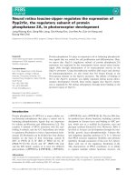
Figure 1 Immunohistochemical analysis of the expression and localisation of hScrib and PP1γ in advanced squamous cervical carcinomas.
(A) Paraffin embedded excised tissues were immunostained with anti-hScrib or anti-PP1γ as indicated, and counterstained with haematoxylin. For the
antibodies, immunostaining was performed according to standard techniques using an autostainer (BenchMark XT; Ventana Medical Systems, Inc.,
Tucson, AZ, USA). Representative experiments for a section of cervical epitheliums from normal cervix and advanced squamous cervical carcinomas
(×200 original magnification). (B) High resolution microscopic images (scale bars: 20 μm) for a section of cervical epitheliums from normal cervix and
HPV-16 positive advanced squamous cervical carcinomas.
Seiki et al. BMC Cancer (2015) 15:230
Immunohistochemistry
All tissue samples were fixed in formalin and embedded
in paraffin (obtained from patients under Institutional
Review Board approval through the University of Tokyo
Hospital). For all antibodies, immunostaining was performed according to standard techniques using an autostainer (BenchMark XT; Ventana Medical Systems, Inc.,
Tucson, AZ, USA). Immunoreactivity was interpreted
based on the negative control, which was incubated
without the primary antibody. Detection of hScrib expression was evaluated based on the existence of basolateral membrane staining as described previously [28].
For PP1γ, the expression was evaluated by nuclear staining. The immunostaining patterns of each sample were
evaluated independently and blindly by pathologists specializing in gynaecological pathology, and cytology.
PCR-based HPV DNA testing
DNA was extracted from cervical smear samples by using
the QIAGEN® DNeasy® Blood & Tissue Kits. PCR-based
HPV DNA testing was performed using the PGMYCHUV assay. Briefly, standard PCR was conducted using
the PGMY09/11 L1 consensus primer sets and HLA-dQ
primer sets. Reverse blotting hybridization was subsequently performed as described previously [33].
Results
Distribution patterns of hScrib and PP1γ in HPV-16
positive cervical intraepithelial neoplasm (CIN) and
cervical carcinoma tissues
Previous studies had highlighted hScrib as a potential
biomarker for HPV-16 induced malignancy [19,28,34].
We reasoned that if PP1γ was also regulated directly by
hScrib, this should be similarly affected in HPV-16 induced malignancy. In order to investigate this we performed IHC analysis of hScrib and PP1γ expression in
HPV-16 positive cervical tumours and control cervix.
The results obtained are shown in Figure 1 and Table 1.
In normal tissue hScrib is found primarily at cell-cell
junctions, with high levels of expression as the cells
begin to differentiate. However, hScrib distribution is altered significantly in all the HPV-16 positive tumours,
with significant redistribution in the pattern of expression in 5/11 tumours and a complete loss of expression
in 6/11 tumours. These results are largely in agreement
with previous studies [28,35]. In the case of PP1γ, this
displays a largely nuclear pattern of expression and this
is present throughout the differentiating epithelium in
the normal cervical tissue. In contrast, in the cervical tumours there is a complete loss of expression of PP1γ in
2/11 cases, with a striking redistribution in the pattern
of expression in the remaining 9 samples, where there
was a shift from a nuclear localisation to a cytoplasmic
pattern of expression.
Page 4 of 9
Table 1 Immunostaining patterns for hScrib and PP1γ in
clinical samples of human uterine cervix
hScrib
Normal
16-positive
Membrane
8
0
Cytoplasm
0
5
Nuclear
0
0
No expression
0
6
Total
8
11
PP1y
HPV negative
16-positive
Membrane
0
0
Cytoplasm
0
9
Nuclear
8
0
No expression
0
2
Total
8
11
We were then interested in investigating whether perturbation in the pattern of PP1γ expression was an early
or late event during HPV-induced neoplastic progression. To do this we repeated the PP1γ IHC analysis on
lesions exhibiting different grades of CIN. The lesions
were classified as CIN1 (n = 4), CIN2 (n = 8), CIN3 (n = 8).
As shown in Figure 2, there is a marked loss in nuclear
PP1γ expression, which is already apparent in CIN2, and
this is more evident in the CIN3 lesion, where there are
also much lower levels of PP1γ expression. Interestingly,
PP1γ positive cells were distributed only in the lower third
of the epithelial layer in CIN1 cases (4/4) and 8/8 of patients with CIN3 had PP1γ positive cells distributed in
the lower, middle, and upper third of the epithelium
(Figure 2B). In the case of hScrib, there is a similar perturbation in the pattern of expression as the lesions develop, but similar to what has been reported previously,
there is a tendency in some lower grade lesions to find
highly overexpressed hScrib in regions of the epithelium.
These results indicate that hScrib and PP1γ, whilst
both being perturbed during the progression to malignancy, are altered in a manner that is not interdependent, suggesting that PP1γ might be an independent
marker for cervical tumour development. Indeed, the
pattern and expression levels of PP1γ declined with an
almost linear relationship from normal tissue, through
increasing grades of CIN lesion, to invasive cancer.
Analysis of PP1γ expression in HPV-16-positive cells
In order to determine whether perturbation of PP1γ expression was a direct result of HPV-16 oncoprotein
function, we proceeded to examine the pattern of PP1γ
expression in cell lines derived from HPV-16 positive
cervical tumours. To do this we analysed the pattern of
PP1γ expression in HPV-16 positive CaSki and SiHa
Seiki et al. BMC Cancer (2015) 15:230
A
Page 5 of 9
hScrib
hScrib
Normal
HPV16 positive CIN2-3
CIN2
CIN3
PP1
PP1
Normal
HPV16 positive CIN2-3
CIN3
B
CIN1
CIN2
CIN3
Figure 2 Immunohistochemical analysis of the expression and localisation of hScrib and PP1γ in various stages of cervical intraepithelial
neoplasms. (A) Paraffin embedded excised tissues were immunostained with anti-hScrib or anti-PP1γ as indicated, and counterstained with
haematoxylin. For the antibodies, immunostaining was performed according to standard techniques using an autostainer (BenchMark XT; Ventana
Medical Systems, Inc., Tucson, AZ, USA). Representative experiments for a section of cervical epitheliums from normal cervix (left) and cervical
intraepithelial neoplasms (CIN) grade 2 and 3 (right) (×200 original magnification). (B) High resolution microscopic images (scale bars: 20 μm) for a
section of cervical epithelia from CIN grade 1 and 3.
cells, and compared this with HPV negative HaCaT cells.
To determine whether any alterations might be HPVspecific, we also transfected the cells with siRNA E6/E7
and siLuc as a control. After 72 hours the cells were harvested and cells fractionated into cytosolic, membrane,
nuclear and cytoskeletal pools, such that the pattern of
PP1γ subcellular distribution could be monitored. The
pattern of PP1γ expression was then ascertained by
western blotting and the results obtained are shown in
Figure 3. PP1γ is found predominantly within the nucleus in HaCaT cells (Figure 3A), whilst in the HPV-16
positive cells it is found weakly re-localised both in nuclear and cytoplasmic locations. However when E6/E7
expression is ablated there is a dramatic redistribution
in the pattern of PP1γ expression, with much higher
levels being found within the nuclear fraction of the cells
(Figure 3B). In contrast, we found no difference in PP1γ
transcript levels after siRNA E6/E7 treatment in HPV-16
positive cells (data not shown).
These results suggest that loss of nuclear PP1γ expression in HPV positive tumour cells is a direct result of
the expression of the HPV E6/E7 oncoproteins.
Seiki et al. BMC Cancer (2015) 15:230
Page 6 of 9
A
F1
HaCaT
F2
F3
F4
F1: cytosol
F2: membrane&organ
F3: nucleus
F4: cytoskeleton
hScrib
PP1
alfa-tubulin
E-Cadherin
p84
Vimentin
B
si luciferase
CaSki
F1
F2
F3
siE6/E7
F4
F1
F2
F3
F4
hScrib
PP1
F1: cytosol
F2: membrane&organ
F3: nucleus
F4: cytoskeleton
p53
E-Cadherin
alfa-tubulin
p84
Vimentin
si luciferase
SiHa
F1
F2
F3
siE6/E7
F4
F1
F2
F3
F4
hScrib
PP1
E-Cadherin
alfa-tubulin
F1:
F2:
F3:
F4:
cytosol
membrane&organ
nucleus
cytoskeleton
p84
Vimentin
Figure 3 PP1γ is mislocalised in HPV-16 positive tumour cells. (A) HaCaT cells, and (B) siE6/E7or siluciferase control transfected CaSki and
SiHa cells were fractioned into cytoplasmic (F1), membrane (F2), nuclear (F3), and cytoskeleton (F4) pools and hScrib and PP1γ were detected by
western blotting. α-tubulin was a loading control for the cytoplasmic fraction, E-Cadherin was a loading control for the membrane fraction, p84
was a loading control for the nuclear fraction, and Vimentin was a loading control for the cytoskeleton fraction.
PP1γ is subject to degradation in HPV-16 positive cells
Interestingly, the fractionation studies indicate that
whilst there is a significant increase in nuclear PP1γ in
the absence of E6/E7, there is not a significant loss of
cytoplasmic PP1γ, suggesting that some of the loss of
nuclear expression may be due to proteasome mediated
degradation. Therefore we were first interested in determining whether E6/E7 expression could affect the total
levels of PP1γ expression. To do this we analysed the
levels of PP1γ expression in total cell extracts from
CaSki and SiHa cells previously transfected with siRNA
E6/E7 or siLuc as a control. After 72 hours the cells
were extracted and the levels of PP1γ expression monitored by western blotting. The results in Figure 4A show
that loss of E6/E7 expression induces a marked increase
in the total levels of PP1γ expression in HPV-16 positive
cells, in a manner similar to that seen for restoration of
p53 levels, which served as a positive control for efficient
ablation of E6/E7 expression. We also monitored the efficiency of E6/E7 knockdown by RT-PCR and found that
Seiki et al. BMC Cancer (2015) 15:230
Page 7 of 9
A
SiHa
CaSKi
Si: si luciferase siE6/E7
Si: si luciferase siE6/E7
hScrib
PP1
p53
Actin
B
pcDNA
16E6
4µg
16E6
8µg
C
16E6
10µg
16E6
5µg
pcDNA
hScrib
hScrib
Lac Z
PP1
PP1
D
HaCaT
-
+
SiHa
-
+
CaSki
-
HeLa
C33A
+
-
+
-
+
MG132
PP1
p53
Actin
Figure 4 PP1γ levels are downregulated in HPV16 positive cells by HPV E6/E7 oncogenes. (A) HPV-16 positive CaSki and SiHa cells were
transfected with siRNAE6/E7 or siLuc as control. Total cell extracts were then made after 72 hours, and hScrib, PP1γ, p53 and Actin were detected
by western blotting. (B) 293 cells were transfected with 4, 8, 10 μg of HPV-16 E6 expression plasmid, and hScrib and PP1γ were analysed by
Western blotting. The middle panel shows the LacZ transfection efficiency and loading control. (C) HaCaT cells were transfected with 5 μg of
HPV-16 E6 expression plasmid, and hScrib and PP1γ were analysed by Western blotting. Tubulin was detected as control. (D) CaSki, SiHa, HeLa,
C33A and HaCaT cells were incubated in the presence of either 10 μM MG132 or solvent before harvesting and analysed by western blotting.
Actin was used as a loading control.
E6/E7 transcripts were reduced by around 60% following
siRNA transfection (data not shown). In contrast to the
change in PP1γ protein levels, we found no difference in
PP1γ transcript levels after siRNA E6/E7 treatment in
HPV-16 positive cells. Furthermore, to determine whether
the cell type or E6 expression contributed to the alterations in PP1γ expression levels, we compared the ability
of E6 to direct the degradation of PP1γ in 293 and HaCaT
cells. First 293 cells were transfected with increasing
amounts of HPV-16 E6, as indicated in Figure 4B.
Then, we performed the same analysis using HaCaT
cells (Figure 4C). The results demonstrated that overexpression of HPV-16 E6 results in a decrease in the level
of PP1γ expression. In order to determine whether the
loss of PP1γ expression was proteasome-mediated HPVpositive SiHa, CaSki and HeLa cells, and HPV-negative
C33A and HaCaT cells were grown in the presence of the
proteasome inhibitor, MG132 for 3 hours, after which the
Seiki et al. BMC Cancer (2015) 15:230
cells were harvested and the levels of PP1γ expression
ascertained by western blotting. As can be seen from
Figure 4D, there are minimal changes in the levels of PP1γ
expression following proteasome inhibition, regardless of
the presence or absence of HPV DNA sequences, whilst
there is efficient rescue of p53 following proteasome inhibition in HPV positive cells. These results indicate that
the effects of E6 upon PP1γ patterns of expression are
most likely proteasome independent.
Discussion
PP1 is a major serine/threonine protein phosphatase,
normally regulating the phosphorylation status of a large
number of important cellular regulatory proteins [36-39].
Important activities include the regulation of chromosome
structure during mitosis and also following DNA damage,
through de-phosphorylation of histones [30,40], and the
control of centrosome disjunction through antagonism of
Nek2A kinase activity [41].
In this study we have identified PP1γ as a potential new
biomarker of HPV-16 induced malignancy. Using HPV-16
positive cervical tumour derived cell lines, IHC analysis of
HPV-16 positive cervical tumours and CIN lesions, we
present compelling evidence that PP1γ expression patterns are perturbed as a result of infection with HPV-16.
We originally considered that the PP1γ/hScrib complex might be a general target for HPV-16 E6, based on
our previous studies showing complex formation between hScrib and PP1γ. However, analysis of the expression patterns of PP1γ and hScrib in cervical tissues
indicate that this is not the case. Most importantly however, this highlights PP1γ as an independent target of
the HPV-16 oncoproteins. In the normal cervix, PP1γ is
expressed throughout the differentiating cervical epithelium, with a predominantly nuclear pattern of expression, which is consistent with previous studies [42]. To
our surprise, we found that in all the HPV-16 positive
cervical tumours analysed, this nuclear localisation of
PP1γ was undetectable. Low levels of PP1γ can still be
found within the cytoplasm of many cells within the majority of the cervical tumours that we analysed, although
in 2/11 cases all PP1γ expression appeared to be lost.
Similarly, perturbation in the pattern of PP1γ expression
is apparent in CIN2 lesions, and this becomes more
marked as the lesions progress to CIN3, suggesting that
perturbation in the pattern of PP1γ expression is an
early event in the development of cervical malignancy.
In order to understand whether these effects on PP1γ
expression patterns were a direct consequence of E6/E7
activity, we then focused our attention on cells derived
from HPV-16 positive cervical tumours. Again we found
striking parallels with the IHC data, with very little PP1γ
expression in the nucleus of HPV-16 positive CaSki or
SiHa cells. In contrast, readily detectable nuclear PP1γ
Page 8 of 9
was observed in HaCaT cells. Most strikingly, siRNA ablation of E6/E7 expression resulted in a dramatic rescue
of PP1γ expression within the nucleus of the HPV-16
positive cells, which appeared very similar to the effects
seen upon the pattern of p53 expression. In contrast to
p53 however, the alteration in the levels and pattern of
PP1γ expression by E6 does not appear to involve the
proteasome in cells derived from cervical tumours. Obviously further studies will be required to elucidate the
precise mechanisms by which HPV-16 targets PP1γ.
Conclusions
Currently we have no information as to whether the
HPV-16 E6/E7 oncoproteins can modulate any of these
phosphorylation events in a PP1γ dependent manner, it
is nonetheless intriguing that all of these pathways are
perturbed to some extent in cells containing the HPV-16
oncoproteins. Future studies will investigate these aspects further, but it is tempting to speculate that targeting of the nuclear forms of PP1γ might contribute
directly towards the generation of genome instability,
chromatin remodeling and tumour progression. The cellular redistribution of PP1γ seems to have an important
role in the development of centrosome abnormalities
and chromosomal instability at early stages of cervical
carcinogenesis. Taken together this study highlights the
potential value of PP1γ as a novel biomarker for HPVinduced cervical neoplasia.
Competing interests
All the authors declare no competing interests.
Authors' contributions
TS performed the experiments and wrote the manuscript. KN (corresponding
author) and LB supervised the experiments and wrote the manuscript. TS,
KN, CK, KK, DM, HK-N, AT, YM, TA, OH-W, KO, SN, TY, MF, LB, YO, and TF
contributed reagents, materials, experimental techniques, and data analysis.
KN, DM, MF contributed pathological evaluation. All authors read and
approved the final manuscript.
Acknowledgement
The authors are grateful to Kei Sakuma (Department of Pathology, Graduate
School of Medicine, The University of Tokyo) for technical support on the
preparation of IHC staining. Additionally, we thank Michihiro Tanikawa, Yuichiro
Miyamoto, Kenbun Sone, Yuriko Uehara, Yuji Ikeda, Aki Miyasaka, Takahiro Koso,
Tomoko Kashiyama, Tomohiko Fukuda, Kanako Inaba, Satoko Kojima, and
Kensuke Tomio for their support and assistance. We also gratefully
acknowledge the particular assistance of all members in Lawrence Banks's lab,
and valuable comments on the manuscript from Miranda Thomas and David
Pim. This work was supported by a Grant-in-Aid for Scientific Research (K.N.)
from the Ministry of Education, Science and Culture, Japan, and in part by a
research grant from the Associazione Italiana per la Ricerca sul Cancro (L.B).
Author details
1
Department of Obstetrics and Gynecology, Faculty of Medicine, The
University of Tokyo, Tokyo 113-8655, Japan. 2International Centre for Genetic
Engineering and Biotechnology, Area Science Park, Padriciano-99, I-34012
Trieste, Italy. 3Department of Pathology, Graduate School of Medicine, The
University of Tokyo, Tokyo 113-8655, Japan. 4Department of Obstetrics and
Gynecology, Graduate School of Medicine, Teikyo University, Tokyo 173-8605,
Japan. 5Department of Obstetrics and Gynecology, National Center for Global
Health and Medicine, Tokyo 162-8655, Japan.
Seiki et al. BMC Cancer (2015) 15:230
Received: 31 May 2014 Accepted: 27 February 2015
References
1. Zur HH. Papillomaviruses and cancer: from basic studies to clinical
application. Nat Rev Cancer. 2002;2:342–50.
2. Bouvard V, Baan R, Straif K, Grosse Y, Secretan B, El Ghissassi F, et al. WHO
international agency for research on cancer monograph working group. A
review of human carcinogens–part B: biological agents. Lancet Oncol.
2009;10:321–2.
3. Butz K, Ristriani T, Hengstermann A, Denk C, Scheffner M, Hoppe-Seyler F.
siRNA targeting of the viral E6 oncogene efficiently kills human
papillomavirus-positive cancer cells. Oncogene. 2003;22:5938–45.
4. Yoshinouchi M. In vitro and in vivo growth suppression of human
papillomavirus 16-positive cervical cancer cells by e6 siRNA. Mol Ther.
2003;8:762–8.
5. McLaughlin-Drubin ME, Meyers J, Munger K. Cancer associated human
papillomaviruses. Curr Opin Virol. 2012;2:459–66.
6. Mantovani F, Banks L. The human papillomavirus E6 protein and its
contribution to malignant progression. Oncogene. 2001;20:7874–87.
7. Huh K, Zhou X, Hayakawa H, Cho J-Y, Libermann TA, Jin J, et al. Human
papillomavirus type 16 E7 oncoprotein associates with the cullin 2 ubiquitin
ligase complex, which contributes to degradation of the retinoblastoma
tumor suppressor. J Virol. 2007;81:9737–47.
8. Scheffner M, Huibregtse JM, Vierstra RD, Howley PM. The HPV-16 E6 and
E6-AP complex functions as a ubiquitin-protein ligase in the ubiquitination
of p53. Cell. 1993;75:495–505.
9. Schlegel R, Phelps WC, Zhang YL, Barbosa M. Quantitative keratinocyte
assay detects two biological activities of human papillomavirus DNA and
identifies viral types associated with cervical carcinoma. EMBO J.
1988;7:3181–7.
10. Barbosa MS, Schlegel R. The E6 and E7 genes of HPV-18 are sufficient for
inducing two-stage in vitro transformation of human keratinocytes. Oncogene
1989:1529-32.
11. Munger K, Phelps WC, Bubb V, Howley PM. The E6 and E7 genes of the
human papillomavirus type 16 together Are necessary and sufficient for
transformation of primary human keratinocytes. J Virol. 1989;63:4417–21.
12. Song S, Liem A, Miller JA, Lambert PF. Human papillomavirus types 16 E6
and E7 contribute differently to carcinogenesis. Virology. 2000;267:141–50.
13. Shai A, Nguyen ML, Wagstaff J, Jiang Y-H, Lambert PF. HPV16 E6 confers
p53-dependent and p53-independent phenotypes in the epidermis of mice
deficient for E6AP. Oncogene. 2007;26:3321–8.
14. Thomas M, Narayan N, Pim D, Tomaić V, Massimi P, Nagasaka K, et al.
Human papillomaviruses, cervical cancer and cell polarity. Oncogene.
2008;27:7018–30.
15. Pim D, Banks L. Interaction of viral oncoproteins with cellular target
molecules: infection with high-risk vs low-risk human papillomaviruses.
APMIS. 2010;118:471–93.
16. Kiyono T, Kiyono T, Hiraiwa A, Fujita M, Hayashi Y, Akiyama T, et al. Binding
of high-risk human papillomavirus E6 oncoproteins to the human
homologue of the Drosophila discs large tumor suppressor protein. Proc
Natl Acad Sci U S A. 1997;94:11612–6.
17. Lee SS, Weiss RS, Javier RT. Binding of human virus oncoproteins to hDlg/
SAP97, a mammalian homolog of the Drosophila discs large tumor
suppressor protein. Proc Natl Acad Sci U S A. 1997;94:6670–5.
18. Gardiol D, Kühne C, Glaunsinger B, Lee SS, Javier R, Banks L. Oncogenic
human papillomavirus E6 proteins target the discs large tumour suppressor
for proteasome-mediated degradation. Oncogene. 1999;18:5487–96.
19. Nakagawa S, Huibregtse JM. Human scribble (Vartul) is targeted for
ubiquitin-mediated degradation by the high-risk papillomavirus E6 proteins
and the E6AP ubiquitin-protein ligase. Mol Cell Biol. 2000;20:8244–53.
20. Glaunsinger BA, Lee SS, Thomas M, Banks L, Javier R. Interactions of the
PDZ-protein MAGI-1 with adenovirus E4-ORF1 and high-risk papillomavirus
E6 oncoproteins. Oncogene. 2000;19:5270–80.
21. Kranjec C, Banks L. A systematic analysis of human papillomavirus (HPV) E6
PDZ substrates identifies MAGI-1 as a major target of HPV type 16 (HPV-16)
and HPV-18 whose loss accompanies disruption of tight junctions. J Virol.
2011;85:1757–64.
22. Bilder D, Li M, Perrimon N. Cooperative regulation of cell polarity and
growth by Drosophila tumor suppressors. Science. 2000;289:113–6.
Page 9 of 9
23. Zhan L. Deregulation of scribble promotes mammary tumorigenesis and
reveals a role for cell polarity in carcinoma. Cell. 2008;135:865–78.
24. Dow LE. Loss of human Scribble cooperates with H-Ras to promote cell
invasion through deregulation of MAPK signalling. Oncogene.
2008;27:5988–6001.
25. Nagasaka K, Pim D, Massimi P, Thomas M, Tomaić V, Subbaiah VK, et al. The
cell polarity regulator hScrib controls ERK activation through a KIM sitedependent interaction. Oncogene. 2010;29:5311–21.
26. Pearson HB, Perez-mancera PA, Dow LE, Ryan A, Tennstedt P, Bogani D, et al.
SCRIB expression is deregulated in human prostate cancer, and its deficiency
in mice promotes prostate neoplasia. J Clin Invest. 2011;121:4257–67.
27. Elsum I a, Yates LL, Pearson HB, Phesse TJ, Long F, O’Donoghue R, Ernst M,
Cullinane C, Humbert PO. Scrib heterozygosity predisposes to lung cancer
and cooperates with KRas hyperactivation to accelerate lung cancer
progression in vivo. Oncogene 2013;:1–11.
28. Nakagawa S, Yano T, Nakagawa K, Takizawa S, Suzuki Y, Yasugi T, et al.
Analysis of the expression and localisation of a LAP protein, human scribble,
in the normal and neoplastic epithelium of uterine cervix. Br J Cancer.
2004;90:194–9.
29. Nagasaka K, Seiki T, Yamashita A, Massimi P, Subbaiah VK, Thomas M, et al.
A novel interaction between hScrib and PP1γ downregulates ERK signaling
and suppresses oncogene-induced cell transformation. PLoS One.
2013;8:e53752.
30. Shimada M, Haruta M, Niida H, Sawamoto K, Nakanishi M. Protein
phosphatase 1γ is responsible for dephosphorylation of histone H3 at Thr
11 after DNA damage. EMBO Rep. 2010;11:883–9.
31. Trinkle-mulcahy L, Andrews PD, Wickramasinghe S, Sleeman J, Prescott A,
Lam YW, et al. Time-lapse imaging reveals dynamic relocalization of PP1γ
throughout the mammalian cell cycle. Mol Biol Cell. 2003;14:107–17.
32. Boukamp P, Petrussevska RT, Breitkreutz D, Hornung J, Markham A, Fusenig
NE. Normal keratinization in a spontaneously immortalized aneuploid
human keratinocyte cell line. J Cell Biol. 1988;106:761–71.
33. Gravitt PE, Peyton CL, Alessi TQ, Wheeler CM, Hildesheim A, Schiffman MH,
et al. Improved amplification of genital human papillomaviruses. J Clin
Microbiol. 2000;38:357–61.
34. Thomas M, Massimi P, Navarro C, Borg J, Banks L. The hScrib/Dlg apicobasal control complex is differentially targeted by HPV-16 and HPV-18 E6
proteins. Oncogene. 2005;24:6222–30.
35. Gardiol D, Zacchi A, Petrera F, Stanta G, Banks L. Human discs large and
scrib are localized at the same regions in colon mucosa and changes in
their expression patterns are correlated with loss of tissue architecture
during malignant progression. Int J Cancer. 2006;119:1285–90.
36. Bollen M. Combinatorial control of protein phosphatase-1. Trends Biochem
Sci. 2001;26:426–31.
37. Cohen PTW. Protein phosphatase 1 – targeted in many directions. J Cell Sci.
2002;115:241–56.
38. Margolis SS, Walsh S, Weiser DC, Yoshida M, Shenolikar S, Kornbluth S. PP1
control of M phase entry exerted through 14-3-3-regulated Cdc25
dephosphorylation. EMBO J. 2003;22:5734–45.
39. Wu JQ, Guo JY, Tang W, Yang CS, Freel CD, Chen C, et al. PP1-mediated
dephosphorylation of phosphoproteins at mitotic exit is controlled by
inhibitor-1 and PP1 phosphorylation. Nat Cell Biol. 2009;11:644–51.
40. Vagnarelli P, Ribeiro S, Sennels L, Sanchez-Pulido L, de Lima AF, Verheyen T,
et al. Repo-Man coordinates chromosomal reorganization with nuclear
envelope reassembly during mitotic exit. Dev Cell. 2011;21:328–42.
41. Mardin BR, Agircan FG, Lange C, Schiebel E. Plk1 controls the Nek2A-PP1γ
antagonism in centrosome disjunction. Curr Biol. 2011;21:1145–51.
42. Andreassen PR, Lacroix FB, Villa-Moruzzi E, Margolis RL. Differential subcellular
localization of protein phosphatase-1 alpha, gamma1, and delta isoforms
during both interphase and mitosis in mammalian cells. J Cell Biol.
1998;141:1207–15.