Detection of morphologic alterations in rectal carcinoma following preoperative radiochemotherapy based on multiphoton microscopy imaging
Bạn đang xem bản rút gọn của tài liệu. Xem và tải ngay bản đầy đủ của tài liệu tại đây (2.9 MB, 7 trang )
Li et al. BMC Cancer (2015) 15:142
DOI 10.1186/s12885-015-1157-5
RESEARCH ARTICLE
Open Access
Detection of morphologic alterations in
rectal carcinoma following preoperative
radiochemotherapy based on multiphoton
microscopy imaging
Lianhuang Li1†, Zhifen Chen2†, Xingfu Wang3†, Hongsheng Li1, Weizhong Jiang2, Shuangmu Zhuo1,
Guoxian Guan2* and Jianxin Chen1*
Abstract
Background: Preoperative radiochemotherapy improves outcomes in patients with locally advanced rectal
carcinoma, and has been used increasingly in patient management. However, there is a strong clinical need to
assess tumor response to neoadjuvant treatment, and a non-invasive technique that allows the precise
identification of morphologic changes in tumors would be of considerable clinical interest.
Methods: In this study, we used multiphoton microscopy (MPM) to detect morphologic alterations in rectal
adenocarcinomas in patients treated with preoperative radiochemotherapy.
Results: MPM was able to identify histopathologic alterations in rectal cancer following preoperative
radiochemotherapy, and allowed the qualitative assessment of treatment efficacy and feasibility in relation
to dose or strategy.
Conclusion: These findings may provide the groundwork for evaluating tumor response to neoadjuvant
treatment, thus allowing the tailoring of effective treatment doses and strategies.
Keywords: Multiphoton microscopy, Preoperative radiochemotherapy, Fibrosis, Two-photon excited
fluorescence, Second harmonic generation
Background
Accumulating evidence has demonstrated that neoadjuvant treatment improves local control and survival in
patients with rectal cancer, and may play an increasing
role, especially in locally advanced disease [1-5]. Preoperative radiochemotherapy has thus been used increasingly in the management of this group of patients.
Pathological assessment based on the histopathological
investigation of resected specimens is important for
* Correspondence: ;
†
Equal contributors
2
Department of Colorectal Surgery, The Affiliated Union Hospital, Fujian
Medical University, Fuzhou 350001, China
1
Institute of Laser and Optoelectronics Technology, Fujian Provincial Key
Laboratory for Photonics Technology, Key Laboratory of OptoElectronic
Science and Technology for Medicine of Ministry of Education, Fujian Normal
University, Fuzhou 350007, China
Full list of author information is available at the end of the article
estimating the prognosis and effect of radiochemotherapy [6,7]. However, pathological examination is associated with several disadvantages, such as crush artifacts,
bleeding, sampling errors, and time-consuming pathological procedures [8,9]. In contrast, multiphoton microscopy (MPM), which is based on intrinsic two-photon
excited fluorescence (TPEF) and second harmonic generation (SHG), offers significant advantages for imaging in
thick tissue and live animals, including greater imaging
penetration depth, reduced out-of-focus photobleaching
and phototoxicity, and the ability to detect the cellular
and subcellular microstructures of biological tissues
[10-12].
This study therefore investigated the treatment-related
morphologic aspects of rectal carcinomas using MPM
imaging, with an emphasis on stromal alterations, changes
in blood vessels and inflammatory cell infiltrate, and
© 2015 Li et al.; licensee BioMed Central. This is an Open Access article distributed under the terms of the Creative Commons
Attribution License ( which permits unrestricted use, distribution, and
reproduction in any medium, provided the original work is properly credited. The Creative Commons Public Domain
Dedication waiver ( applies to the data made available in this article,
unless otherwise stated.
Li et al. BMC Cancer (2015) 15:142
residual tumor cells. The study aimed to provide a detailed
morphologic description of rectal carcinoma in patients
treated with preoperative radiochemotherapy, and to identify patterns of morphologic alterations with prognostic
significance. We also aimed to determine the efficacy of
radiochemotherapy and the appropriateness of the treatment dose and strategy by monitoring theses morphological changes.
Methods
MPM imaging system
The MPM imaging system used in this study has previously been described in detail [13,14]. Briefly, MPM images were acquired using a LSM 510 META system
(Zeiss, Jena, Germany) coupled to a Ti:sapphire laser
(Mira 900-F, Coherent Inc., Santa Clara, CA, USA). An
oil immersion objective (Plan-Apochromat 63×, N.A.1.4)
was used to focus the excitation beam into samples and
to collect the back-scattered TPEF/SHG signals. Two
different channels were selected to obtain high-contrast
images of collagen and fluorescence components, respectively. One channel corresponded to wavelengths of
387–419 nm to reveal the collagen microstructure using
SHG signals, while the other channel covered wavelengths 430–716 nm to reveal the morphology of fluorescence components using TPEF signals at an excitation
wavelength of 810 nm. The contrast of the SHG/TPEF
images was increased by color-coding the SHG images
green and the TPEF images red.
Specimen preparation
This investigation was approved by the Institutional Review Board of the Affiliated Union Hospital, Fujian Medical University and conformed with the institutional
rules governing clinical investigations of human subjects
in biomedical research. Prior to study participation, all
patients signed an informed consent form. All patients
underwent long-term preoperative radiochemotherapy
(45 Gy/25 fractions followed by a 5.4 Gy boost, for a
total of 50.4 Gy, and oral capecitabine 825 mg/m2 twice
daily during radiotherapy). According to the Chinese
guidelines for colorectal cancer treatment, radical surgery
was performed about 8 weeks after the end of radiotherapy. Patients with different responses to therapy were selected, and patients who failed to respond were excluded.
Tumor response to therapy was judged macroscopically in
harvested specimens.
Seven fresh tumor samples were obtained from seven
patients undergoing rectum resection after preoperative
radiochemotherapy at the Affiliated Union Hospital of
Fujian Medical University. Normal specimens were
collected 6 cm outside the cancer margin. Patient ages
ranged from 38–67 years (53 ± 10 years) and the male/
female ratio was 2.5. Detailed information, including
Page 2 of 7
cancer classification and clinical stage, was shown in
Table 1. Five serial tissue slices (10 μm thick) were cut
from each specimen and the middle slice was processed
for histological examination with hematoxylin and eosin
(H&E) stain, according to standard histology procedures.
The other tissue sections were sandwiched between a
microscope slide and glass coverslip for MPM imaging.
The tissue slices were imaged with the coverslip facing the
microscope objective. Phosphate-buffered saline solution
was dripped into the specimen during imaging to avoid
dehydration or shrinkage during the imaging process.
Histology
H&E-stained sections were reviewed by a certified pathologist. Images were obtained by standard bright-field
light microscopy (Eclipse Ci-L, Nikon Instruments Inc.,
Japan) with a charge-coupled device (Nikon, DS-Fi2).
Finally, the MPM and corresponding H&E-stained images (40×) were analyzed and compared by a certified
pathologist.
Quantification of morphological features
Quantitative changes in fibrotic tissue, cellular architecture, and blood vessels during rectal carcinoma progression following preoperative radiochemotherapy were
assessed by calculating the collagen density, nuclear area,
and vessel wall thickness, respectively. For each blood vessel, three random positions were selected and the vessel
wall thickness was determined as the mean thicknesses of
the three positions. Collagen density was defined as the ratio of SHG to all pixels in each image. The nuclear area
was calculated by measuring the area enclosed by the nuclear boundary. Values were expressed as means and
standard deviations (mean ± SD). The standard deviation
signified the change in nucleus size or vessel wall thicknesses, respectively.
Results
Stromal alteration
Representative MPM and corresponding H&E-stained
images showed stromal alterations in rectal carcinomas
Table 1 Patient information including cancer
classification and clinical stage
Patients
Cancer classification
Clinical stage
1
Adenocarcinoma
cT2N + M0, Stage III
2
Adenocarcinoma
cT3N + M0, Stage III
3
Adenocarcinoma
cT3N + M0, Stage III
4
Adenocarcinoma
cT3N0M0, Stage II
5
Adenocarcinoma
cT3N + M0, Stage III
6
Adenocarcinoma
cT3N + M0, Stage III
7
Adenocarcinoma
cT3N + M0, Stage III
Li et al. BMC Cancer (2015) 15:142
Page 3 of 7
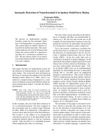
induced by preoperative radiochemotherapy with cancerous cell invasion into the muscularis propria (Figure 1).
MPM images clearly revealed stromal changes in rectal
cancer patients undergoing preoperative radiochemotherapy. Consecutive muscular tissues were disrupted
and collagen fibers were abundant but disordered, as
demonstrated by SHG signals (green) (Figure 1(a)).
This may be interpreted as former tumor infiltration
leading to the destruction of muscular tissues, and
post-treatment tumor regression represented by fibrosis or fibroinflammatory changes replacing neoplastic
glands [15,16]. These fibrotic tissues also produced
comparable TPEF signals (red) (Figure 1(b)), and overlaid TPEF/SHG images therefore appear yellowish
(Figure 1(c)). The details revealed by MPM correlated
well with the H&E-stained images (Figure 1(d)).
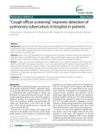
in the serosa after preoperative radiochemotherapy
(Figure 2). The blood vessels showed significant alterations with thickening and fibrosis of the intima and
media, as shown by TPEF signals in MPM (Figure 2(c))
(blue arrows) [7]. TPEF signals also revealed inflammatory cell infiltration and large numbers of inflammatory cells infiltrating into the stroma (Figure 2(c))
(white arrows). The tumor-associated inflammatory
reaction has long been considered as a type of host response and an important factor in tumor progression
[16]. Previous studies also demonstrated lower recurrence rates and better outcomes in patients with rectal
cancer who had abundant inflammatory cells in the
stroma post-irradiation [17,18]. These qualitative morphological variations were consistent with the paired
histological sections in the current study (Figure 2(d)).
Changes in blood vessels and inflammatory cell infiltrate
Residual tumor cells
MPM and H&E staining revealed obvious radiogenic
blood vessel changes and inflammatory cell infiltration
MPM and H&E-stained images showed residual tumors in the muscularis propria after preoperative
Figure 1 Representative TPEF/SHG images of stromal alterations in rectal carcinoma after preoperative radiochemotherapy characterized
by fibrosis or fibroinflammatory changes. Scale bar = 100 μm. (a) SHG image (green); (b) TPEF image (red); (c) overlay of SHG/TPEF images; and
(d) corresponding H&E-stained image (40× magnification).
Li et al. BMC Cancer (2015) 15:142
Page 4 of 7
Figure 2 Representative TPEF/SHG images of blood vessel changes with thickening and fibrosis of the intima and media, and
inflammatory cell infiltration. Scale bar = 100 μm. (a) SHG image (green); (b) TPEF image (red); (c) overlay of SHG/TPEF images; and (d)
corresponding H&E-stained image (40× magnification).
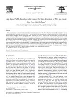
radiochemotherapy (Figure 3). Tumor cells in the rectal
carcinoma may show marked posttreatment changes, such
as nuclear atypia, and these altered tumor cells may retain
a glandular growth pattern or become more solid [15,16].
In the current study, the dominant tumor morphologic
pattern remained similar in treated and untreated rectal
adenocarcinomas (white arrows); the tumors were surrounded by fibrosis with minimal inflammatory cells,
while some tumor cells became solid (blue arrow). Furthermore, MPM allowed the differentiation of cellular
features such as nuclear pleomorphism, which is an
important biologic characteristic reflecting tumor
grade, degree of differentiation, and proliferation. The
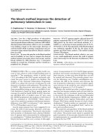
region of interest within the white box in Figure 3(c) is
magnified in Figure 4 to show the ultrastructure of the
residual cancerous cells more clearly. These alterations
following preoperative radiochemotherapy are common and might be clinically meaningful. The details of
the morphological changes revealed by MPM correlated well with those shown in H&E-stained images
(Figure 3(d)).
Quantitative analysis of morphological features
Changes in the morphological features of rectal carcinomas treated by radiochemotherapy were described
quantitatively by analyzing collagen density, vessel wall
thickness, and nuclear area (Table 2).
*P < 0.0001 (collagen density between the submucosa,
serosa and fibrosis), and P > 0.05 (collagen density between the submucosa and serosa).
The collagen density in stromal fibrosis (0.97 ± 0.03)
was significantly higher compared with normal submucosa (0.77 ± 0.06) and serosa (0.71 ± 0.05) (Figure 5)
(P < 0.0001 between the submucosa, serosa, and fibrosis:
one-way ANOVA test; SPSS 15.0). The vessel wall
thickness was 117.23 ± 36.46 μm, and the relatively
large SD indicates considerable variation in blood
vessel size. The nuclear area was 83.28 ± 57.02 μm2,
and again the large SD demonstrates that carcinomatous cells in post-treatment rectal carcinoma displayed
marked nuclear atypia. A combination of collagen
density, vessel wall thickness, and nuclear area may
thus be useful for quantitatively monitoring the development of rectal carcinomas in patients undergoing preoperative radiochemotherapy.
Discussion
Preoperative radiochemotherapy has been shown to be
useful for reducing tumor size and increasing operability. More importantly, preoperative radiotherapy in
Li et al. BMC Cancer (2015) 15:142
Page 5 of 7
Figure 3 Representative TPEF/SHG images of residual tumors surrounded by fibrosis with minimal inflammatory cells. Scale bar = 100 μm.
(a) SHG image (green); (b) TPEF image (red); (c) overlay of SHG/TPEF images; and (d) corresponding H&E-stained image (40× magnification).
combination with surgery has been shown to decrease
the rate of local recurrence in rectal cancer patients
[17-19]. Preoperative radiochemotherapy can modify
the histologic appearance of rectal cancer in terms of
fibrosis, colloid response (data not shown), blood vessel
hyperplasia, and inflammatory reaction, and these morphologic alterations have shown clinically meaningful correlations with patient outcome [7,16]. Although these
changes can be determined by histological examination of
resection specimens, the procedure is troublesome and
time-consuming.
Fortunately, fibrous tissues consist mainly of collagen
fibers and emit strong SHG signals, whereas blood
vessels, residual carcinomatous cells, and inflammatory
cells contain abundant elastin and generate TPEF
signals [20,21]. These signals make it possible to detect
histopathologic changes using MPM. The current
study employed MPM to provide a detailed morphologic description of rectal carcinomas in patients
undergoing preoperative radiochemotherapy. The results demonstrated that MPM imaging was able to
identify blood vessel hyperplasia and tumor regression by
Figure 4 Magnification of region of interest in Figure 3(c) (white box) and corresponding H&E-stained image. (a) MPM image; and
(b) H&E-stained image.
Li et al. BMC Cancer (2015) 15:142
Page 6 of 7
Table 2 Morphological features of rectal carcinoma after preoperative radiochemotherapy
Patient no.
Morphologic features
1
2
3
4
5
6
7
Average
Collagen density of normal submucosa
0.78
0.81
0.85
0.75
0.79
0.67
0.71
0.77 ± 0.06
Collagen density of normal serosa
0.69
0.67
0.75
0.70
0.79
0.63
0.74
0.71 ± 0.05
Collagen density of fibrosis associate with carcinoma
0.97
0.96
1.00
0.92
0.99
0.95
0.98
0.97 ± 0.03
Nuclear area (μm ) of carcinoma
197.51
89.18
74.63
103.86
42.67
44.34
30.77
83.28 ± 57.02
Vessel wall thickness (μm) of carcinoma
71.50
89.15
130.69
162.96
150.50
135.65
80.13
117.23 ± 36.46
2
the disappearance of carcinoma cells and their replacement by fibrous or fibroinflammatory tissues. Furthermore, the high resolution of MPM to approximately the
cellular/sub-cellular level [22,23], enables this technique
to identify residual carcinomatous cells and distinguish
between tumor cells and other cells with different morphologies, such as adipose cells [24].
There is currently no validated method for directly
monitoring the efficacy of radiochemotherapy, and the
appropriateness of the treatment dose and strategy. The
current study showed that radiochemotherapy caused
morphological changes in rectal carcinomas, including
changes in nuclear shape, collagen density, and vessel
wall thickness, and these changes could be identified
and quantitatively described by MPM. These results suggest that MPM may be a useful tool for evaluating tumor
response to neoadjuvant treatment by enabling monitoring of the morphological changes and subsequent tailoring
of the effective treatment dose or strategy.
Radiochemotherapy is a standard approach in advanced
solid human tumors. Tumors often develop in the mucosal layer and gradually infiltrate to the lamina propria,
submucosal layer, and deeper layer of the bowel wall. The
cancer is therefore most advanced in the superficial layer,
and the curative effect of radiochemotherapy can be evaluated qualitatively by monitoring treatment-related morphological changes in the superficial layer. MPM has been
reported to penetrate to depths of millimeters [25]. MPM
may therefore be useful for determining if radiochemotherapy has a curative effect, and if the treatment dose
Figure 5 Collagen density in stromal fibrosis, normal
submucosa, and serosa. Error bars indicate SD.
and strategy are appropriate regardless of cancer stage.
Subsequent treatments can then be tailored to meet the
specific clinical needs of different patients.
There are some drawbacks associated with this technique, and some limitations of the current study. There
are three major factors limiting the in vivo application of
MPM. First, the limited field of view makes examination
of large areas or volumes problematic, and may lead to interobserver variability. Second, the depth of imaging is
limited, though this limitation may be overcome by the
continuous advancement of a gradient index lens-based
MPM. Finally, the technique is expensive, though it is possible that the cost may be reduced by using a combination
of fiber laser and MPM. The study limitations included
the small patient number and lack of comparison of preand post-therapeutic samples. However, these factors do
not affect the conclusion that MPM can be used to monitor tumor response to preoperative radiochemotherapy.
The importance of the impact of histopathological
factors on prognosis and an awareness of the role of
morphologic alterations in ensuring an accurate pathologic assessment [16,26,27] indicate the urgent need for
a new, label-free, real-time, noninvasive technique for
differentiating among the various morphologic alterations induced by radiochemotherapy. The results of the
present study suggest that MPM might provide a realtime, label-free technique for evaluating tumor response
following preoperative radiochemotherapy and for noninvasive, in vivo pathophysiological analysis. The advantages of MPM indicate that it may be useful for the
in vivo assessment of treatment effect, dose, and strategy
in patients with rectal carcinoma receiving preoperative
radiochemotherapy.
Conclusions
MPM can be used to differentiate morphologic changes
in rectal carcinomas in patients undergoing preoperative
radiochemotherapy. The advancement of clinically miniaturized MPM and multiphoton probes to allow MPM
to be combined with standard endoscopes will allow the
real-time in vivo evaluation of tumor response to neoadjuvant therapy and the subsequent tailoring of effective
treatment doses and strategies.
Li et al. BMC Cancer (2015) 15:142
Competing interests
The authors declare that they have no competing interests.
Authors’ contributions
LHL, XFW and JXC are responsible for conception and design. Data were
obtained by LHL and HSL. ZFC, XFW, WZJ, GXG and JXC provided technical
support. All authors contributed to the analysis and interpretation of data,
wrote, reviewed and approved the final manuscript.
Authors’ information
Initials: Lianhuang Li (LHL), Zhifen Chen (ZFC), Xingfu Wang (XFW),
Hongsheng Li (HSL), Weizhong Jiang (WZJ), Guoxian Guan (GXG), Jianxin
Chen (JXC).
Acknowledgments
The project was supported by the Program for Changjiang Scholars and
Innovative Research Team in University (Grant No. IRT1115), the National
Natural Science Foundation of China (Grant No. 81271620), the Natural
Science Foundation for Distinguished Young Scholars of Fujian Province
(Grant No. 2014 J06016), and the Youth Scientific Research Foundation of
Fujian Provincial Department of Health (2013-2-36), National Clinical Key
Specialty Construction Project (General Surgery).
Author details
1
Institute of Laser and Optoelectronics Technology, Fujian Provincial Key
Laboratory for Photonics Technology, Key Laboratory of OptoElectronic
Science and Technology for Medicine of Ministry of Education, Fujian Normal
University, Fuzhou 350007, China. 2Department of Colorectal Surgery, The
Affiliated Union Hospital, Fujian Medical University, Fuzhou 350001, China.
3
Department of Pathology, The First Affiliated Hospital, Fujian Medical
University, Fuzhou 350001, China.
Received: 4 June 2014 Accepted: 3 March 2015
References
1. Schrag D, Weiser MR, Goodman KA, Gon̈en M, Hollywood E, Cercek A, et al.
Neoadjuvant chemotherapy without routine use of radiation therapy for
patients with locally advanced rectal cancer: a pilot trial. J Clin Oncol.
2014;32:1–6.
2. Hwang MR, Park JW, Park S, Yoon H, Kim DY, Chang HJ, et al. Prognostic
impact of circumferential resection margin in rectal cancer treated with
preoperative chemoradiotherapy. Ann Surg Oncol. 2014;21:1345–51.
3. Wheeler JMD, Warren BF, Jones AC, Mortensen NJM. Preoperative
radiotherapy for rectal cancer: implications for surgeons, pathologists and
radiologists. Brit J Surg. 1999;86:1108–20.
4. Nagtegaal ID, Marijnen CAM, Kranenbarg EK, Mulder-Stapel A, Hermans J,
van de Velde CJH, et al. Short term preoperative radiotherapy interferes with
determination of pathological parameters in rectal cancer. J Pathol.
2002;197:20–7.
5. Hartley A, Ho KF, Mcconkey C, Geh JI. Pathological complete response
following pre-operative chemoradiotherapy in rectal cancer: analysis of
phase I/III trials. Brit J Radiol. 2005;78:934–8.
6. Thies S, Langer R. Tumor regression grading of gastrointestinal carcinomas
after neoadjuvant treatment. Front Oncol. 2013;3:262.
7. Dworak O, Keilholz L, Hoffmann A. Pathological features of rectal cancer
after preoperative radiochemotherapy. Int J Color Dis. 1997;12:19–23.
8. Ying MG, Zhuo SM, Chen G, Zhuo CH, Lu JP, Zhu WF, et al. Real-time
noninvasive optical diagnosis for colorectal cancer using multiphoton
microscopy. Scanning. 2012;34:181–5.
9. Wu XF, Chen G, Lu JP, Zhu WF, Qiu JT, Chen JX, et al. Label-free detection
of breast masses using multiphoton microscopy. Plos One. 2013;88:e65933.
10. Chen Y, Guo HC, Gong W, Qin LY, Aleyasin H, Ratan RR, et al. Recent
advances in two-photon imaging: technology developments and
biomedical applications. Chin Opt Lett. 2013;11:011703.
11. Hoover EE, Squier JA. Advances in multiphoton microscopy technology. Nat
Photonics. 2013;7:93–101.
12. Zipfel WR, Williams RM, Christie R, Nikitin AY, Hyman BT, Webb WW. Live
tissue intrinsic emission microscopy using multiphoton-excited native
fluorescence and second harmonic generation. PNAS. 2003;100:7075–80.
Page 7 of 7
13. Chen JX, Xu J, Kang DY, Xu MF, Zhuo SM, Zhu XQ, et al. Multiphoton
microscopic imaging of histological sections without hematoxylin and eosin
staining differentiates carcinoma in situ lesion from normal oesophagus.
Appl Phys Lett. 2013;103:183701.
14. Zhuo SM, Yan J, Chen G, Shi H, Zhu XQ, Lu JP, et al. Label-free imaging of
basement membranes differentiates normal, precancerous, and cancerous
colonic tissues by second-harmonic generation microscopy. Plos One.
2012;7:e38655.
15. Micev M, Micev-Ćosić M, Todorović V, Krsmanović M, Krivokapić Z, Popović
M, et al. Histopathology of residual rectal carcinoma following preoperative
radiochemotherapy. Acta Chir Iugosl. 2004;51:99–108.
16. Shia J, Guillem JG, Moore HG, Tickoo SK, Qin J, Ruo L, et al. Patterns of
morphologic alteration in residual rectal carcinoma following preoperative
chemoradiation and their association with long-term outcome. Am J Surg
Pathol. 2004;28:215–23.
17. Nagtegaal ID, Marijnen CAM, Kranenbarg EK, Mulder-Stapel A, Hermans J,
van de Velde CJH, et al. Local and distant recurrences in rectal cancer patients
are predicted by the nonspecific immune response; specific immune response
has only a systemic effect-a histopathological and immunohistochemical study.
BMC Cancer. 2001;1:7.
18. García-Aguilar J, De Anda EH, Sirivongs P, Lee SH, Madoff RD, Rothenberger
DA. A pathologic complete response to preoperative chemoradiation is
associated with lower local recurrence and improved survival in rectal
cancer patients treated by mesorectal excision. Dis Colon Rectum.
2003;46:298–304.
19. Bouzourene H, Bosman FT, Matter M, Coucke P. Predictive factors in locally
advanced rectal cancer treated with preoperative hyperfractionated and
accelerated radiotherapy. Hum Pathol. 2003;34:541–8.
20. Monic M. Cell and tissue autofluorescence research and diagnostic
applications. Biotechnol Annu Rev. 2005;11:227–56.
21. Konig K. Multiphoton microscopy in life sciences. J Microsc. 2000;200:83–104.
22. Rivera DR, Brown CM, Quzounov DG, Webb WW, Xu C. Multifocal
multiphoton endoscope. Opt Lett. 2012;37:1349–51.
23. Wang TJ, Li QY, Xiao P, Ahn J, Kim YE, Park Y, et al. Gradient index lens based
combined two-photon microscopy and optical coherence tomography.
Opt Express. 2014;22:12962–70.
24. Huang ZF, Zhuo SM, Chen JX, Chen R, Jiang XS. Multiphoton microscopic
imaging of adipose tissue based on second-harmonic generation and
two-photon excited fluorescence. Scanning. 2008;30:452–6.
25. Helmchen F, Denk W. Deep tissue two-photon microscopy. Nat Methods.
2005;2:932–40.
26. Wheeler JMD, Warren BF, Mortensen NJMCC, Ekanyaka N, Kulacoglu H,
Jones AC, et al. Quantification of histologic regression of rectal cancer after
irradiation. Dis Colon Rectum. 2002;45:1051–6.
27. Bozzetti F, Baratti D, Andreola S, Zucali R, Schiavo M, Spinelli P, et al.
Preoperative radiation therapy for patients with T2-T3 carcinoma of the
middle-to-low rectum. Cancer. 1999;86:398–404.
Submit your next manuscript to BioMed Central
and take full advantage of:
• Convenient online submission
• Thorough peer review
• No space constraints or color figure charges
• Immediate publication on acceptance
• Inclusion in PubMed, CAS, Scopus and Google Scholar
• Research which is freely available for redistribution
Submit your manuscript at
www.biomedcentral.com/submit