Oridonin stabilizes retinoic acid receptor alpha through ROS-activated NF-κB signaling
Bạn đang xem bản rút gọn của tài liệu. Xem và tải ngay bản đầy đủ của tài liệu tại đây (1.93 MB, 12 trang )
Cao et al. BMC Cancer (2015) 15:248
DOI 10.1186/s12885-015-1219-8
RESEARCH ARTICLE
Open Access
Oridonin stabilizes retinoic acid receptor alpha
through ROS-activated NF-κB signaling
Yang Cao1†, Wei Wei2†, Nan Zhang1, Qing Yu1, Wen-Bin Xu1, Wen-Jun Yu1, Guo-Qiang Chen3, Ying-Li Wu3*
and Hua Yan1*
Abstract
Background: Retinoic acid receptor alpha (RARα) plays an essential role in the regulation of many biological
processes, such as hematopoietic cell differentiation, while abnormal RARα function contributes to the
pathogenesis of certain diseases including cancers, especially acute promyelocytic leukemia (APL). Recently,
oridonin, a natural diterpenoid isolated from Rabdosia rubescens, was demonstrated to regulate RARα by
increasing its protein level. However, the underlying molecular mechanism for this action has not been
fully elucidated.
Methods: In the APL cell line, NB4, the effect of oridonin on RARα protein was analyzed by western blot and
real-time quantitative RT-PCR analyses. Flow cytometry was performed to detect intracellular levels of reactive
oxygen species (ROS). The association between nuclear factor-kappa B (NF-κB) signaling and the effect of
oridonin was assessed using specific inhibitors, shRNA gene knockdown, and immunofluorescence assays. In
addition, primary leukemia cells were treated with oridonin and analyzed by western blot in this study.
Results: RARα possesses transcriptional activity in the presence of its ligand, all-trans retinoic acid (ATRA).
Oridonin remarkably stabilized the RARα protein, which retained transcriptional activity. Oridonin also moderately
increased intracellular ROS levels, while pretreatment with the ROS scavenger, N-acetyl-l-cysteine (NAC), dramatically
abrogated RARα stabilization by oridonin. More intriguingly, direct exposure to low concentrations of H2O2 also
increased RARα protein but not mRNA levels, suggesting a role for ROS in oridonin stabilization of RARα protein. Further
investigations showed that NAC antagonized oridonin-induced activation of NF-κB signaling, while the
NF-κB signaling inhibitor, Bay 11–7082, effectively blocked the oridonin increase in RARα protein levels. In line with this,
over-expression of IκΒα (A32/36), a super-repressor form of IκΒα, or NF-κB-p65 knockdown inhibited oridonin or H2O2induced RARα stability. Finally, tumor necrosis factor alpha (TNFα), a classical activator of NF-κB signaling, modulated
the stability of RARα protein.
Conclusions: Oridonin stabilizes RARα protein by increasing cellular ROS levels, which causes activation of the
NF-κB signaling pathway.
Keywords: RARα, Oridonin, ROS, NF-κB
* Correspondence: ;
†
Equal contributors
3
Department of Pathophysiology, Chemical Biology Division of Shanghai
Universities E-Institutes, Key Laboratory of Cell Differentiation and Apoptosis
of National Ministry of Education, Shanghai Jiao-Tong University School of
Medicine, Shanghai, China
1
Department of Hematology, Rui-Jin Hospital, Shanghai Jiao-Tong University
School of Medicine, Shanghai, China
Full list of author information is available at the end of the article
© 2015 Cao et al.; licensee BioMed Central. This is an Open Access article distributed under the terms of the Creative
Commons Attribution License ( which permits unrestricted use, distribution, and
reproduction in any medium, provided the original work is properly credited. The Creative Commons Public Domain
Dedication waiver ( applies to the data made available in this article,
unless otherwise stated.
Cao et al. BMC Cancer (2015) 15:248
Background
Retinoid receptors are retinoid ligand-activated transcription factors that are divided into retinoic acid receptors (RARs) and retinoid X receptors (RXRs). Both
RARs and RXRs have three isoforms, including RARα/β/
γ and RXRα/β/γ. These proteins are encoded by distinct
loci and exist as alternatively spliced variants [1]. Active
retinoid receptors consist of RAR/RXR heterodimers,
which activate the transcription of many target genes by
binding retinoic acid responsive elements in promoter
and/or enhancer regions. They exert many essential and
potent biological functions with respect to the regulation
of cell proliferation, differentiation, apoptosis, and autophagy [2-4]. Accordingly, retinoids and their receptors
are also widely involved in the pathogenesis of many diseases, especially cancers [5]. A typical example is that of
acute promyelocytic leukemia (APL), a unique subtype
of acute myeloid leukemia (AML). Almost all APL patients carry chromosome translocations involving RARα,
most of which are t(15;17). This causes fusion of the
promyelocytic leukemia (PML) gene to the RARα gene
and expression of a PML-RARα fusion gene, leading to
impaired retinoid signaling and pathogenesis of APL.
Importantly, all-trans retinoic acid (ATRA) and arsenic
trioxide target the PML-RARα fusion protein to induce
differentiation and/or apoptosis of leukemia-initiating cells
[6-10]. Besides APL, some other types of cancer also
present with aberrant expression of RARs. For example,
the expression of RARα/β and RXRα/β are downregulated in pancreatic ductal adenocarcinoma, which is
associated with poor patient survival outcomes [11].
The mechanisms regulating the expression of RARs
are not fully understood. ATRA can directly target
RARα to ubiquitin-proteasome degradation in APL and
non-APL cells [12], while activation of c-Jun N-terminal
kinase (JNK) can contribute to RAR dysfunction by
phosphorylating RARα at Thr181, Ser445, and Ser461.
This induces RAR degradation through the ubiquitinproteasome pathway, pointing to JNK as a key mediator
of aberrant retinoid signaling in lung cancer cells [13].
Additionally, JNK activation by oxidative stress also suppresses retinoid signaling through proteasomal degradation of RARα in hepatic cells [14]. More recently,
pharicin B, a novel natural ent-kaurene diterpenoid derived from Isodon pharicus leaves, was reported to rapidly stabilize RARα protein in various AML cell lines
and primary leukemic cells from AML patients [15].
Oridonin, another ent-kaurene diterpenoid isolated
from Rabdosia rubescens, has a variety of biological
effects, such as anti-inflammatory, anti-viral, and antibacterial functions, as well as anti-tumor effects on different cancers including liver [16], prostate [17], breast
[18], and leukemia [19]. Accumulating evidence illustrates that oridonin has extensive anti-tumor effects
Page 2 of 12
involving regulation of the cell cycle, apoptosis, autophagy,
and differentiation [20-22]. Previously, we reported that
oridonin could induce ROS-initiated apoptosis and enhance ATRA-induced differentiation in APL cells. Interestingly, the differentiation-enhancing effect of oridonin
was accompanied by increased levels of RARα protein
[23]. In this work, we further investigated the mechanisms
underlying oridonin stabilization of RARα protein.
Methods
Cells
NB4/GFP and NB4/GFP-MAD cells were generous gifts
from F. Besancon (Hôpital St. Louis, Paris, France). Construction of the two cell lines was described previously
by Komura et al. [24]. NB4, NB4/GFP, and NB4/GFPMAD cells were cultured in RPMI 1640 medium
(Sigma-Aldrich, St. Louis, MO, USA), supplemented
with 10% (v/v) heat-inactivated fetal bovine serum (FBS;
HyClone, Logan, UT, USA). COS-7 and 293 T cells were
cultured in Dulbecco’s modified Eagle’s medium (Life
Technologies, USA), supplemented with 10% FBS in a
humidified incubator at 37°C with 5% CO2/95% air (v/v).
Reagents and antibodies
Oridonin (purity >99.5%) was purchased from Xi’an
Haoxuan Biotechnique, China. It was dissolved in dimethyl sulfoxide (DMSO) at a stock concentration of
10 mM and stored at −20°C. Both N-acetyl-l-cysteine
(NAC) and ATRA were purchased from Sigma-Aldrich.
Recombinant human tumor necrosis factor (TNFα) was
obtained from Peprotech (Rocky Hill, NJ, USA). Cycloheximide was purchased from Sigma-Aldrich. ERK inhibitor PD98059, p38 inhibitor SB203580, JNK inhibitor
SP600125, and NF-κB inhibitor Bay 11–7082 were purchased from Santa Cruz Biotechnology (Santa Cruz, CA,
USA). When cells were treated with these reagents,
matching concentrations of vehicle were used as the
control and the final concentration of DMSO was kept
at or below 0.1% in all experiments.
Antibodies recognizing p65, IκBα, and RARα were purchased from Santa Cruz Biotechnology. Antibodies recognizing phospho-IκBα (Ser32/Ser36), phospho-p65, IκB
kinase beta (IKKβ), phospho-IKKα/β, phospho-ERK1/
ERK2, ERK1/ERK2, phospho-p38, p38, phospho-JNK, and
JNK were purchased from Cell Signaling Technology
(Beverly, MA, USA).
Western blot
Equal amounts of protein extracts were loaded onto a
sodium dodecyl sulfate-polyacrylamide gel electrophoresis (SDS-PAGE) system, electrophoresed, and transferred to nitrocellulose membranes (Amersham). After
blocking with 5% (w/v) nonfat milk in PBS for 2 hours
at room temperature, the membranes were incubated
Cao et al. BMC Cancer (2015) 15:248
Page 3 of 12
with specific antibodies overnight, followed by incubation with horseradish peroxidase-linked secondary antibody (Cell Signaling Technology) for 1 hour at room
temperature. The signals were detected by the chemiluminescence phototope-HRP kit (Millipore), according to
the manufacturer’s instructions. β-actin was probed as
an internal control. All experiments were repeated three
times, and similar results were obtained.
The cells were incubated with 2′,7′-dichlorodihydrofluorescein diacetate (H2DCFDA) (Molecular Probes/Invitrogen) in PBS for 30 min at 37°C while protected from light.
The fluorescence intensity, which resulted from the oxidation of the dye, was measured by fluorescence-activated
cell sorting (FACS) to determine the level of ROS. The experiments were performed in triplicate.
RNA extraction and real-time quantitative RT-PCR
Plasmid construction and transfection
The cells were lysed, and total RNA was isolated using a
TRIzol kit (Invitrogen). Then, the RNA was treated with
DNase (Promega). Complementary DNA was synthesized according to the manufacturer’s instructions. Realtime quantitative RT-polymerase chain reactions (PCRs)
for RARα, retinoic acid receptor beta (RARβ), CCAAT/
enhancer binding protein-beta (C/EBP-β), retinoic acidinduced genes E (RIG-E) and interferon regulatory factor 1
(IRF-1), were performed with SYBR Green PCR Master
Mixture Reagents (Applied Biosystems) on the Applied Biosystems 7300 real-time RT-PCR system. The specific
primers used as follows: 5′-TCTGTGAGAAACGACCG
AAAC-3′ and 5′-TGAGGGTGGT GAAGCCG-3′ for
RARα gene, 5′-AGTTTGATGGAGTTGGG TGGAC-3′
and 5′-GATGCTGCCATTCGGTTTG-3′ for RARβ, 5′-T
CAGCACCC TGCGGAACTT-3′ and 5′-AAGTGCCCCA
GTGCCAAAG-3′ for C/EBPβ, 5′-AGG GAGACCGTG
TCAGTA GGG-3′ and 5′-CGGAAGTGGCAGAAACC
CC-3′ for RIG-E, and 5′-ATGAGACCCTGGCTAGA
G-3′ and 5′-AAGCATCCGGTAC ACTCG-3′ for IRF-1.
The primers were synthesized by Sangon Biotech
(Shanghai, China). All experiments were performed in
triplicate. Data were normalized to the housekeeping
gene β-actin, and the relative abundance of transcripts
was calculated by the comparative ΔΔCT method.
Pairs of complementary shRNA oligonucleotides against
catalase (5′-AGATGATCTACT CAGAAAT-3′), p65
(5′-GATGAGATCTTCCTACTGT-3′), and non-targeting
control NC (5′-TCCCGTGAATTGGAATCCT-3′) were
synthesized by Sangon Biotech (Shanghai, China),
annealed, and ligated into the pSIREN-RetroQ Vector
(Clontech Laboratories) between the BamHI and EcoRI
sites. A full-length cDNA of human RARα was amplified
from NB4 cells by PCR and cloned into the virus expression vector, pMSCV-puro (Clontech Laboratories). shRNA/
cDNA-carrying retroviruses were produced in 293 T cells
and used to infect NB4 or COS-7 cells. Forty-eight hours
after transfection, cells were selected with puromycin
(Sigma-Aldrich).
Redox diagonal electrophoresis
The samples were prepared in 1× SDS sample buffer without any reducing agent and loaded onto 10% SDS-PAGE
gels. After the first dimension, non-reducing electrophoresis, the entire lane containing the separated proteins was
excised and soaked for 20 min in SDS sample buffer containing 100 mM dithiothreitol to reduce any disulfide
bonds present between proteins or within proteins. The
gel lane was then rotated 90 degrees and placed horizontally on top of a large-format, 1.5-mm-thick 10% acrylamide gel. Under these conditions, the proteins that do
not form disulfide bond electrophorese identically in both
dimensions and form a diagonal after the second dimension. In contrast, proteins that contain intra-chain
disulfide bond lie above this diagonal, while those that
form inter-disulfide bond fall below the diagonal. Finally, immunoblot was performed to identify the dots
containing RARα.
Detection of intracellular ROS level
Immunofluorescence assay
The cells, which were treated as described in the text,
were collected onto slides and fixed with 4% paraformaldehyde. After permeabilization with methanol and
blocking with 2% (w/v) bovine serum albumin in PBS,
the cells were incubated overnight with the antibody
against p65. Then, the cells were stained with FITClabeled anti-rabbit IgG for 1 hour. The cell nuclei were
stained with 4′,6-diamidino-2-phenylindole (DAPI,
Molecular Probes, Eugene, OR). The stained cells were
visualized by fluorescence microscopy (Olympus BX51;
Olympus, Tokyo, Japan).
Patient samples
Patient samples were collected after obtaining informed
consent under a procurement protocol that was approved by the Ethics Committee of Rui-Jin Hospital affiliated to Shanghai Jiao Tong University School of
Medicine, Shanghai, China. Mononuclear cells were isolated from bone marrow of AML patients using FicollHypaque liquid (Pharmacia, Piscataway, NJ, USA) and
standard procedures.
Statistical analysis
Results were derived from at least three independent experiments and expressed as the mean ± standard deviation. The Student’s t-test was used for statistical analysis.
P < 0.05 was considered to be statistically significant.
Cao et al. BMC Cancer (2015) 15:248
Results
Oridonin-induced stabilization of RARα protein in
leukemia cells
Previously, we reported that oridonin increased RARα
protein levels and antagonized ATRA-induced RARα
loss in leukemia cell lines [23]. To further investigate
this, we used oridonin to treat primary leukemia cells
and the APL cell line, NB4. The effect of oridonin in increasing the levels of RARα protein could be clearly seen
in primary leukemia cells from the bone marrow of three
AML patients (Figure 1A). Clinical information of patients is shown in Figure 1B. In NB4 cells, oridonin increased RARα protein levels in a dose-dependent
manner (left panel, Figure 1C). When 10 μM of oridonin
was applied for 12 h, the levels of RARα protein became
significantly increased (right panel, Figure 1C). More
interestingly, oridonin failed to modulate the levels of
RARα mRNA in NB4 cells (Figure 1D). Moreover, we
stably transfected RARα-expressing plasmids into COS7 cells, and found that oridonin could also increase
levels of the ectopically expressed RARα protein (data
not shown). These data suggest that oridonin regulates
RARα at the post-transcriptional level. In line with this
notion, oridonin delayed the degradation of RARα protein in NB4 cells treated with oridonin plus cycloheximide (CHX) compared with cells treated with CHX
alone for different times (Figure 1E). We also determined the mRNA levels of four known RARα-targeted
genes, RARβ, C/EBP-β, RIG-E, and IRF-1, in NB4 cells
with or without oridonin (10 μM) and/or ATRA (10 nM)
treatment. Consistent with previous reports [25-28],
ATRA treatment alone increased the expression of all four
of these genes, and this expression was significantly enhanced by oridonin (Figure 1F). Of note, oridonin alone
also slightly, but significantly, increased the expression of
RIG-E and IRF-1 but not of RARβ and C/EBPα (Figure 1F).
Overall, our results indicate that oridonin can stabilize
RARα protein, which shows transcriptional activity in the
presence of its ligand, ATRA.
Involvement of ROS in oridonin-induced RARα
stabilization
Many studies have shown that oridonin can induce oxidative stress [29,30]. Indeed, oridonin rapidly and transiently increased intracellular reactive oxygen species
(ROS) levels to a moderate but statistically significant
degree in NB4 cells, as assessed by flow cytometric
measurement of the ROS probe, H2DCFDA (Figure 2A).
To investigate whether the increased levels of ROS were
involved in oridonin-induced RARα stabilization, we
treated NB4 cells with 10 μM oridonin for an additional
12 h after pretreatment with or without the ROS scavenger NAC for 1 h, which totally inhibited oridonininduced ROS accumulation (left panel, Figure 2B). Of
Page 4 of 12
great importance, NAC pretreatment also dramatically
abrogated RARα stabilization by oridonin (right panel,
Figure 2B). This was also true in primary AML cells
(Figure 2C).
We then used H2O2 to treat NB4 cells to determine
the potential role of ROS in RARα stabilization. Intriguingly, direct exposure of a low concentration of H2O2
obviously increased RARα protein (Figure 2D–E) but
not mRNA levels (Figure 2F) in a dose- and timedependent manner. Furthermore, CHX experiments also
demonstrated that H2O2 delayed the degradation of RARα
protein (Figure 2G). In addition, the specific shRNAmediated knockdown of catalase, a key antioxidant enzyme that eliminates H2O2 [31], increased endogenous
ROS levels in NB4 cells (left panel, Figure 2H). Accordingly, it also increased the abundance of RARα protein
(right panel, Figure 2H). Together, these data indicate that
a moderately increased level of ROS mediates RARα
stabilization.
Activation of multiple cellular signaling pathways by
oridonin
Next, we addressed how ROS accumulation increases
RARα stabilization. We tested whether ROS cause the
oxidation of RARα protein by treating NB4 cells with
5 μM of H2O2 for 4 h, followed by redox diagonal electrophoresis [32]. The results showed that H2O2 did not
directly target RARα protein to cause its oxidative modification (Figure 3A). However, converging lines of evidence indicate that ROS, especially H2O2, can actually
function as signaling messengers and drive several aspects of cellular signaling [33-35]. We showed that oridonin could activate mitogen-activated protein kinases
such as ERK1/ERK2 and p38, as well as JNK1 and JNK2,
as assessed by their increased phosphorylation (Figure 3B). Of note, levels of phosphorylated ERK1/ERK2
rapidly increased 6 h after oridonin treatment, and then
declined after 12 h, indicating that oridonin activates
ERK1/ERK2 over a short time. More interestingly, oridonin could also induce phosphorylation of some important components of NF-κB signaling, such as inhibitor
kappa B alpha (IκBα) and IKKα/β, indicating that this
compound can activate NF-κB signaling (Figure 3B). In
addition, oridonin also induced phosphorylation of NFκB-p65 itself (Figure 3B). Consistently, immunofluorescence staining demonstrated that oridonin treatment
induced nuclear localization of NF-κB-p65 (Figure 3C),
supporting the idea that oridonin activates NF-κB
signaling.
Suppression of oridonin-induced RARα stability by
chemical inhibition of NF-κB signaling
To figure out which pathway(s) mediate oridonininduced RARα stability, we used specific inhibitors to
Cao et al. BMC Cancer (2015) 15:248
Page 5 of 12
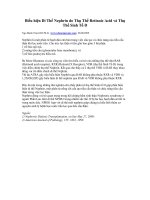
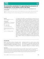
Figure 1 Oridonin stabilizes RARα protein in leukemia cells. (A) Primary leukemia cells from three newly diagnosed AML patients were
treated with 10 μM oridonin for 12 h, followed by detection of RARα protein with β-actin as a loading control. (B) Clinical data of the three AML
patients. (C) NB4 cells were treated with the indicated concentrations of oridonin for 12 h (left panel) or with 10 μM oridonin for the indicated
times (right panel), followed by western blot analysis of the RARα protein with β-actin as a loading control. The symbol * denotes a non-specific
protein. (D) NB4 cells were treated as described in panel C, followed by the quantification of RARα mRNA by real-time RT-PCR. (E) NB4 cells were
incubated with 5 μg/mL CHX alone or with 10 μM oridonin for the indicated times. Increased amounts of cell lysates compared with panel A
were loaded and then blotted for the RARα protein with β-actin as a loading control. (F) NB4 cells were treated with 10 μM oridonin and/or 10
nM ATRA for 48 h, and the mRNA levels of the indicated genes were measured by real-time RT-PCR. The data are represented as fold changes
against the control. The symbols * and # represent P values less than 0.05 and 0.01, respectively. All experiments were replicated three times and
gave consistent results.
Cao et al. BMC Cancer (2015) 15:248
Page 6 of 12
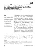
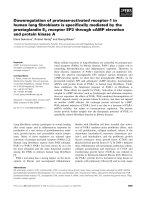
Figure 2 ROS are involved in oridonin-induced RARα stabilization. (A) NB4 cells were treated with the indicated concentrations of oridonin
for 12 h (left panel) or with 10 μM oridonin for the indicated times (right panel), followed by detection of ROS levels by flow cytometry. The
symbols * and # represent P values less than 0.05 and 0.01, respectively. (B) After pretreatment with or without 2 mM NAC for 1 h, NB4 cells were
incubated with 10 μM oridonin for 12 h, followed by detection of ROS levels by flow cytometry (left panel) and western blot detection for RARα
protein with β-actin as loading control (right panel). The symbol # represents a P value less than 0.01. (C) Primary AML cells were treated as NB4
cells in the panel B, and the levels of RARα protein were measured. (D, E) NB4 cells were treated with the indicated concentrations of H2O2 for
2 h (D) or with 5 μM H2O2 for the indicated times (E), then the level of RARα protein was assessed. (F) NB4 cells were treated with 5 μM H2O2 for
the indicated times, and RARα mRNA levels were evaluated by real-time RT-PCR. (G) NB4 cells were incubated with 5 μg/mL CHX alone or in
combination with 5 μM H2O2, followed by western blot detection of RARα protein with β-actin as loading control. (H) NB4 cells were infected
with pSIREN-RetroQ-derived retroviruses carrying shRNA specifically against catalase (sh-CAT) or non-specific scrambled shRNA as a control (NC).
Infected cells were assayed for ROS production (left panel) and western blotted for the indicated proteins. The symbol # represents P values less
than 0.01, respectively. All experiments were repeated three times and gave consistent results.
pretreat NB4 cells for 1 h, followed by oridonin incubation for an additional 12 h. As shown in Figure 4A, pretreatment with PD98059 (ERK inhibitor) or SB203580
(p38 inhibitor) did not influence oridonin-induced RARα
stability. In contrast, the JNK inhibitor, SP600125, could
slightly enhance oridonin-increased RARα protein levels.
The effects of these three kinase inhibitors ruled out the
involvement of these pathways in oridonin stabilization
of RARα. However, use of the NF-κB signaling inhibitor,
Bay 11–7082, significantly inhibited oridonin-induced
phosphorylation of IκBα and NF-κB-p65. Interestingly,
pre-incubation with Bay 11–7082 antagonized oridoninincreased RARα protein levels in NB4 cells, which indicated that activation of the NF-κB pathway is required
for oridonin-induced RARα stability (Figure 4B). Similar
results were achieved in AML patient samples (Figure 4C).
In addition, NAC preincubation also blocked oridonininduced phosphorylation of IKKα/β, IκBα and NF-κB-p65
(Figure 4D), consistent with its inhibitory effect on
oridonin-stabilized RARα (Figure 2B and C). These data
suggested that oridonin stabilized RARα protein via the
ROS-activated NF-κB pathway.
Cao et al. BMC Cancer (2015) 15:248
Page 7 of 12
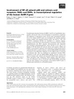
Figure 3 Oridonin activates multiple cellular signaling pathways. (A) NB4 cells were treated with 5 μM H2O2 for 4 h. RARα protein levels
were examined by redox diagonal electrophoresis, followed by western blot analysis for RARα. (B) NB4 cells were treated with 10 μM oridonin
for the indicated times, and cell lysates were western blotted for the proteins indicated. (C) NB4 cells were treated with 10 μM oridonin for the
indicated times. The intracellular localization of p65 was analyzed by indirect immunofluorescence using anti-p65 antibodies (green). Nuclear DAPI
staining (blue) is also shown. Scale bars represent 20 μm. All experiments were repeated three times and gave consistent results.
Figure 4 NF-κB inhibitor blocks oridonin-induced RARα stability. After pretreatment with and without PD98059, SB203580, SP600125 (A), Bay
11–7082 (B, C), or NAC (D) for 1 h, NB4 cells or primary AML cells were treated with 10 μM oridonin for 12 h, followed by western blot analysis
of proteins as indicated. All experiments were repeated three times and gave consistent results.
Cao et al. BMC Cancer (2015) 15:248
Essential role of activation and nuclear translocation of
NF-κB for oridonin-induced RARα stability
To confirm that oridonin stabilizes RARα through the
NF-κB pathway, we used NB4/GFP-MAD cells to perform further experiments. This engineered cell line stably expresses the GFP-tagged super-repressor form of
IκΒα, namely IκΒα (A32/36), which confers cellular
Page 8 of 12
resistance to signal-induced phosphorylation and subsequent proteasome-mediated degradation of IκΒα, resulting in the constitutive suppression of NF-κB activation
by sequestering it in the cytoplasm [36]. As shown in
Figure 5A, the over-expression of IκΒα (A32/36) blocked
oridonin-induced nuclear translocation of p65. As expected, both oridonin- and H2O2-induced RARα stability
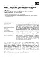
Figure 5 Oridonin-induced RARα stability requires the activation and nuclear translocation of p65. (A) NB4/GFP and NB4/GFP-MAD cells
were treated with 10 μM oridonin for 12 h. The intracellular localization of p65 was analyzed using anti-p65 antibodies (red) with DAPI staining
(blue) for nuclei. Scale bars represent 20 μm. (B, C) NB4/GFP and NB4/GFP-MAD cells were treated with 10 μM oridonin for 12 h (B) or treated
with 5 μM H2O2 for the times indicated (C), and the cell lysates were western blotted for the indicated proteins. (D) NB4 cells were infected with
pSIREN-RetroQ-derived retroviruses carrying shRNA for p65 or scrambled shRNA as a control, and the cell lysates were western blotted for the
indicated proteins. (E, F) NB4-NC and NB4-sh-p65 cells were treated with 10 μM oridonin for 12 h (E) or with 5 μM H2O2 for the times indicated
(F), and the cell lysates were western blotted for proteins as indicated. All experiments were repeated three times and gave consistent results.
Cao et al. BMC Cancer (2015) 15:248
Page 9 of 12
were inhibited in NB4/GFP-MAD cells compared with
NB4/GFP cells (Figure 5B and C). Furthermore, we stably transfected NB4 cells with shRNA specifically against
the p65 subunit of the NF-κB family, which effectively
silenced the expression of p65 but not p50 (Figure 5D).
Notably, p65 knockdown prevented oridonin and H2O2induced RARα stability in NB4 cells (Figure 5E and F).
Collectively, these results suggest that the activation and
nuclear translocation of p65 is essential for oridonin to
stabilize RARα.
results showed that TNFα treatment also resulted in a
strong increase in RARα expression together with activation and nuclear translocation of NF-κB-p65 in NB4
cells (Figure 6A and B). This TNFα-induced RARα stability could be inhibited by p65 knockdown (Figure 6C).
In addition, the over-expression of IκΒα (A32/36)
blocked the nuclear translocation of p65 and RARα stability induced by TNFα (Figure 6D and E). All these data
support the idea that translocation of p65 nuclear induces RARα stability.
Promotion of p65 nuclear translocation increases
RARα stability
Discussion
In this study, we report that the natural diterpenoid, oridonin, induces a moderate production of cellular ROS
that activates upstream of the NF-κB signaling pathway
to cause nuclear translocation of p65, which is responsible for oridonin-stabilized RARα protein. These findings indicate that moderate oxidative stress induced by
It is well known that TNFα is a classical activator of NFκB signaling; therefore, we investigated the consequence
of TNFα treatment on RARα expression to address
whether oridonin-induced RARα stability is mediated
specifically by ROS-activated NF-κB activation. Our
Figure 6 TNFα stabilizes RARα protein by activating NF-κB. (A) NB4 cells were treated with 10 ng/mL TNFα for the times indicated, followed
by western blotting for proteins as indicated. (B) NB4 cells were treated with 10 ng/mL TNFα for 0.5 h, followed by immunofluorescent staining
using anti-p65 antibodies. (C) NB4 cells with NC or sh-p65 infection were treated with 10 ng/mL TNFα for 12 h, followed by western blot analysis
for proteins as indicated. (D) NB4/GFP and NB4/GFP-MAD cells were treated with 10 ng/mL TNFα for 0.5 h, and then the intracellular localization
of p65 was analyzed by immunofluorescence. (E) NB4/GFP and NB4/GFP-MAD cells were treated with 10 ng/mL TNFα for 12 h. The cell lysates
were western blotted for the indicated proteins. All experiments were repeated three times and gave consistent results.
Cao et al. BMC Cancer (2015) 15:248
oridonin may change the intrinsic mechanisms that
regulate RARα protein stability through the NF-κB signaling pathway, which provides a new perspective of oridonin as a candidate anti-neoplastic drug.
The modulation of RARα by ATRA during APL treatment has stimulated considerable interest in RARα metabolism and its potential therapeutic mechanism [37]. ATRA
activates RARα signaling with subsequent effects on differentiation, while at the same time steady-state RARα protein levels are markedly reduced [12]. RARα, as the
receptor for ATRA, is required for its action; therefore,
RARα degradation is thought to be an inbuilt resetting
mechanism to make ATRA signaling self-limiting. Therefore, it is possible that stabilizing the RARα protein can
optimize this signaling, which indicates that RARα could
be a potential target for cancer therapeutics. Recently, several studies have demonstrated that some compounds,
such as lithium chloride (LiCl) [38], granulocyte-colony
stimulating factor [38], STI571 [39], di-tert-butyl-benzohydroquinone [40], Pharicin B [15], and oridonin [23], which
are capable of attenuating ATRA-induced loss of RARα
protein, have been shown to enhance ATRA-induced differentiation. However, the underlying mechanism of RARα
accumulation has not been fully described. In this work,
we used oridonin as a probe to show that a moderate level
of oxidative stress can stabilize RARα protein through the
nuclear translocation of p65. Further investigation is
needed to test whether this mechanism can be extended
to other small molecules with similar RARα-stabilizing ability. In addition, because RARα is an essential transcriptional
and homeostatic regulator of a plethora of physiological
processes, numerous investigations have established correlations between down-regulation of RARα and malignant
progression. In addition to APL, this has been observed
in cervical carcinoma [41], skin tumors [42], motor
neuron disease [43], and breast cancer [44]. In this context, stabilizing RARα may permit optimized use of retinoids in cancer prevention and treatment, which
warrants further investigation.
It is now widely accepted that a moderate degree of
ROS can play an important role in determining cell fate
through the modulation of cellular signaling and gene
expression [45,46]. For example, elevated but sub-lethal
levels of ROS can modulate the differentiation of various
types of cells, such as hematopoietic cells [47,48], neurons [49], embryonic stem cells [50], osteoclasts [51],
and cardiac stem cells [52]. However, little is known regarding the molecular targets of ROS. Here, we found
that moderately increased levels of ROS are crucial for
oridonin-induced RARα stabilization, which may account for the anti-neoplastic mechanism of oridonin. It
is tempting to suggest that this newly identified mechanism may underlie similar differentiation effects of some
natural diterpenoids. Nevertheless, attention should be
Page 10 of 12
paid to the cell type, as well as to the extent and duration of ROS increase, as these factors can determine
the precise consequences of the cellular response to oxidative stress. For instance, a relatively high concentration of H2O2 (0.1 mM) can suppress retinoid signaling
through the proteasomal degradation of RARα [14].
The NF-κB family is a group of transcriptional factors
consisting of p65 (RelA), RelB, c-Rel, p50/p105, and
p52/p100. In the classical NF-κB signaling pathway, the
p50/p65 dimer is sequestered in the cytoplasm by IκΒα.
After stimulation, IκΒα is phosphorylated and consequently degraded through the proteasomal pathway. Thus,
the p50/p65 dimer is released, translocates to the nucleus,
and activates target genes [53]. In this report, we revealed
that oridonin stabilizes RARα protein by inducing nuclear
translocation of p65, which was evidenced by the use of
the ROS scavenger, NAC, the NF-κB inhibitor, Bay 11–
7082, IκΒα (A32/36) over-expression, and p65 knockdown. Moreover, we tested whether TNFα, a classical
activator of NF-κB signaling, modulates stability of RARα
protein. As expected, TNFα treatment also strongly increased RARα expression, which may account, at least in
part, for TNFα-induced differentiation in some leukemia
cells [54,55]. Previous studies indicated that oridonin
mainly activates the upstream of the NF-κB signaling
pathway, while its inhibitory effect is due to the direct
interference of NF-κB DNA binding activity [56-59].
Leung et al. demonstrated that oridonin decreased the
DNA binding activity of NF-κB without interfering with
p65 translocation [59]. Of note, the exact mechanisms by
which activated NF-κB stabilizes RARα protein require
further investigation.
Conclusions
Our results indicate that oridonin stabilizes RARα protein by increasing the levels of cellular ROS, followed by
activation of the NF-κB signaling pathway. Accordingly,
the NF-κB activator, TNFα, can also increase the stability
of RARα protein. These findings suggest a new mechanism underlying the regulation of RARα protein stability
and shed new light on understanding potential therapeutic roles of oridonin in leukemia and other RARαrelated diseases.
Abbreviations
AML: Acute myeloid leukemia; APL: Acute promyelocytic leukemia; ATRA:
All-trans retinoic acid; CHX: cycloheximide; H2O2: Hydrogen peroxide; IκB: Inhibitor
of NF-κB; IKK: IκB kinase; LiCl: Lithium chloride; NAC: N-acetyl-l-cysteine;
NF-κB: Nuclear factor-kappa B; PML: Promyelocytic leukemia; RARα: Retinoic acid
receptor alpha; ROS: Reactive oxygen species; RXRs: Retinoid X receptors;
shRNAs: Short hairpin interfering RNAs.
Competing interests
The authors declare that they have no competing interest.
Cao et al. BMC Cancer (2015) 15:248
Authors’ contributions
Conceived and designed the experiments: HY, YLW. Performed all the
experiments and analyzed the data: YC, WW. Contributed reagents/materials/
analysis tools: NZ, QY, WBX, WJY. Wrote the manuscript: YC, WW, GQC, YLW.
Revised the manuscript: GQC, HY, YLW. All authors read and approved the
final manuscript.
Acknowledgements
This work was supported in part by grants from the National Basic Research
Program of China (973 Program) (NO. 2010CB912104, 2015CB910403),
National Natural Science Foundation of China (81170509, 81272886,
91313303), and Science and Technology Committee of Shanghai
(11JC1406500).
Author details
1
Department of Hematology, Rui-Jin Hospital, Shanghai Jiao-Tong University
School of Medicine, Shanghai, China. 2Department of Hematology, Xinhua
Hospital, Shanghai Jiao-Tong University School of Medicine, Shanghai, China.
3
Department of Pathophysiology, Chemical Biology Division of Shanghai
Universities E-Institutes, Key Laboratory of Cell Differentiation and Apoptosis
of National Ministry of Education, Shanghai Jiao-Tong University School of
Medicine, Shanghai, China.
Received: 15 July 2014 Accepted: 19 March 2015
References
1. Garattini E, Bolis M, Garattini SK, Fratelli M, Centritto F, Paroni G, et al.
Retinoids and breast cancer: From basic studies to the clinic and back
again. Cancer Treat Rev. 2014;40:739–49.
2. Collins SJ. Retinoic acid receptors, hematopoiesis and leukemogenesis.
Curr Opin Hematol. 2008;15:346–51.
3. Lee YS, Jeong WI. Retinoic acids and hepatic stellate cells in liver disease.
J Gastroenterol Hepatol. 2012;27 Suppl 2:75–9.
4. Orfali N, McKenna SL, Cahill MR, Gudas LJ, Mongan NP. Retinoid receptor
signaling and autophagy in acute promyelocytic leukemia. Exp Cell Res.
2014;324:1–12.
5. Tang XH, Gudas LJ. Retinoids, retinoic acid receptors, and cancer. Annu Rev
Pathol. 2011;6:345–64.
6. Zhang XW, Yan XJ, Zhou ZR, Yang FF, Wu ZY, Sun HB, et al. Arsenic trioxide
controls the fate of the PML-RARalpha oncoprotein by directly binding PML.
Science. 2010;328:240–3.
7. Wang ZY, Chen Z. Acute promyelocytic leukemia: from highly fatal to highly
curable. Blood. 2008;111:2505–15.
8. Dos Santos GA, Kats L, Pandolfi PP. Synergy against PML-RARa: targeting
transcription, proteolysis, differentiation, and self-renewal in acute
promyelocytic leukemia. J Exp Med. 2013;210:2793–802.
9. Zheng XM, Seshire A, Ruester B, Bug G, Beissert T, Puccetti E, et al. Arsenic
but not all-trans retinoic acid overcomes the aberrant stem cell capacity of
PML/RAR alpha-positive leukemic stem cells. Haematologica. 2007;92:323–31.
10. Chen GQ, Shi XG, Tang W, Xiong SM, Zhu J, Cai X, et al. Use of arsenic
trioxide (As2O3) in the treatment of acute promyelocytic leukemia (APL): I.
As2O3 exerts dose-dependent dual effects on APL cells. Blood.
1997;89:3345–53.
11. Bleul T, Ruhl R, Bulashevska S, Karakhanova S, Werner J, Bazhin AV. Reduced
retinoids and retinoid receptors’ expression in pancreatic cancer: A link to
patient survival. Mol Carcinog 2014 Apr 11. doi: 10.1002/mc.22158
12. Zhu J, Gianni M, Kopf E, Honore N, Chelbi-Alix M, Koken M, et al. Retinoic
acid induces proteasome-dependent degradation of retinoic acid receptor
alpha (RARalpha) and oncogenic RARalpha fusion proteins. Proc Natl Acad
Sci U S A. 1999;96:14807–12.
13. Srinivas H, Juroske DM, Kalyankrishna S, Cody DD, Price RE, Xu XC, et al.
c-Jun N-terminal kinase contributes to aberrant retinoid signaling in lung
cancer cells by phosphorylating and inducing proteasomal degradation of
retinoic acid receptor alpha. Mol Cell Biol. 2005;25:1054–69.
14. Hoshikawa Y, Kanki K, Ashla AA, Arakaki Y, Azumi J, Yasui T, et al. c-Jun
N-terminal kinase activation by oxidative stress suppresses retinoid signaling
through proteasomal degradation of retinoic acid receptor alpha protein in
hepatic cells. Cancer Sci. 2011;102:934–41.
Page 11 of 12
15. Gu ZM, Wu YL, Zhou MY, Liu CX, Xu HZ, Yan H, et al. Pharicin B stabilizes
retinoic acid receptor-alpha and presents synergistic differentiation
induction with ATRA in myeloid leukemic cells. Blood. 2010;116:5289–97.
16. Chen G, Wang K, Yang BY, Tang B, Chen JX, Hua ZC. Synergistic antitumor
activity of oridonin and arsenic trioxide on hepatocellular carcinoma cells.
Int J Oncol. 2012;40:139–47.
17. Li X, Li X, Wang J, Ye Z, Li JC. Oridonin up-regulates expression of P21 and
induces autophagy and apoptosis in human prostate cancer cells. Int J Biol
Sci. 2012;8:901–12.
18. Wang S, Zhong Z, Wan J, Tan W, Wu G, Chen M, et al. Oridonin induces
apoptosis, inhibits migration and invasion on highly-metastatic human
breast cancer cells. Am J Chin Med. 2013;41:177–96.
19. Zhou GB, Kang H, Wang L, Gao L, Liu P, Xie J, et al. Oridonin, a diterpenoid
extracted from medicinal herbs, targets AML1-ETO fusion protein and shows
potent antitumor activity with low adverse effects on t(8;21) leukemia
in vitro and in vivo. Blood. 2007;109:3441–50.
20. Li CY, Wang EQ, Cheng Y, Bao JK. Oridonin: An active diterpenoid targeting
cell cycle arrest, apoptotic and autophagic pathways for cancer
therapeutics. Int J Biochem Cell Biol. 2011;43:701–4.
21. Hu AP, Du JM, Li JY, Liu JW. Oridonin promotes CD4+/CD25+ Treg
differentiation, modulates Th1/Th2 balance and induces HO-1 in rat splenic
lymphocytes. Inflamm Res. 2008;57:163–70.
22. Ren KK, Wang HZ, Xie LP, Chen DW, Liu X, Sun J, et al. The effects of
oridonin on cell growth, cell cycle, cell migration and differentiation in
melanoma cells. J Ethnopharmacol. 2006;103:176–80.
23. Gao F, Tang Q, Yang P, Fang Y, Li W, Wu Y. Apoptosis inducing and
differentiation enhancement effect of oridonin on the all-trans-retinoic
acid-sensitive and -resistant acute promyelocytic leukemia cells. Int J Lab
Hematol. 2010;32:e114–22.
24. Komura E, Tonetti C, Penard-Lacronique V, Chagraoui H, Lacout C,
Lecouedic JP, et al. Role for the nuclear factor kappaB pathway in
transforming growth factor-beta1 production in idiopathic myelofibrosis:
possible relationship with FK506 binding protein 51 overexpression.
Cancer Res. 2005;65:3281–9.
25. De The H, Vivanco-Ruiz MM, Tiollais P, Stunnenberg H, Dejean A. Identification
of a retinoic acid responsive element in the retinoic acid receptor beta gene.
Nature. 1990;343:177–80.
26. Duprez E, Wagner K, Koch H, Tenen DG. C/EBPbeta: a major PML-RARAresponsive gene in retinoic acid-induced differentiation of APL cells. EMBO
J. 2003;22:5806–16.
27. Mao M, Yu M, Tong JH, Ye J, Zhu J, Huang QH, et al. RIG-E, a human
homolog of the murine Ly-6 family, is induced by retinoic acid during the
differentiation of acute promyelocytic leukemia cell. Proc Natl Acad Sci U S A.
1996;93:5910–4.
28. Matikainen S, Ronni T, Hurme M, Pine R, Julkunen I. Retinoic acid activates
interferon regulatory factor-1 gene expression in myeloid cells. Blood.
1996;88:114–23.
29. Wang H, Ye Y, Yu ZL. Proteomic and functional analyses demonstrate the
involvement of oxidative stress in the anticancer activities of oridonin in
HepG2 cells. Oncol Rep. 2014;31:2165–72.
30. Zang L, He H, Xu Q, Yu Y, Zheng N, Liu W, et al. Reactive oxygen species
H2O2 and *OH, but not O2*(−) promote oridonin-induced phagocytosis of
apoptotic cells by human histocytic lymphoma U937 cells. Int
Immunopharmacol. 2013;15:414–23.
31. Nicholls P. Classical catalase: ancient and modern. Arch Biochem Biophys.
2012;525:95–101.
32. Zuo Y, Xiang B, Yang J, Sun X, Wang Y, Cang H, et al. Oxidative modification
of caspase-9 facilitates its activation via disulfide-mediated interaction with
Apaf-1. Cell Res. 2009;19:449–57.
33. Rhee SG, Kang SW, Jeong W, Chang TS, Yang KS, Woo HA. Intracellular
messenger function of hydrogen peroxide and its regulation by
peroxiredoxins. Curr Opin Cell Biol. 2005;17:183–9.
34. Sies H. Role of Metabolic H2O2 Generation: REDOX SIGNALING AND
OXIDATIVE STRESS. J Biol Chem. 2014;289:8735–41.
35. Liu CX, Yin QQ, Zhou HC, Wu YL, Pu JX, Xia L, et al. Adenanthin targets
peroxiredoxin I and II to induce differentiation of leukemic cells. Nat Chem
Biol. 2012;8:486–93.
36. Traenckner EB, Pahl HL, Henkel T, Schmidt KN, Wilk S, Baeuerle PA.
Phosphorylation of human I kappa B-alpha on serines 32 and 36 controls I
kappa B-alpha proteolysis and NF-kappa B activation in response to diverse
stimuli. EMBO J. 1995;14:2876–83.
Cao et al. BMC Cancer (2015) 15:248
37. Maire A, Alvarez S, Shankaranarayanan P, Lera AR, Bourguet W, Gronemeyer
H. Retinoid receptors and therapeutic applications of RAR/RXR modulators.
Curr Top Med Chem. 2012;12:505–27.
38. Finch RA, Li J, Chou TC, Sartorelli AC. Maintenance of retinoic acid receptor
alpha pools by granulocyte colony-stimulating factor and lithium chloride
in all-trans retinoic acid-treated WEHI-3B leukemia cells: relevance to the
synergistic induction of terminal differentiation. Blood. 2000;96:2262–8.
39. Gianni M, Kalac Y, Ponzanelli I, Rambaldi A, Terao M, Garattini E. Tyrosine
kinase inhibitor STI571 potentiates the pharmacologic activity of retinoic
acid in acute promyelocytic leukemia cells: effects on the degradation of
RARalpha and PML-RARalpha. Blood. 2001;97:3234–43.
40. Launay S, Gianni M, Diomede L, Machesky LM, Enouf J, Papp B.
Enhancement of ATRA-induced cell differentiation by inhibition of calcium
accumulation into the endoplasmic reticulum: cross-talk between RAR alpha
and calcium-dependent signaling. Blood. 2003;101:3220–8.
41. Geisen C, Denk C, Gremm B, Baust C, Karger A, Bollag W, et al. High-level
expression of the retinoic acid receptor beta gene in normal cells of the uterine
cervix is regulated by the retinoic acid receptor alpha and is abnormally
down-regulated in cervical carcinoma cells. Cancer Res. 1997;57:1460–7.
42. Darwiche N, Scita G, Jones C, Rutberg S, Greenwald E, Tennenbaum T, et al.
Loss of retinoic acid receptors in mouse skin and skin tumors is associated
with activation of the ras(Ha) oncogene and high risk for premalignant
progression. Cancer Res. 1996;56:4942–9.
43. Corcoran J, So PL, Maden M. Absence of retinoids can induce motoneuron
disease in the adult rat and a retinoid defect is present in motoneuron
disease patients. J Cell Sci. 2002;115:4735–41.
44. Han QX, Allegretto EA, Shao ZM, Kute TE, Ordonez J, Aisner SC, et al.
Elevated expression of retinoic acid receptor-alpha (RAR alpha) in
estrogen-receptor-positive breast carcinomas as detected by
immunohistochemistry. Diagn Mol Pathol. 1997;6:42–8.
45. Bae YS, Oh H, Rhee SG, Yoo YD. Regulation of reactive oxygen species
generation in cell signaling. Mol Cells. 2011;32:491–509.
46. Ray PD, Huang BW, Tsuji Y. Reactive oxygen species (ROS) homeostasis and
redox regulation in cellular signaling. Cell Signal. 2012;24:981–90.
47. Jang YY, Sharkis SJ. A low level of reactive oxygen species selects for
primitive hematopoietic stem cells that may reside in the low-oxygenic
niche. Blood. 2007;110:3056–63.
48. Abdel-Wahab O, Levine RL. Metabolism and the leukemic stem cell. J Exp
Med. 2010;207:677–80.
49. Tsatmali M, Walcott EC, Makarenkova H, Crossin KL. Reactive oxygen species
modulate the differentiation of neurons in clonal cortical cultures. Mol Cell
Neurosci. 2006;33:345–57.
50. Ji AR, Ku SY, Cho MS, Kim YY, Kim YJ, Oh SK, et al. Reactive oxygen species
enhance differentiation of human embryonic stem cells into
mesendodermal lineage. Exp Mol Med. 2010;42:175–86.
51. Lee NK, Choi YG, Baik JY, Han SY, Jeong DW, Bae YS, et al. A crucial role for
reactive oxygen species in RANKL-induced osteoclast differentiation. Blood.
2005;106:852–9.
52. Sauer H, Wartenberg M. Reactive oxygen species as signaling molecules in
cardiovascular differentiation of embryonic stem cells and tumor-induced
angiogenesis. Antioxid Redox Signal. 2005;7:1423–34.
53. Hayden MS, Ghosh S. Signaling to NF-kappaB. Genes Dev. 2004;18:2195–224.
54. Secchiero P, Milani D, Gonelli A, Melloni E, Campioni D, Gibellini D, et al.
Tumor necrosis factor (TNF)-related apoptosis-inducing ligand (TRAIL) and
TNF-alpha promote the NF-kappaB-dependent maturation of normal and
leukemic myeloid cells. J Leukoc Biol. 2003;74:223–32.
55. Mudipalli A, Li Z, Hromchak R, Bloch A. NF-kappaB (p65/RelA) as a regulator
of TNFalpha-mediated ML-1 cell differentiation. Leukemia. 2001;15:808–13.
56. Zhang Y, Wu Y, Wu D, Tashiro S, Onodera S, Ikejima T. NF-kappab facilitates
oridonin-induced apoptosis and autophagy in HT1080 cells through a
p53-mediated pathway. Arch Biochem Biophys. 2009;489:25–33.
57. Zang L, He H, Ye Y, Liu W, Fan S, Tashiro S, et al. Nitric oxide augments
oridonin-induced efferocytosis by human histocytic lymphoma U937 cells
via autophagy and the NF-kappaB-COX-2-IL-1beta pathway. Free Radic Res.
2012;46:1207–19.
Page 12 of 12
58. Ikezoe T, Yang Y, Bandobashi K, Saito T, Takemoto S, Machida H, et al.
Oridonin, a diterpenoid purified from Rabdosia rubescens, inhibits the
proliferation of cells from lymphoid malignancies in association with blockade
of the NF-kappa B signal pathways. Mol Cancer Ther. 2005;4:578–86.
59. Leung CH, Grill SP, Lam W, Han QB, Sun HD, Cheng YC. Novel mechanism
of inhibition of nuclear factor-kappa B DNA-binding activity by diterpenoids
isolated from Isodon rubescens. Mol Pharmacol. 2005;68:286–97.
Submit your next manuscript to BioMed Central
and take full advantage of:
• Convenient online submission
• Thorough peer review
• No space constraints or color figure charges
• Immediate publication on acceptance
• Inclusion in PubMed, CAS, Scopus and Google Scholar
• Research which is freely available for redistribution
Submit your manuscript at
www.biomedcentral.com/submit