Homozygous deletions of UGT2B17 modifies effects of smoking on TP53-mutations and relapse of head and neck carcinoma
Bạn đang xem bản rút gọn của tài liệu. Xem và tải ngay bản đầy đủ của tài liệu tại đây (640.88 KB, 9 trang )
Mafune et al. BMC Cancer (2015) 15:205
DOI 10.1186/s12885-015-1220-2
RESEARCH ARTICLE
Open Access
Homozygous deletions of UGT2B17 modifies
effects of smoking on TP53-mutations and relapse
of head and neck carcinoma
Aki Mafune1,5, Takanori Hama1,2*, Toshihito Suda2, Yutaka Suzuki4, Masahiro Ikegami3, Chikako Sakanashi1,
Satoko Imai1, Akio Nakashima1,5, Takashi Yokoo5, Kota Wada2,6, Hiromi Kojima2 and Mitsuyoshi Urashima1
Abstract
Background: Smoking induces oncogenic TP53-mutations in head and neck squamous cell carcinomas (HNSCCs).
Disruptive mutations of TP53-gene and expression of p16 protein [p16 (+)] in tumor tissue associate with worse
and better prognosis, respectively. UDP-glucuronosyltransferase 2 family, polypeptide B17 (UGT2B17) detoxifies
smoking-related metabolites. Differences among ethnic groups in UGT2B17 are extremely high. Homozygous
deletions of UGT2B17 gene (UGT2B17-deletion) are a common copy number variant (CNV) among Japanese,
but not a common CNV among Africans and Europeans. Thus, we examined Japanese patients with HNSCC
to explore if UGT2B17-deletion and/or p16 (+) modify effects of smoking on TP53-mutations and affect relapse.
Methods: We conducted a posthoc analysis of a prospective cohort. Polymerase chain reaction, immunohistochemistry,
and direct sequencing were used to determine UGT2B17-deletion, p16 (+), and detailed TP53-mutations, respectively.
Results: UGT2B17-deletion was observed in 80% of this study population. For this 80%, TP53-mutations were
significantly more common among smokers than non-smokers (P = 0.0016), but this difference between
smokers and nonsmokers was not significant for the 20% with UGT2B17. In patients with UGT2B17-deletion
and p16 (+), simultaneously, TP53-mutations were much more common among smokers than among non-smokers
(81% versus 17%; P = 0.0050). Patients with both UGT2B17-deletion and disruptive TP53-mutations had higher relapse
rates than other patients (hazard ratio, 2.22; 95% confidence interval, 1.30 to 3.80, P = 0.004) in a stepwise method.
Conclusions: These results suggest that UGT2B17-deletion interacting with p16 (+) may modify effects of smoking on
TP53-mutations and may further interact with the disruptive TP53-mutations to raise relapse rates among Japanese
patients with HNSCC.
Keywords: UGT2B17, TP53, HNSCC (head and neck squamous-cell carcinoma) and smoking
Background
Tobacco smoking is associated with 5 million deaths per
year worldwide and is regarded as one of the leading causes
of premature death [1]. Nicotine, a natural ingredient in tobacco leaves, is so addictive that people smoke habitually,
which in turn results in exposure to a diverse array of carcinogens. Metabolites of nicotine, including cotinine and
other compounds, are further catabolized and detoxified
* Correspondence:
1
Division of Molecular Epidemiology, Jikei University School of Medicine,
Tokyo, Japan
2
Department of Oto-Rhino-laryngology, Jikei University School of
Medicine, 3 – 25 - 8 Nishi-shimbashi, Minato-ku, Tokyo 105-8461, Japan
Full list of author information is available at the end of the article
via CYP2A6 [2] and the UDP-glucuronosyltransferase
(UGT) family of enzymes. One UGT gene, UDPglucuronosyltransferase 2 family, polypeptide B17
(UGT2B17) enzyme decreases the abundance nicotinerelated metabolites via glucuronidation [3]. Consequently,
UGT2B17 gene deletions may reduce detoxification rates
of carcinogens in tobacco and tobacco smoke [4]. Therefore, this UGT2B17-deletion may increase an individual’s susceptibility to tobacco-related cancers, e.g., lung
cancer [5].
Copy number variants (CNVs) of UGT2B17 gene, known
to vary greatly among ethnic populations; for example,
homozygous deletion of UGT2B17 (0 copy) is not a
© 2015 Mafune et al.; licensee BioMed Central. This is an Open Access article distributed under the terms of the Creative
Commons Attribution License ( which permits unrestricted use, distribution, and
reproduction in any medium, provided the original work is properly credited. The Creative Commons Public Domain
Dedication waiver ( applies to the data made available in this article,
unless otherwise stated.
Mafune et al. BMC Cancer (2015) 15:205
common CNV among Africans or Europeans e.g., 14%
of Nigerians, but it is common among East Asian populations, e.g., 92% of Japanese [6]. Smoking is a major risk factor for head and neck squamous cell carcinoma (HNSCC)
[7], by inducing oncogenic mutations of the TP53 oncosuppressor gene [8] and of other genes [9,10]. In particular,
disruptive mutations in TP53 were associated with reduced
survival in patients with HNSCC [11].
Therefore, we hypothesized that smoking may increase
the risk of TP53-mutations among patients with homozygous for UGT2B17 deletions (defined as “UGT2B17deletion” in this study) to a greater extent than among
patients with one or two copies of UGT2B17 (defined as
“UGT2B17-presence” in this study). Because UGT2B17
deletion is common among Japanese, the power to detect
interacting effects between smoking and UGT2B17deletion on TP53-mutations can be enhanced by focusing on Japanese patients with HNSCC. In addition to
TP53-mutations, overexpression of p16-protein [defined
as “p16 (+)” in this study] in tumors, which is encoded
by CDKN2A, increases survival time in cases of oropharyngeal cancer [12,13]. We reported that heavy alcohol
consumption triggered previously known and unknown
somatic copy number alterations (SCNAs) including
CDKN2A, but that smoking induced TP53-mutations [14].
Using this cohort of Japanese patients with HNSCC as post
hoc analysis, we newly explored if UGT2B17-deletion
modify effects of smoking on TP53-mutations, in combination with p16 (+). Furthermore, we studied if combinations among UGT2B17-deletion, p16 (+), and disruptive
TP53-mutations affect cancer relapse.
Methods
Study design
We conducted a cohort study at Jikei University Hospital
from March 2006 to November 2012. The study protocol
was reviewed and approved by the Ethics Committee for
Biomedical Research of the Jikei Institutional Review
Board. The entire process of study design, data monitoring, and data analyses were performed in the Division
of Molecular Epidemiology. Eligible participants were
Japanese patients with HNSCC (oropharyngeal, hypopharyngeal, laryngeal, oral and nasal cancer) aged 20 years
or older who had newly diagnosed or recurrent disease. A
total of 262 patients provided written informed consent to
participate in this study. Of these 262 patients, 28 patients
were excluded because pathological diagnosis was not
squamous cell carcinoma or because the primary tumor
site was unknown. 27patient received in combination with
chemotherapy or radiotherapy after surgery for close surgical margin and/or extracapsular spread of metastatic
node. All of them were stage IV. Clinical data from the
remaining 234 patients were used. Clinical information
was obtained from clinical and surgical charts. Tumor
Page 2 of 9
node metastasis (TNM) classification and cancer stages
were determined according to the 6th Union for International Cancer Control TNM classification and stage
groupings. Tumor grade with regard to cell differentiation
was classified into three categories—well differentiated,
moderately differentiated, or poorly differentiated—by a
pathologist (M.I.). Of these 234 patients, nine patients
were unknown of cell differentiation.
Smoking and alcohol drinking
A history of current or past cigarette smoking was obtained based on a questionnaire completed by each patient
at surgery. The age at which they started smoking and the
number of cigarettes smoked per day was recorded. For
past smokers, the age at which the patient ceased smoking
was also recorded. The extent of previous smoking was
quantified in pack-years (PYs); 10 PYs is any equivalent to
smoking 1 pack including 20 cigarettes/day for 10 years
(e.g., 2 packs/day for 5 years). Patients were classified as
smokers if they had smoked at for least 10 PYs within the
20 years preceding diagnosis of HNSCC. Non-smokers
were defined as patients who had never smoked, had not
smoked in the 20 years preceding diagnosis, or smoked
less than 10 PY prior to surgical resection of HNSCC. Of
these 234 patients, two patients were unknown of smoking status.
The following three categories were used to classify
patients based upon average daily alcohol consumption
during the 20 years preceding diagnosis of HNSCC: 1)
non-drinkers were defined as patients who did not consume alcohol or consumed less than one drink per day;
2) moderate drinkers were defined as patients who consumed at least one, but less than two, drinks per day,
and 3) heavy drinkers were defined as patients who consumed two or more drinks per day. One drink was defined as containing approximately 10 g of alcohol, which
is equal to 30 ml of hard liquor, 100 ml of wine containing 12% alcohol, or 360 ml of beer.
Samples
With each patient’s consent, peripheral blood samples
and tumor tissue were collected during the operation.
QIAamp DNA Micro Kits 50 (Qiagen, Tokyo, Japan)
were used to purify extracted DNA, and NanoVue plus
(General Electric healthcare Japan, Tokyo, Japan) was used
to measure DNA concentration in each sample; samples
were then frozen at -80°C until use.
Array-based comparative genome hybridization (CGH)
An Agilent Enzymatic Labeling Kit was used according
to the manufacturer’s instructions to label 0.5 μg of genomic DNA for each CGH array. Labeled DNA was hybridized to an Agilent-022060 SurePrint G3 Human CGH
Microarray 4x180K (Agilent Technologies, Inc., Santa
Mafune et al. BMC Cancer (2015) 15:205
Clara, CA, USA); the Agilent Microarray Scanner and
Feature Extraction v.10.7.3.1 (Agilent Technologies), were
used according to manufacturer’s instruction to scan probed
arrays. Control DNA was obtained from one Japanese individual who is an author (MU) of this study. We focused
only on previously reported SCNAs of CDKN2A [14] and
on CNVs of UGT2B17 that are associated with metabolism of nicotine [15]. The data described in this article have
been deposited in NCBIs Gene Expression Omnibus
(GEO) [16] and are accessible through GEO series accession number GSE47443.
TaqMan Real-time PCR
We also performed real-time polymerase chain reaction
(PCR) to confirm the microarray data. The TaqMan-based
real-time PCR method for comparative quantification
was performed with extracted DNA according to Life
Technologies’ protocol. Genomic sequences of UGT2B17
were used to generate the specific target sequence.
Primers for UGT2B17 (Taqman Copy Number Assays
No. 186891217) and a probe for RNase P (Taqman copy
number Reference Assay RNase P No. 4401631) were used
(Life Technologies Corp.). Reactions (20 μL) were performed in 96-well plates using Brilliant III Ultra-Fast
QPCR Master Mix, Reference Dye (30 nM), nuclease-free
water (8 μL), DNA sample (1 μL), and UGT2B17 primer
(1 μL) (Applied Biosystems) or TaqMan Copy Number
Reference Assay RNase P (1 μL); reaction mixtures were
subjected to 40 cycles of 95°C for 3 min, 95°C for 10 s,
and 60°C for 30 s. For the precise and accurate amplification of DNA, each assay with each primer pairs was run
in duplicate. Comparative quantification was calculated
using a sample from the same person (MU) who provided
the control samples for the CGH array. A MX 3005P
Real-Time QPCR System with Mx Pro Software version
4.10 (Agilent Technologies) was used to measure the
product of each real-time PCR assay.
The method of measurement was based on the comparative cycle threshold (Ct) method for the target sequence (UGT2B17) and a reference sequence (RNase P).
The RNase P gene was co-amplified with UGT2B17 and
served as an internal standard. The PCR amplification
efficiencies of RNase P and UGT2B17 were 100% and
99%; these were calculated by using the comparative
ΔΔCt methods as described by Pfaffl et al. [17]. The fold
changes in copy numbers of the gene were log2 transformed and determined to be gene positive or gene negative (over two copies or not). Finally, 97% of array results
were consistent with real-time PCR.
PCR to differentiate between one and two copies of
UGT2B17
In 3% of samples, array and real-time PCR results were
conflicted and could not differentiate between one and
Page 3 of 9
two copies of the UGT2B17 gene, To determine the absence or presence of the UGT2B17 gene, we further performed PCR as follows. Because a high level of sequence
identity exists between the UGT2B17 and UGT2B15
genes, we used gene-specific PCR primers to distinguish
UGT2B17 from UGT2B15 and to distinguish between
one and two copies of the UGT2B17 gene: Marker D
(Forward primer 5’-TCACAAGTCAATCTCCCATCC-3’,
Reverse primer 5’-CTGCAGAATATGTCAATAATTGG
C-3’) is positive for one copy and two copies (100 bp),
Marker J (Forward primer 5’-TGCACAGAGTTAAGA
AATGGAGAGATGTG-3’, Reverse primer 5’-GATCAT
CCTATATCCTGACAGAATT-3’) is positive for only
one copy (900 bp) [18,19]. PCR reactions were carried
out in 25-μl mixtures containing 1 μg of genomic DNA,
2.5 μL of 10xLA PCR buffer II, 2 μL of dNTP (400 μM),
0.25 μL of LA Taq (Takara Bio Inc., Shiga, Japan),
18.25 μL of nuclease-free water, and 0.5 μL of each of
the two primers (100 pmol/μL). Each reaction mixture
was incubated at 94°C for 3 min and then subjected to
30 cycles of 94°C for 20 s, 60°C for 30 s, and 72°C
for 90 s; each reaction was then incubated at 16°C
until analysis.
TP53-mutations
The quality or quantity of DNA samples from 14 patients was not adequate to assess TP53 mutational status;
therefore, only 234 samples were analyzed with regard to
TP53-mutations.
Exons 2 thru 11 of the TP53 gene were each independently amplified by PCR using purchased primers following the manufacturer’s protocol (NIPPON GENE Co.
Ltd., Chiyoda-ku, Tokyo, Japan). Each resulting PCR
product was cloned and then sequenced with the ABI
PRISM 3700 Genetic Analyzer (Life Technologies Corp.).
The following 10 single-nucleotide polymorphisms —
V31I, P36P, P47S, P72R, R158R, R213R, V217M, P222P,
T312S, and G360A—are reportedly each caused by a
single nucleotide polymorphism [20], and thus excluded
from total TP53-mutations. Disruptive TP53-mutations
were defined as non-conservative mutations located inside the key DNA-binding domain (L2-L3 region) or as
stop codons in any region [9]. Sites containing cytidine
phosphate guanosine (CpG) dinucleotides were determined according to the database of WHO’s International
Agency for Research on Cancer and based on the work
by Petitjean et al. [21].
p16 immunohistochemistry
Formalin-fixed, paraffin-embedded tumor specimens were
evaluated for p16 overexpression with a rabbit monoclonal
antibody that recognizes p16 (Anti-CDKN2A/p16INK4a
antibody [EPR1473]: Abcam Plc, Science Park, Cambridge,
England). In this study, positive p16-protein expression
Mafune et al. BMC Cancer (2015) 15:205
(designated p16 (+)) determined via immunohistochemistry (IHC) was defined as strong and diffuse nuclear, cytoplasmic staining or both in at least 70% of tumor cells. Any
other pattern of p16 expression was classified as p16 (-).
Statistical analysis
To evaluate significant differences between groups, the
unpaired t test and the Mann-Whitney test were used to
analyze ages and PYs, respectively. The chi-square test
was used to assess categorical variables. Interaction effects between smoking and each of ten sub-groupings—
age (< vs. ≥ 65 years), gender, drinking status, primary
sites of tumor, tumor grades, stages, UGT2B17-CNV,
CDKN2A-SCNA, p16-ICH, and UGT2B17-CNV and
p16-ICH combined—were assessed with respect to any type
of TP53-mutations; potential interactions were assessed
by a Pinteraction term. Then, for each sub-grouping, risks
for any kind of TP53-mutations were compared between
smokers and non-smokers using a risk ratio (RR) with a
95% confidence interval (95% CI).
In survival analyses, the time from surgery to relapse
was used to calculate relapse-free rates. Patients were
considered as “censored”, when follow-ups were stopped
at the time of a patient’s death by causes other than
HNSCC relapse or the last outpatient clinic visit. The
Cox proportional hazard model was used to calculate
each hazard ratio (HR) with a 95% CI. To distinguish
significant prognostic factors from non-significant factors, a stepwise backward elimination method was applied
to all 13 factors identified—age, gender, smoker (10PYs≤),
heavy drinker, primary sites of tumor, CDKN2A-SCNA,
p16-ICH, disruptive TP53-mutations, UGT2B17-deletion,
interaction between disruptive TP53-mutations and
UGT2B17-deletion, interaction between disruptive TP53mutations and p16 (+), stages, tumor grades— with a cutoff point of P = 0.05. The Kaplan–Meier survival curves
were drawn based on relapse-free rates; log-rank tests
were used to compare these rates differentiated by
p16 (+), UGT2B17-deletion and disruptive TP53mutations. Each P < 0.05 was considered statistically
significant. However, the Bonferroni correction was
used to correct for multiple testing, and each pairwise
interaction among the 10 subgroups was considered
significant when Pinteraction was less than 0.005. All
statistical analyses were performed using STATA 13.1
(STATA Crop., College Station, TX).
Results
Patient characteristics
Patient characteristics were compared between nonsmokers and smokers and between patients with wild-type
TP53 and those with any type of TP53-mutations in the
primary tumors (Table 1). Tumors with TP53-mutations
were significantly more common among smokers (67%)
Page 4 of 9
than among non-smokers (52%) (RR: 1.29, 95% CI: 1.00 to
1.65, P = 0.030), which we have already reported [14]. Men
(P < 0.001) and alcohol-drinkers (P < 0.001) were also significantly more common among smokers than among
non-smokers. Oral cancer was more frequent among nonsmokers than smokers compared with other primary
tumor sites (P = 0.030). Well differentiated histology was
less common among smokers than non-smokers. Heterozygous and homozygous deletions of the CDNK2A-gene
were significantly more prevalent among patients with
TP53-mutations than those with wild-type TP53 (P =
0.035). Additionally, we found that 80% of this study
population harbored UGT2B17-deletions. However, nonsmokers did not differ significantly from smokers with regard to p16 (+) or UGT2B17-CNVs; similarly, patients
with wild-type TP53 did not differ significantly from those
with TP53-mutations with regard to p16 (+) or UGT2B17CNVs.
Then, we focused more closely on TP53 status of tumors. Of the 234 tumor samples analyzed, 86 samples
had no TP53 mutation, 84 had one mutation, 27 had
two mutations, 20 had three, 7 had four, 9 had five, and
1 had six. The frequencies of specific base-pair changes
among these 234 patients were as follows: A:T > C:G, 1
(0.4%); A:T > G:C, 13 (5.6%), A:T > T:A, 5 (2%); G:C > A:T,
60 (26%); G:C > C:G, 19 (8%); G:C > T:A, 82 (35%). The
frequencies of other types of mutations were as follows:
deletion, 10 (4%); insertion, 4 (2%); nonsense, 63 (27%);
missense, 69 (30%); frameshift, 14 (6%). In non-smokers, 9
in 37 (24%; 95% CI, 12 to 41%) TP53-mutations occurred
at CpG sites, but in smokers, 13 in 108 (12%; 95% CI, 7 to
20%) did.
Effects modifiers of smoking on TP53-mutations
Interactions between smoking and each of 11 variables—
age, gender, alcohol drinking status, the primary sites of
tumors, tumor grades, stages, the number of lymph node
metastasis, UGT2B17-deletion, CDKN2A-SCNAs, p16
(+), and a combination of UGT2B17-deletion and p16
(+)—were assessed (Table 2). In variables of the primary
sites of tumors, CDKN2A-SCNAs, p16 (+), and a combination of UGT2B17-deletion and p16 (+), interactions
were analyzed except for HPV-positive patients. Smoking
interacted significantly with four factors—stages,
UGT2B17-deletion, p16 (+), and the combination of
UGT2B17-deletions and p16 (+)—to induce TP53mutations, but not with age (P = 0.55), gender (P = 0.22),
drinking status (P = 0.90), primary tumor sites (P = 0.09),
tumor grades (P = 0.30), the number of lymph node metastasis (P = 0.51) or CDKN2A-SCNAs (P = 0.08). Restricting to patients with UGT2B17-deletion, TP53-mutations
were more prevalent among smokers than among nonsmokers (P = 0.0016), but restricting to patients with
UGT2B17-presence, differences between smokers and
Mafune et al. BMC Cancer (2015) 15:205
Page 5 of 9
Table 1 Patient*1 characteristics assessed based on smoking status and TP53-mutations
Total
Smokers*2
Non-smokers*2
(N = 161: 69%)
(N = 71: 31%)
p-value
Mutant TP53
(N = 147: 63%)
Wild-type
TP53
p-value
(N = 87: 37%)
Smoking status – PYs
25%/50%/75%
0/25/40
25/40/46
0/0/0
Smokers – no. (%)
161 (69)
-
-
TP53-mutations – no. (%)
147 (63)
108 (67)
37 (52)
Age, years – yr. mean ± s.d.
63.2 ± 10.9
63.5 ± 10.2
62.6 ± 12.6
Men – no. (%)
*6
187 (80)
*7
152 (94)
33 (46)
Drinking status – no. (%)*6
*7
<0.0001*3
8/30/40
0/20/40
0.085*3
-
108 (74)
53 (61)
0.030*4
64.1 ± 10.4
61.6 ± 11.5
0.082*5
122 (83)
65 (75)
0.13*4
*4
0.030
0.56*5
*4
<0.001
<0.001*4
0.043*4
Non-drinker
89 (38)
35 (22)
53 (75)
50 (34)
39 (45)
Moderate drinker
74 (32)
64 (40)
9 (13)
55 (37)
19 (22)
Heavy drinker
71 (30)
62 (39)
9 (13)
42 (29)
29 (33)
Primary site of tumor – no. (%)
*6
*4
0.13*4
0.030
Oropharyngea
63 (27)
47 (29)
16 (23)
37 (25)
26 (30)
Hypopharyngeal
64 (27)
47 (29)
16 (23)
49 (33)
15 (17)
Laryngeal
29 (12)
24 (15)
5 (7)
17 (12)
12 (14)
Oral
57 (24)
32 (20)
25 (35)
32 (22)
25 (29)
Nasal
21 (9)
11 (7)
9 (13)
12 (8)
9 (10)
Cell differentiation – no. (%)*6
0.023*4
39 (25)
29 (43)
0.94*4
Well differentiated
69 (31)
Moderately differentiated
111 (49)
84 (54)
26 (39)
70 (49)
41 (50)
Poorly differentiated
45 (20)
33 (21)
12 (18)
28 (20)
17 (21)
Stages – no. (%)*6
45 (31)
24 (29)
0.12*4
0.97*4
I
12 (5)
11 (7)
1 (1)
8 (5)
4 (5)
II
48 (21)
34 (21)
14 (20)
29 (20)
19 (22)
III
48 (21)
28 (18)
20 (29)
30 (21)
18 (21)
IV
124 (53)
87 (54)
35 (50)
79 (54)
45 (52)
27 (12)
22 (13)
4 (6)
15 (10)
12 (14)
Anticancer therapy – no. (%)
Radiotherapy ± Chemotherapy
CDKN2A-SCNAs – no. (%)
*6
0.13*4
*4
0.035*4
0.73
Norma
174 (77)
117 (76)
55 (80)
99 (72)
75 (86)
Heterozygous deletion
39 (17)
29 (19)
10 (14)
29 (21)
10 (11)
Homozygous deletion
12 (5)
8 (5)
4 (6)
10 (7)
2 (2)
p16 (+) – no. (%)
47 (20)
28 (17)
19 (27)
24 (16)
23 (26)
UGT2B17 CNVs – no. (%)*6
0.10*4
0.60*4
Homozygous deletions: 0 copy
181 (80)
124 (81)
55 (80)
Heterozygous deletion: 1 copy
42 (19)
28 (18)
14 (20)
Normal: 2 copies
2 (1)
2 (1)
0 (0)
0.60*4
0.41*4
0.062*4
0.95*4
111 (80)
70 (80)
26 (19)
16 (18)
1 (1)
1 (1)
0.95*4
*1
Smoking history was unavailable for two of the 234.
Non-smokers were defined as having a <10-PYs history; smokers were defined as having a ≥10-PYs history.
Mann-Whitney test was used to calculate the p-value.
*4 2
χ test was used to calculate the p-value. *5Student’s t test was used to calculate the –value.
*6
Because of rounding, total values are not always 100%. *7RR, 1.29; 95% CI, 1.00 to 1.65.
*2
*3
non-smokers were not significant. Similarly, restricting to
patients with p16 (+) tumors, smoking increased the risk
of TP53-mutations up to 3.48-fold in comparison with
non-smoking, but not in restricting to patients with p16
(-) tumors. In restricting to patients with UGT2B17deletion and had p16 (+), smokers had significantly higher
Mafune et al. BMC Cancer (2015) 15:205
Page 6 of 9
Table 2 Effects modifiers of smoking on TP53-mutations in tumors*1
Outcome: any type of TP53-mutations
Pinteraction *2
Primary sites of tumor – no. (%)*6
0.09
Smokers N = 167
Non-smokers N = 65
RR
95% CI
p-value
Oropharyngeal
26 (76)
4 (80)
0.96
0.59 to 1.54
0.86
Hypopharyngeal
37 (79)
11 (69)
1.15
0.80 to 1.64
0.42
Laryngeal
14 (58)
3 (60)
0.97
0.44 to 2.15
0.95
Oral
15 (48)
16 (67)
0.73
0.46 to 1.15
0.18
10 (91)
1 (11)
8.18
1.28 to 52.4
0.0004
Nasal
Stages – no. (%)
0.0019
I
7 (64)
1 (100)
0.64
0.41 to 0.99
0.46
II
19 (56)
10 (71)
0.78
0.50 to 1.22
0.32
III
21 (75)
9 (45)
1.67
0.98 to 2.83
0.034
IV
60 (69)
17 (49)
1.42
0.98 to 2.05
0.035
85 (69)
24 (44)
1.57
1.14 to 2.17
0.0016
16 (53)
11 (79)
0.68
0.44 to 1.05
0.11
Normal: 2 copies
68 (65)
22 (51)
1.27
0.91 to 1.75
0.12
Heterozygous deletion: 1 copy
21 (78)
7 (70)
1.11
0.71 to 1.75
0.62
6 (75)
4 (100)
0.75
0.50 to 1.12
0.27
p16 (-)
85 (68)
33 (66)
1.03
0.82 to 1.30
0.80
p16 (+)
17 (77)
2 (22)
3.48
1.00 to 12.1
0.0043
13 (57)
10 (91)
0.62
0.42 to 0.93
0.045
UGT2B17-deletions & p16 (-)
67 (69)
21 (57)
1.22
0.89 to 1.66
0.18
UGT2B17-presence & p16 (+)
2 (50)
1 (33)
1.50
0.23 to 9.80
0.66
UGT2B17-deletions & p16 (+)
13 (81)
1 (17)
4.88
0.80 to 29.6
0.0050
UGT2B17 CNVs*5 – no. (%)
0.0016
UGT2B17-deletion: 0 copy
UGT2B17-presence: 1 copy or 2 copies
CDKN2A SCNA*3*6 – no. (%)
0.08
Homozygous deletions: 0 copy
p16-ICH in tumor*4*6 – no. (%)
0.043
UGT2B17-CNVs & p16-ICH – no. (%)
*6
UGT2B17-presence & p16 (-)
0.0080
Any type of TP53-mutations observed in the tumor genome was used as the outcome.
*2
Pinteraction was calculated as interaction effect between a factor and smoking on the risk of TP53-mutations in a tumor. With the Bonferroni correction, p < 0.005
was considered as statistically significant.
*3
CDKN2A SCNA: Copy number alterations of CDKN2A, which encodes p16, were determined by CGH array.
*4
p16 overexpression in tumor samples was determined via ICH and classified as positive (+) or negative (-).
*5
UGT2B17 CNV: Copy number variants of UGT2B17 were screened via CGH array and confirmed by real-time PCR and PCR to differentiate between one and two
copies of UGT2B17-gene.
*6
Interaction was analyzed except for HPV-positive patients.
*1
risk of TP53-mutations than did non-smokers (RR, 4.88;
95% CI, 0.80 to 29.6; P = 0.0050), but not in other
combinations: UGT2B17-presence and p16 (-), UGT2B17deletion and p16 (-), and UGT2B17 presence and p16
(+) (Table 2).
Prognostic factors
Using backward elimination for 13 candidate prognostic
factors (Table 3), we found that disruptive TP53-mutations
and UGT2B17-deletion interacted to significantly increase
the risk of relapse (HR, 2.22; 95% CI, 1.30 to 3.80, P =
0.004); however, either TP53-mutations or UGT2B17deletion alone did not significantly affect the risk. Notably,
p16 (+) was a better prognostic factor than p16 (-) (HR,
0.53; 95% CI, 0.29 to 0.99, P = 0.047). Thus, we analyzed
three grouping of the 234 patients based on combinations
of three factors—p16 (+) tumors, presence of disruptive
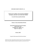
TP53-mutations, and UGT2B17-deletions. During a median follow-up period of 1.5 years (interquartile range, 1.0
to 2.5 years), relapse occurred in 89 of 232 patients (38%)
in this study. Based on Kaplan-Meier curves, patients harboring both UGT2B17-deletion and a disruptive TP53mutation in the primary tumors had the highest relapse
rates among the three groups, and the group comprising
patients with p16 (+) tumors and lacking any disruptive
TP53-mutation in the primary tumors had the lowest
relapse rates (Figure 1). Relapse was occurred in 21 of 35
patients in the group of both UGT2B17-deletion and a
disruptive TP53-mutation in the primary tumors (indicated as green-colored line in Figure 1), 16 of 59 patients
Mafune et al. BMC Cancer (2015) 15:205
Page 7 of 9
Table 3 Cox proportional hazard model as determined with backward eliminated via a stepwise method*1
Outcome: any type of TP53-mutations in a tumor
HR
95% CI
p-value
Having both disruptive TP53-mutations and UGT2B17-deletions
2.22
1.30 to 3.80
0.004
p16-positive tumor
0.53
0.29 to 0.99
0.047
Stage IV
2.32
1.44 to 3.74
0.001
Poorly differentiated tumor grade
1.66
1.01 to 2.74
0.047
By backward elimination from age, gender, smoker (10 PYs ≤), heavy drinker, primary site of tumor, CDKN2A-SCNAs, p16 (+), disruptive TP53-mutations,
UGT2B17-deletion, interaction effect between disruptive TP53-mutations and UGT2B17-deletion, interaction between disruptive TP53-mutations and p16 (+),
stages, tumor grade of cell differentiation.
*1
in the group of both p16 (+) and no disruptive TP53mutation in the primary tumors (indicated as bluecolored line in Figure 1) and 48 of 131 patients in the
other groups (indicated as red-colored line in Figure 1).
We also analyzed overall survival by Kaplan-Meier curves.
Although patients with p16 (+) tumors and lacking any
disruptive TP53-mutation in the primary tumors had the
highest survival rates than the other groups (P = 0.0190,
figure was not shown), there was no significant effect
among these three factors; status of disruptive TP53mutation, p16 and UGT2B17.
Discussion
The prevalence of copy number variants (CNVs) of UGT2B17
gene is quite different among ethnic populations. The
frequency of UGT2B17-deletion was only about 10 to
15% among general Caucasian population or Caucasian
with lung cancer [5,22]. In contrast, the frequency of
UGT2B17-deletion among Japanese athletes was 74.5% in
male and 60.2% in female [23] and 92% among those contributing to the Japanese HapMap [6]. We first confirmed
that homozygous deletion of UGT2B17 is highly prevalent
among this cohort of Japanese patients. Of the 234 patients
examined, 80% were homozygous for UGT2B17-deletions;
19% carried one copy of UGT2B17, and only 1% had two
copies; these findings were within the range of previous
reports for the Japanese or East Asian [6,23]. Therefore,
Japanese patients with HNSCC constitute a valid study
population for examination of the influence of UGT2B17deletion on TP53 mutation ratio and relapse rates.
Figure 1 Kaplan-Meier curves of relapse-free rates in 234 patients with HNSCC. Differences in time until relapse were compared among
combinations of p16 (+) tumors, disruptive TP53-mutations, and homozygous UGT2B17 deletions. The group of both UGT2B17-deletion and a
disruptive TP53-mutation in the primary tumors is indicated as green-colored line, the group of both p16 (+) and no disruptive TP53-mutation in
the primary tumors is indicated as blue-colored line and the other groups are indicated as red-colored line. p16 (+): p16-positive tumor; p16 (-):
p16- negative tumor; dTP53 (+): presence of disruptive TP53-mutations; dTP53 (-): no disruptive TP53-mutations or wild-type TP53; UGT2B17 (+):
UGT2B17-deletion: homozygous deletion of UGT2B17; UGT2B17 (-): UGT2B17-presence: one or two copies of UGT2B17.
Mafune et al. BMC Cancer (2015) 15:205
We next found that 63% of all tumors had some type
of mutation within exon-2 to exon-11 of TP53. There
were significantly more TP53-mutant tumors among
smokers than non-smokers, which we have already reported [14]. Moreover, the frequency of TP53-mutations
at CpG sites was 2-fold higher among non-smokers than
among smokers. These findings were consistent with previous findings from whole-exome sequencing studies [9].
Thirdly, we found a significant interaction effect between UGT2B17-deletion and smoking on TP53 mutation rate (Pinteraction = 0.0016). Specifically, restricting to
patients with UGT2B17-deletion, TP53-mutations were
significantly more common in tumors from smokers
than those from non-smokers, but not for patients with
UGT2B17-presence; to our knowledge, these and following findings have not been reported previously. UGT2B17deletion may 1) reduce a person’s ability to detoxify
smoking-associated metabolites, 2) allow cellular DNA to
become exposed to high levels of carcinogens in cigarettes
and cigarette smoke, and 3) raise the probability of DNA
injury, and thus increase the risk of TP53-mutations,
resulting in tumor development. Moreover, p16 expression also interacted with smoking to increase the risk of
TP53-mutations. Specifically, restricting to patients with
p16 (+) tumors, the frequency of TP53-mutations was
3.48-fold higher among smokers than non-smokers. Overexpression of p16 can result from disruption of the negative feedback loop that normally operates among p16,
cyclin-dependent kinases, cyclins, and phosphorylation of
retinoblastoma protein; several causes—including human
papillomavirus infection—can disrupt this loop [24], and
such disruption may facilitate cell cycle progression and
make cells more susceptible to the carcinogens in cigarettes and cigarette smoke.
We then assessed a combination of these two variables—
UGT2B17-deletion and p16 (+)—to assess potential interactions between them. Restricting to patients with
UGT2B17-deletion and p16 (+) tumors, TP53-mutations
were significantly more common among tumors from
smokers (81%) than those from non-smokers (17%) (RR,
4.88; 95% CI, 0.80 to 29.6; P = 0.0050). On the other hand,
patients with UGT2B17-presence and p16 (-) tumors,
TP53-mutations were significantly less common among
tumors from smokers (57%) than those from non-smokers
(91%) (RR, 0.62; 95% CI, 0.42 to 0.93; P = 0.045). From
these findings, together UGT2B17-deletion and p16 (+)
synergistically enhanced the risk of TP53-mutations occurring in tumors, because UGT2B17-deletion reduced
the metabolism and detoxification of metabolites from
cigarette smoke and p16-overexpression reflected abnormal cell cycle progression and increased cellular susceptibility to carcinogens.
In survival analyses, we confirmed that patients survival
were significantly associated with stages, cell differentiation
Page 8 of 9
levels and the number of lymph node metastasis before
adjustment; these results were consistent with previous
studies. Then we analyzed using stepwise elimination and
survival analysis by adjusting for stages, cell differentiation
levels and others.
Fourth, we found a significant interaction between disruptive TP53-mutations and UGT2B17-deletion. Patients
with TP53-mutant tumors and UGT2B17-deletion were
more than twice as likely to relapse as all other patients;
this finding was novel and striking. In contrast, patients
with p16 (+) tumors and wild-type TP53 were half as
likely to relapse as those with other patterns of tumor
mutation; this finding was consistent with a previous
finding about oropharyngeal cancer [12].
There were four main limitations in this study. Only
two patients had two copies of UGT2B17; therefore, we
mainly compared the effects of homozygosity with those
of heterozygosity with regard to UGT2B17-deletions.
Among 262 participants, TP53-mutations could not be
measured in 28 samples, because of too small size of
resected tumors to use for this study. Third is we analyzed relapse-free survival within the patients who had
newly diagnosed or recurrent disease. It appears that
one of the causes for no significant differences in the
over survival may be the effect by the curative treatment
to recurrence. Fourth is the most patients were advanced
stage III to IV (74%) in this study. In spite of limited to
the patients with early stage I to II, the patients harboring
both UGT2B17-deletion and a disruptive TP53-mutation
in the primary tumors had the highest relapse rates among
the three groups using Kaplain-Meier curves (Log-rank
test, P = 0.0071, figure was not shown).
Conclusions
In conclusions, homozygous UGT2B17-deletion may interact with smoking and p16-protein expression to increase
the risk of TP53-mutations, and may further interact with
disruptive TP53-mutations to raise relapse rates among
Japanese patients with HNSCC.
Abbreviation
HNSCC: Head and neck squamous cell carcinoma; UGT2B17: UDPglucuronosyltransferase 2 family, polypeptide B17; CNV: Common copy
number variant; UGT: UDP-glucuronosyltransferase; SCNA: Somatic copy
number alterations; TNM: Tumor node metastasis; PY: Pack-year;
CGH: comparative genome hybridization; PCR: Polymerase chain reaction;
CpG: Cytidine phosphate guanosine; IHC: Immunohistochemistry; RR: Risk
ratio; 95% CI: 95% confidence interval; HR: Hazard ratio.
Competing interests
The authors declare that they have no competing interests.
Authors’ contributions
YS, TH,TK, HK and MU designed the study. TH KWand TS contributed to
collecting the tissue samples and clinical data. MI contributed to
pathological examination. AM, CS, SI and AN have carried out molecular
studies. AM and MU performed analysis and interpretation of data. AM, TH
and MU participate in drafting the manuscript. All authors have read and
approved the final manuscript.
Mafune et al. BMC Cancer (2015) 15:205
Acknowledgements
We would also like to thank Mr. Hiroaki Suga and Mr. Takeshi Mimura for
arranging samples. This research was supported by the Ministry of Education,
Culture, Sports, Science and Technology in the Japan-Supported Program
for the Strategic Research Foundation at Private Universities the Ministry of
Education, Science, Sports and Culture, Grant-in-Aid for Scientific Research
(C) and The Jikei University Research Fund. All authors read and
approved the final manuscript.
Author details
1
Division of Molecular Epidemiology, Jikei University School of Medicine,
Tokyo, Japan. 2Department of Oto-Rhino-laryngology, Jikei University
School of Medicine, 3 – 25 - 8 Nishi-shimbashi, Minato-ku, Tokyo
105-8461, Japan. 3Department of Pathology, Jikei University School of
Medicine, Tokyo, Japan. 4Department of Surgery, International University
of Health and welfare, Tochigi, Japan. 5Division of Nephrology and
Hypertension, Department of Internal Medicine, Jikei University School of
Medicine, Tokyo, Japan. 6Department of Otorhinolaryngology, Toho
University, Tokyo, Japan.
Received: 19 October 2014 Accepted: 19 March 2015
References
1. Hatsukami DK, Stead LF, Gupta PC. Tobacco addiction. Lancet.
2008;371:2027–38.
2. Hukkanen J, Jacob 3rd P, Benowitz NL. Metabolism and disposition kinetics
of nicotine. Pharmacol Rev. 2005;57:79–115.
3. Caldwell WS, Greene JM, Byrd GD, Chang KM, Uhrig MS, de Bethizy JD, et al.
Characterization of the glucuronide conjugate of cotinine: a previously
unidentified major metabolite of nicotine in smokers’ urine. Chem Res
Toxicol. 1992;5:280–5.
4. Lazarus P, Zheng Y, Runkle EA, Muscat JE, Wiener D. Genotype-phenotype
correlation between the polymorphic UGT2B17-gene deletions and NNAL
glucuronidation activities in human liver microsomes. Pharmacogenet
Genomics. 2005;15:769–78.
5. Gallagher CJ, Muscat JE, Hicks AN, Zheng Y, Dyer AM, Chase GA, et al. The
UDP-glucuronosyltransferase 2B17 gene deletions polymorphism: sex-specific
association with urinary 4-(methylnitrosamino)-1-(3-pyridyl)-1-butanol
glucuronidation phenotype and risk for lung cancer. Cancer Epidemiol
Biomarkers Prev. 2007;16:823–8.
6. Xue Y, Sun D, Daly A, Yang F, Zhou X, Zhao M, et al. Adaptive evolution of
UGT2B17 copy-number variation. Am J Hum Genet. 2008;83:337–46.
7. LLubin JH, Purdue M, Kelsey K, Zhang ZF, Winn D, Wei Q, et al. Total
exposure and exposure rate effects for alcohol and smoking and risk of
head and neck cancer: a pooled analysis of case-control studies. Am J
Epidemiol. 2009;170:937–47.
8. Brennan JA, Boyle JO, Koch WM, Goodman SN, Hurban RH, Eby YJ, et al.
Association between cigarette smoking and mutation of the p53 gene in
squamous-cell carcinoma of the head and neck. N Engl J Med.
1995;332:712–7.
9. Stransky N, Egloff AM, Tward AD, Kostic AD, Cibulskis K, Sivachenko A, et al.
The mutational landscape of head and neck squamous cell carcinoma.
Science. 2011;333:1157–60.
10. Ang KK, Harris J, Wheeler R, Weber R, Rosenthal DI, Nguyen-Tân PF, et al.
Exome sequencing of head and neck squamous cell carcinoma reveals
inactivating mutations in NOTCH1. Science. 2011;333:1154–7.
11. Poeta ML, Manola J, Goldwasser MA, Forastiere A, Benoit N, Califano JA,
et al. TP53-mutations and survival in squamous-cell carcinoma of the head
and neck. N Engl J Med. 2007;357:2552–61.
12. Rischin D, Young RJ, Fisher R, Fox SB, Le QT, Peters LJ, et al. Prognostic
significance of p16INK4A and human papillomavirus in patients with
oropharyngeal cancer treated on TROG 02.02 phase III trial. J Clin Oncol.
2010;28:4142–8.
13. Ang KK, Harris J, Wheeler R, Weber R, Rosenthal DI, Nguyen-Tân PF, et al.
Human papillomavirus and survival of patients with oropharyngeal cancer.
N Engl J Med. 2010;363:24–35.
14. Urashima M, Hama T, Suda T, Suzuki Y, Ikegami M, Sakanashi C, et al.
Distinct effects of alcohol consumption and smoking on genetic alterations
in head and neck carcinoma. PLoS One. 2013;8:e80828.
Page 9 of 9
15. Chen G, Giambrone Jr NE, Dluzen DF, Muscat JE, Berg A, Gallagher CJ, et al.
Glucuronidation genotypes and nicotine metabolic phenotypes: importance
of functional UGT2B10 and UGT2B17 polymorphisms. Cancer Res.
2010;70:7543–52.
16. NCBIs Gene Expression Omnibus (GEO). [ />17. Pfaffl MW. A new mathematical model for relative quantification in real-time
RT-PCR. Nucleic Acids Res. 2001;29(9):e45.
18. Yang TL, Chen XD, Guo Y, Lei SF, Wang JT, Zhou Q, et al. Genome-wide
copy-number-variation study identified a susceptibility gene, UGT2B17, for
osteoporosis. Am J Hum Genet. 2008;83:663–74.
19. Chew S, Mullin BH, Lewis JR, Spector TD, Prince RL, Wilson SG. Homozygous
deletion of the UGT2B17 gene is not associated with osteoporosis risk in
elderly Caucasian women. Osteoporos Int. 2011;22:1981–6.
20. International Agency for Research on Cancer (IARC) TP53 Database.
[ />21. Petitjean A, Mathe E, Kato S, Ishioka C, Tavtigian SV, Hainaut P, et al. Impact
of mutant p53 functional properties on TP53 mutation patterns and tumor
phenotype: lessons from recent developments in the IARC TP53 database.
Hum Mutat. 2007;28:622–9.
22. Gruber M, Le T, Filipits M, Gsur A, Mannhalter C, Jäger U, et al.
UDP-glucuronosyltransferase 2B17 genotype and the risk of lung cancer
among Austrian Caucasians. Cancer Epidemiol. 2013;37:625–8.
23. Okano M, Ueda T, Nishitani Y, Kano H, Ikekita A, Kageyama S.
UDP-glucuronosyltransferase 2B17 genotyping in Japanese athletes
and evaluation of the current sports drug testing for detecting testosterone
misuse. Drug Test Anal. 2013;5:166–81.
24. Dyson N, Howley PM, Münger K, Harlow E. The human papilloma virus-16
E7 oncoprotein is able to bind to the retinoblastoma gene product. Science.
1989;243(4893):934–7.
Submit your next manuscript to BioMed Central
and take full advantage of:
• Convenient online submission
• Thorough peer review
• No space constraints or color figure charges
• Immediate publication on acceptance
• Inclusion in PubMed, CAS, Scopus and Google Scholar
• Research which is freely available for redistribution
Submit your manuscript at
www.biomedcentral.com/submit