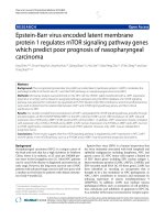
NQO1 protein expression predicts poor prognosis of non-small cell lung cancers
Bạn đang xem bản rút gọn của tài liệu. Xem và tải ngay bản đầy đủ của tài liệu tại đây (2.17 MB, 9 trang )
Li et al. BMC Cancer (2015) 15:207
DOI 10.1186/s12885-015-1227-8
RESEARCH ARTICLE
Open Access
NQO1 protein expression predicts poor prognosis
of non-small cell lung cancers
Zhenling Li1†, Yue Zhang2†, Tiefeng Jin1, Jiguang Men3, Zhenhua Lin1, Peng Qi3, Yingshi Piao1,4* and Guanghai Yan1,3*
Abstract
Background: High-level expression of NAD(P)H: quinoneoxidoreductase 1 (NQO1) has been correlated with many
types of human cancers, suggesting that NQO1 plays important roles in tumor occurrence and progression. This
study attempted to explore the role of NQO1 in tumor progression and prognostic evaluation of non-small cell lung
cancer (NSCLC).
Methods: Total 164 tissue samples, including 150 NSCLC paired with the adjacent non-tumor tissues and 14 normal
lung tissues, were picked-up for immunohistochemical (IHC) staining of the NQO1 protein, and immunofluorescence
(IF) staining was also performed to detect the subcellular localization of the NQO1 protein in A549 human lung cancer
cells. The correlation between NQO1 expression and clinicopathological characteristics were evaluated by Chi-square
test and Fisher’s exact tests. The disease-free survival (DFS) and overall survival (OS) rates of NSCLC patients were
calculated by the Kaplan-Meier method, and univariate and multivariate analyses were performed using the Cox
proportional hazards regression model.
Results: The NQO1 protein showed a mainly cytoplasmic staining pattern in lung cancer cells, including
adenocarcinoma and squamous cell carcinoma (SCC). Both positive rate and strongly positive rate of NQO1 protein
expression were significantly higher in NSCLC (59.3% and 28.0%) than that in adjacent non tumor (8.0% and 1.3%) and
normal lung tissues (0%). The positive rate of NQO1 was related with clinical stage and lymph node metastasis, and the
strongly positive rate of NQO1 protein was significantly correlated with tumor size, poor differentiation, advanced
clinical stage and lymph node metastasis in NSCLC. Additionally, survival analyses showed that the patients with NQO1
positive expression had lower OS rates compared with those with NQO1 negative expression in the groups of T1-2,
T3-4, without LN metastasis and stage I-II of NSCLC, respectively; however, in the groups of patients with LN metastasis
or III-IV stages, OS rate was not correlated with NQO1 expression status. Moreover, multivariate analysis suggested that
NQO1 emerged as a significant independent prognostic factor along with tumor size, differentiation, lymph node
metastasis and clinical stage in patients with NSCLC.
Conclusions: NQO1 is upregulated in NSCLC, and it may be a useful poor prognostic biomarker and a potential
therapeutic target for patients with NSCLC.
Keywords: Non-small cell lung cancer, NQO1, Immunohistochemistry, Prognosis, Survival analysis
Background
Non-small cell lung cancer (NSCLC) accounted for approximately 85% of all lung cancers, and it is the most
common cause of death in both men and women [1].
Currently, molecular target therapy is one of the promising field of NSCLC treatment, and its target includes
* Correspondence: ;
†
Equal contributors
1
Department of Pathology & Cancer Research Center, Yanbian University
Medical College, Yanji 133002, China
Full list of author information is available at the end of the article
epidermal growth factor receptor (EGFR) and echinoderm microtubule associated protein like4-anaplastic
lymphoma kinase (EML4-ALK). EGFR tyrosine kinase
inhibitor (EGFR TKI, such as gefitinib and erlotinib) and
EML4/ALK inhibitor (Crizotinib) have achieved better
results in the clinical therapy of advanced NSCLC [2,3].
Despite progress in the multimodality treatment of lung
cancer, prognosis is still poor, with 10-15% 5-year survival rates. More than 90% of deaths from NSCLC are
attributable to metastases [1,4].
© 2015 Li et al.; licensee BioMed Central. This is an Open Access article distributed under the terms of the Creative Commons
Attribution License ( which permits unrestricted use, distribution, and
reproduction in any medium, provided the original work is properly credited. The Creative Commons Public Domain
Dedication waiver ( applies to the data made available in this article,
unless otherwise stated.
Li et al. BMC Cancer (2015) 15:207
NAD(P)H: quinone oxidoreductase 1 (NQO1, EC
1.6.99.2) is well known as DT-diaphorase, and it can
protect cells against radiation and chemical-induced
oxidative stress. NQO1 is a cytosolic flavoenzyme that
catalyzes the obligatory two-electron reduction of a variety of quinone substrates by using NADH or NADPH
as electron donors [5]. And several functions of NQO1
have been found, such as xenobiotic detoxification,
superoxide scavenging, modulation of p53, maintenance
of endogenous antioxidants, and proteasomal degradation [6]. Due to the ability of NQO1, it is imaginable
that NQO1 may play an important role in protecting
normal cells against oxidative damage and electrophilic
attack [7,8]. Recent studies reported that NQO1 is
mainly expressed in cytosol, and low expression levels
have been found in the nucleus. Moreover, NQO1 was
found to be expressed at high levels in many human
cancers, including liver, colon, pancreas and cholangiocarcinoma [9-12]. Garate et al. [13] indicated that the
expression of NQO1 protein significantly induced cell
cycle progression and led to the proliferation of melanoma cells by the up-regulation of cyclin A2, B1 and D1.
However, the role of NQO1 in progression of lungcancer cells remains unidentified, and the correlation between
NQO1 expression and NSCLC has not been adequately
elucidated yet.
To determine whether NQO1 is important in the
tumorigenesis of NSCLC and investigate the prognostic
value of NQO1 expression level, total 150 cases of
NSCLC paired with the adjacent non-tumor tissues and
14 of normal lung tissues were selected for NQO1 IHC
staining. Our data uncover that NQO1 is frequently
upregulated in NSCLC compared with the normal
counterpart, and suggest that NQO1 may be an independent biomarker for prognostic evaluation of patients with
NSCLC.
Methods
Ethic statement
This research complied with the Helsinki Declaration
and was approved by the Human Ethics Committee and
the Research Ethics Committee of Yanbian University
Medical College. Patients were informed that the resected
specimens were stored by the hospital and potentially
used for scientific research, and that their privacy would
be maintained. Follow-up survival data were collected
retrospectively through medical record analyses.
Clinical samples
Total 164 tissue samples were used for this study, including 150NSCLC paired with the adjacent non-tumor
tissues and 14 normal lung tissues (from autopsy cases).
All of these tissues were collected from Shanghai Outdo
Biotech Co. Ltd. (Outdo Biotech) and Tissue Bank of
Page 2 of 9
Yanbian University Medical College. All tissues were
routinely fixed in 10% buffered formalin and embedded
in paraffin blocks. The study protocol was approved by
the institutional review board of Yanbian University
Medical College. The pathological parameters, including
gender, age, tumor size, clinical stage, differentiation,
nodal metastasis and survival data, were carefully reviewed in all 150 NSCLC cases.
The patients with NSCLC including 112 males and 38
females, and ranging from 43 to 76 years with a mean
age of 62 years. A total of 150 patients, 99 cases were
60 years old or over, and 51 cases were below 60 years
old. All cases were confirmed with NSCLC by pathological examination. TNM staging was assessed according to the staging system established by the American
Joint Committee on Cancer (AJCC). Of the 150 NSCLC,
98 cases were stages I-II while 52 cases were stages III-IV,
and for the tumor sizes, 119 cases were defined as T1-T2
and 31 cases were T3-T4. In addition, 34 cases were
defined as well differentiated, while 89 cases as moderately
and 27 cases as poorly differentiated. Additionally, 96
cases have lymph node (LN) metastasis, and 54 cases have
no LN metastasis. None of the patients received radiochemotherapy before surgery. The 150 patients with
NSCLC had been followed for eight years or until death.
In this study, 150 cases of adjacent non-tumor lung tissues
from the cancer resection margin and 14 cases of normal
lung tissues were also included.
Immunofluorescence (IF) staining for NQO1 protein in
A549 lung cancer cells
Lung cancer cell line A549 was grown on coverslips to
70% confluence, then all cells were fixed with 4% paraformaldehyde for 10 minutes and permeabilized with
0.5% TritonX-100 for 10 minutes after 24 hours. Blocking was performed with 3% Albumin Bovine V (A8020,
Solarbio, Beijing, China) for 1 hour at the room temperature (RT). After washing with PBS, cells were incubated
with antibody against NQO1 (1:200, Cell Signaling Technology, Boston, USA) for 2 hours at 37°C, and followed
the incubation by Alexa Fluor®488 goat anti-rabbit IgG
(H + C) (A11008, Invitrogen, USA) respectively, for 1 hour
at RT. After washing with PBS, cells were counterstained with 49-6-diamidino-2-phenylindole (DAPI)
(C1006, Beyotime, Shanghai, China) and the coverslips
were mounted with Antifade Mounting Medium (P0126,
Beyotime, Shanghai, China). Finally, the immunofluorescence signals were visualized and recorded by Leica SP5II
confocal microscope.
Immunohistochemistry (IHC) for NQO1 protein in
paraffin-embedded tissues
IHC analysis was performed using the DAKO LSAB kit
(DAKO A/S, Glostrup, Denmark). Briefly, to eliminate
Li et al. BMC Cancer (2015) 15:207
Page 3 of 9
endogenous peroxidase activity, 4 μm thick tissue sections were deparaffinized, rehydrated and incubated with
3% H2O2 in methanol for 15 min at RT. The antigen
was retrieved at 95°C for 20 min by placing the slides in
0.01 M sodium citrate buffer (pH 6.0). The slides were
then incubated with NQO1 antibody (1:600, BD Biosciences Pharmingen, CA, USA) at 4°C overnight. After incubation with biotinylated secondary antibody at RT for
30 min, the slides were incubated with streptavidinperoxidase complex at RT for 30 min. IHC staining was
developed by using 3,3′-diaminobenzidine, and Mayer’s
hematoxylin was used for counterstaining. In addition,
the positive tissue sections were processed with omitting
of the primary antibody as negative controls.
Statistical analysis
Evaluation of IHC staining
Results
All specimens were examined by two investigators (Jin T &
Lin Z) who did not possess knowledge of the clinical data.
In case of discrepancies, a final score was established by
reassessment on a double-headed microscope. Briefly, the
IHC staining for NQO1 was semi-quantitatively scored
as ‘-’ (negative, no or less than 5% positive cells), ‘+’ (550% positive cells), and ‘++’ (more than 50% positive
cells, considered as strongly positive). Only the cytoplasmic expression pattern was considered as positive
staining.
High expression of NQO1 protein in NSCLC
Statistical analyses were performed using the SPSS software program for windows, version 17.0 (SPSS, Inc.,
Chicago, IL, USA). Correlation between NQO1 expression and clinicopathological characteristics were evaluated by Chi-square test and Fisher’s exact tests. The
survival rates after tumor removal were calculated by
the Kaplan-Meier method, and differences in survival
curves were analyzed by the Log-rank tests. Multivariate
survival analysis was performed on all the significant
characteristics measured by univariate survival analysis
through the Cox proportional hazard regression model.
P-values less than 0.05 were considered statistically
significant.
IF staining indicated that NQO1 protein was mainly
located in the cytoplasm of A549 lung cancer cells
(Figure 1). IHC staining consistently showed that the
NQO1 protein was located in the cytoplasm of lung SCC
and adenocarcinoma (Figure 2B & D). The positive rate of
the NQO1 protein expression was 59.3% (89/150) in
NSCLC tissues, which was significantly higher than that
in adjacent non-tumor (8.0%, 12/150), and the expression
were all negative in normal lung tissues (P < 0.01). Similarly,
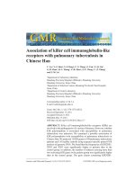
Figure 1 IF staining for NQO1 protein in A549 human lung cancer cells. NQO1 protein located in the cytoplasm of A549 cells (Red for NQO1,
Green for Actin, and Blue for DAPI).
Li et al. BMC Cancer (2015) 15:207
Page 4 of 9
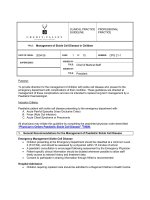
Figure 2 IHC staining for NQO1 protein expression in lung tissues. (A) NQO1 protein was negative in normal lung tissues. (B) NQO1 protein
was showed diffuse and strong positive staining in cytopalsm of lung SCC cells with LN metastasis. (C) NQO1 was weakly positive in lung SCC
without LN metastasis. (D) Diffuse and strong positive NQO1 protein signal in lung adenocarcinoma. (E & F) NQO1 protein staining is negative or
weakly positive in lung adenocarcinoma. (Original magnification, 200× in A-F).
the strongly positive rate of NQO1 expression was 28.0%
(42/150) in NSCLC, which was also significantly
higher than that in adjacent non-tumor (1.3%, 2/150)
(P < 0.01) (Table 1).
Clinicopathological significance of NQO1 expression in
patients with NSCLC
The relationship between NQO1 protein and the clinicopathological parameter of NSCLC was analyzed. The
positive rate of NQO1 protein was related with clinical
stage and lymph node metastasis. Moreover, the strongly
positiverate of NQO1 protein was significantly higher in
NSCLC with T3-4 (>5 cm) tumor size than in cases with
T1-2 (≤5 cm) tumor size (P = 0.005). Similarly, we found
that the strongly positive rate of NQO1 protein was significantly higher in stages III-IV (36.54%, 19/52) than
those in stages I-II (23.47%, 23/98) (P = 0.003). Also, it
was higher in poorly differentiated NSCLC (55.56%, 15/
27) than in moderately (31.46%, 28/89) and well differentiated NSCLC (26.47%, 9/34) (P = 0.012). Additionally,
it was also higher in NSCLC patients with lymph node
metastasis (50.00%, 27/54) than in cases without metastasis (15.63%, 15/96) (P = 0.000). However, there was
no significant correlations between high-level NQO1
expression and gender, and age of patients with
NSCLC (P > 0.05, respectively) (Table 2).
To further substantiate the importance of NQO1
expression in NSCLC progression, we analyzed the relationships between NQO1 positive expression rate and
DFS and OS in 150 lung cancer cases using the KaplanMeier method, and found that patients with NQO1 positive expression had lower DFS (Log-rank = 13.899, P <
0.001) and OS (Log-rank = 10.146, P = 0.001) rates than
those with NQO1 negative expression (Figure 3A & B).
Similarly, we also analyzed the association between
the NQO1 expression and tumor size, lymph node
metastasis, and clinical stages of NSCLC. The patients
with NQO1 positive expression had lower OS rates
compared with those with NQO1 negative expression
in the groups of T1-2 (Log-rank = 9.931, P = 0.002),
T3-4 (Log-rank = 9.387, P = 0.002) (Figure 4A & B),
without LN metastasis (Log-rank = 9.274, P = 0.002)
and stage I-II of NSCLC (Log-rank = 5.770, P = 0.016)
(Figure 4C & E), however, in the groups of patients
with LN metastasis or III-IV stages, OS rate was not
correlated with NQO1 expression status (Log-rank =
0.919, P = 0.553 and Log-rank = 0.572, P = 0.050, respectively) (Figure 4D & F).
Table 1 NQO1 protein expression in NSCLC
Diagnosis
No. of
cases
NQO1 protein expression
-
+
++
42
Positive rate
(+ ~ ++)
Strongly positive
rate(++)
59.3%**
28.0%**
NSCLC
150
61
47
Adjacent non tumor
150
138
10
2
8.0%
1.3%
Normal lung tissues
14
14
0
0
0
0
**P < 0.01compared with normal lung tissues and adjacent non tumor tissues.
Li et al. BMC Cancer (2015) 15:207
Page 5 of 9
Table 2 Correlation between NQO1 expression and clinicopathological features of NSCLC
Variables
Case no.
NQO1 positive(+ ~ ++)
n (%)
Gender
NQO1 strongly positive(++)
P value
n (%)
0.579
Male
112
65(58.04)
30(26.79)
Female
38
24(63.16)
12(31.58)
≧60
99
57(57.58)
<60
51
32(62.75)
Age
0.541
Tumor size
0.914
28(28.28)
14(27.45)
0.567
0.005**
T1-2
119
72(60.50)
27(22.69)
T3-4
31
17(54.84)
15(48.39)
I-II
98
51(52.04)
III-IV
52
38(73.08)
Stage
0.013*
Differentiation
0.003**
23(23.47)
19(36.54)
0.085
0.012*
Well
34
15(44.12)
9(26.47)
Moderately
89
54(60.67)
28(31.46)
Poorly
27
20(74.07)
LN metastasis
Negative Positive
P value
0.570
15(55.56)
0.039*
0.000**
96
51(53.13)
15(15.63)
54
38(70.37)
27(50.00)
*P < 0.05, **P < 0.01.
NQO1 expression is an independent prognostic
biomarkerin NSCLC by Cox proportional hazardsregression
model
Univariate analysis demonstrated that the NSCLC patients
with NQO1 positive expression had significant lower OS
rate (HR: 1.442, 95% CI: 1.036-2.007, P = 0.030) than
those with NQO1 negative expression. Additionally, age
(HR: 1.498, 95% CI: 1.062-2.113, P = 0.021), tumor size
(HR: 5.566, 95% CI: 3.499-8.857, P = 0.000), differentiation (HR: 1.426, 95% CI: 1.101-1.847, P = 0.007), lymph
node metastasis (HR: 2.300, 95% CI: 1.607-3.292, P =
0.000)and clinical stage (HR: 3.720, 95% CI: 2.526-5.477,
P = 0.000) were all significantly associated with OS rates
of NSCLC patients. Then, multivariate analysis was performed using the Cox proportional hazards model for
all of the significant variables, which were examined in
the univariate survival analysis. We found that NQO1
expression emerged as a significant independent prognostic factor for OS rates in patients with NSCLCs (HR:
1.514, 95% CI: 1.066-2.151, P = 0.020) along with tumor
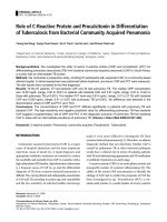
Figure 3 Kaplan-Meier analysis of DFS and OS rates in 150 NSCLC patients in relation to NQO1 protein expression. Patients of NSCLC
with NQO1 positive expression had lower DFS (A, P < 0.001) and OS (B, P < 0.001) rates than those with NQO1 negative expression (+, positive
expression; −, negative expression).
Li et al. BMC Cancer (2015) 15:207
Page 6 of 9
Figure 4 Kaplan-Meier analysis of OS rates in patients with or without NQO1 expressed NSCLC in prognostic factors. OS was assessed in
NSCLC patients with T1-2 (A, P = 0.002), T3-4 (B, P = 0.002), LN metastasis (−) (C, P = 0.002), LN metastasis (+) (D, P = 0.553), I-II stage (E, P = 0.016),
and III-IV stage (F, P = 0.050) concomitant with either positive- or negative-expression of NQO1.
size (HR: 5.545, 95% CI: 3.283-9.366, P = 0.000), differentiation (HR: 1.369, 95% CI: 1.055-1.775, P = 0.018),
lymph node metastasis (HR: 1.962, 95% CI: 1.334-2.884,
P = 0.001) and clinical stage (HR: 2.192, 95% CI: 1.4033.425, P = 0.001) (Table 3).
Discussion
NQO1, known as NAD(P)H: quinone oxidoreductase-1,
was first identified by Ernster and Navazio in 1958 [14].
NQO1 is a homodimericflavoprotein and many functions
have been proposed, such as xenobiotic detoxification,
superoxide scavenging, modulation of p53, maintenance
of endogenous antioxidants, and proteasomal degradation
[6]. Several studies have indicated that the phase II
enzyme NQO1 catalyzes the metabolic detoxification of
quinones and protects cells against chemical-induced oxidative stress and cancer [15,16]. Nagata et al. [17] and
Malik et al. [18] reported that the C609T polymorphism
in the NQO1 gene affects the translation of the NQO1
protein, and have been reported to be associated with
an increased risk of cancers death. Moreover, NQO1
polymorphism that leads to the enzyme inactivity has
been found to be a strong prognostic and predictive factor in the poor outcome of breast cancer [19]. NQO1
has also been shown to act as a chaperone, thereby
stabilizing various proteins, including the tumor suppressor protein p53 [20] and other short-lived proteins
such as ornithine decarboxylase [21]. These studies suggested that NQO1 activities may be essential for cancer
progression.
Li et al. BMC Cancer (2015) 15:207
Page 7 of 9
Table 3 Univariateand multivariate analysis of clinicopathological factors for the overall survival rate of 150 patients
with NSCLC
Characteristics
Univariate analysis HR (95% CI)
P value
Multivariate analysis HR (95% CI)
P value
Gender
1.047(0.724-1.513)
0.809
1.023(0.700-1.496)
0.907
Age
1.498(1.062-2.113)
0.021*
1.050(0.728-1.516)
0.793
Tumor size
5.566(3.499-8.857)
0.000**
5.545(3.283-9.366)
0.000**
Differentiation
1.426(1.101-1.847)
0.007**
1.369(1.055-1.775)
0.018*
LN metastasis
2.300(1.607-3.292)
0.000**
1.962(1.334-2.884)
0.001**
Stage
3.720(2.526-5.477)
0.000**
2.192(1.403-3.425)
0.001**
NQO1
1.442(1.036-2.007)
0.030*
1.514(1.066-2.151)
0.020*
LN: lymph node; HR: hazard ratio; CI: confidence interval.
*P <0.05, **P <0.01.
Accumulating studies showed that NQO1 was
expressed at relatively high levels in many solid tumors.
For example, our previous study [22] demonstrated that
NQO1 protein expression was significantly elevated in
breast cancer tissues compared with hyperplasia or
adjacent non-tumor tissues, indicating that NQO1 upregulation may occur in the initiation stage of breast
cancer progression. Similarly, compared with normal
cervical epithelia, the strongly positive rate of NQO1
protein expression was also significantly higher in cervical
SCC and intraepithelial neoplasia tissues, indicating that
NQO1 expression might be related to tumorigenesis of
cervical cancer [23]. Furthermore, we also found that
NQO1 protein was frequently high-expressed in gastric
adenocarcinoma compared with the gastric dysplasia and
adjacent non-tumor tissues, indicating that NQO1 was a
significant prognostic or predictive maker of gastric
adenocarcinoma [24]. Consistently, Awadallah et al. [25]
and Lyn-Cook et al. [26] reported that NQO1 protein was
up-regulated in pancreatic ductal adenocasinoma, and also
considered that NQO1 may represent a role of useful biomarker for pancreatic cancer. Malkinson et al. [27] found
that NQO1 gene was observed to be high-expressed in
human lung cancer tissues, and Rosvold et al. [28] and
Heller et al. [29] also indicated that the gene encoding
NQO1 is a promising candidate in the pathogenesis of
lung cancer. However, to date, the clinicopathological
significance of NQO1 protein expression in NSCLC has
not been elucidated.
Thus, here we performed IF and IHC staining in 150
NSCLC paired with the adjacent non-tumor tissues and
14 normal lung tissues, and found that NQO1 protein
localized in the cytoplasm of A549 lung cancer cells and
NSCLC tissues. Both positive and strongly positive rates
of NQO1 protein expression were significantly higher
than both in adjacent non-tumor and normal lung tissues.
These results indicate that NQO1 played an important
role in the progression of lung cancer. Mikami K et al.
[30] reported that the expression and enzyme activity of
NQO1 was up-regulated in colon cancer cell lines and
colorectal tumors, and moreover significantly higher in
tumors with LN metastases than those without metastasis.
Here we analyzed the correlation between NQO1 expression and clinicopathological parameters of NSCLC, and
the results showed that NQO1 expression and highexpression was all significantly associated with LN metastasis and clinical stage. Moreover, the strongly positive rate
of NQO1 protein was higher in NSCLCs with larger
tumor size (>5 cm) than in cases with smaller (≤5 cm),
and it was also significantly higher in poorly differentiated NSCLC than in moderately and well differentiated
NSCLC. These results indicated that NQO1 might be a
predictive biomarker for poor prognostic evaluation of
NSCLCs, and NQO1 protein maybe participated in the
tumorigenesis and malignant progression of NSCLC.
In regard to survival, we previously found that high
expression of NQO1 protein was strongly associated
with advanced stage, lymph node metastasis, Her2 overexpression and shortened survival of patients with breast
cancer [22]. Moreover, Buranrat et al. [12] also reported
a significant association between high level of NQO1
expression and short overall survival time of cholangiocarcinoma patients, which raised the exciting possibility
of using NQO1 as a tumor marker. However, Kim et al.
[31] reported that there was no correlation between
NQO1 and prognosis of small-cell lung cancer. Here we
found that NSCLC patients with NQO1 protein positiveexpression had a lower DFS and OS rates than those
with NQO1 protein negative-expression. Additionally,
age, tumor size, differentiation, lymph node metastasis,
clinical stage and NQO1 expression were all significantly
associated with OS rates of NSCLC patients (P < 0.05).
Furthermore, multivariate survival analysis demonstrated
that NQO1 positive-expression was an independent prognostic factor along with tumor size, differentiation, lymph
node metastasis and clinical stage. These findings indicated that NQO1 might be a potentially predictive biomarker of poor prognosis, especially in patients with poor
differentiation, lymph node metastasis and clinical stage of
NSCLC.
Li et al. BMC Cancer (2015) 15:207
Recently, NQO1 has been used as the target enzyme
in tumor cells to exemplify the ‘enzyme directed’ approach to anticancer drug development [32]. Park et al.
[33] and Kung et al. [34] demonstrated that NQO1
bioactivatable drugs (β-Lapachone or deoxynyboquinone
[DNQ]) can effectively kill the cancer cells. Huang et al.
[35] reported that the potency and NQO1-dependent
therapeutic window of DNQ and its apparent reduced
metabolism by one-electron oxidoreductases make this
drug (or derivatives) very promising. Therefore, the further study will be significant to verify if the NQO1 inhibitor could be used for the therapy of patients with
NSCLC.
Page 8 of 9
7.
8.
9.
10.
11.
Conclusions
In conclusion, NQO1 is frequently upregulated in NSCLC,
and it may be a useful poor prognostic biomarker and a
potential therapeutic target for patients with NSCLC.
12.
Abbreviations
NQO1: NAD(P)H quinone oxidoreductase 1; NSCLC: Non-small cell lung
cancer; SCC: Squamous cell carcinoma; EGFR: Epidermal growth factor
receptor; EML4-ALK: Echinoderm microtubule associated protein
like4-anaplastic lymphoma kinase; DNQ: Deoxynyboquinone.
14.
Competing interests
The authors declare that they have no competing interests.
16.
Authors’ contributions
ZL, YZ, TJ and YP participated in the study conception, design, case selection
and experiments. ZL, JM and PQ carried out data collection. GY, SP and ZL
performed the data analysis and wrote the manuscript. All authors read and
approved the final manuscript.
Acknowledgments
This study was supported by grants from National Natural Science Funds of
China (81260665), and The Projects of Research & Innovation of Jilin Youth
Leader and Team, China (20140519013JH).
Author details
1
Department of Pathology & Cancer Research Center, Yanbian University
Medical College, Yanji 133002, China. 2Department of TCM, Jilin Cancer
Hospital, Changchun 130012, China. 3Department of Anatomy and Histology
and Embryology, Yanbian University Medical College, Yanji 133002, China.
4
Department of Pathophysiology, Yanbian University Medical College, Yanji
133002, China.
13.
15.
17.
18.
19.
20.
21.
22.
Received: 7 September 2014 Accepted: 19 March 2015
23.
References
1. Siegel R, Naishadham D, Jemal A. Cancer statistics, 2013. CA Cancer J Clin.
2013;63(1):11–30.
2. Chen X, Liu Y, Røe OD, Qian Y, Guo R, Zhu L, et al. Gefitinib or erlotinib as
maintenance therapy in patients with advanced stage non-small cell lung
cancer: a systematic review. PLoS One. 2013;8(3):e59314.
3. Gaughan EM, Costa DB. Genotype-driven therapies for non-small cell lung
cancer: focus on EGFR, KRAS and ALK gene abnormalities. Ther Adv Med
Oncol. 2011;3(3):113–25.
4. Jin T, Jin J, Shang Y, Zhang S, Yu K, Piao Y, et al. Prognostic implications of
ezrin and phosphorylated ezrin expression in non-small cell lung cancer.
BMC Cancer. 2014;14:191.
5. Vasilious V, Rose D, Nebert DW. Update of the NAD(P)H: quinone
oxidoreductase (NQO) gene family. Hum Genomic. 2006;2(5):329–35.
6. Buranrat B, Prawan A, Kukongviriyapan U, Kongpetch S, Kukongviriyapan V.
Dicoumarol enhances gemcitabine-induced cytotoxicity in high
24.
25.
26.
27.
NQO1-expressing cholangiocarcinoma cells. World J Gastroenterol.
2010;16(19):2362–70.
Siegel D, Gustafson DL, Dehn DL, Han JY, Boonchoong P, Berliner LJ, et al.
NAD(P)H:quinone oxidoreductase 1: role as a superoxide scavengar.
Mol Pharmacol. 2004;65(5):1238–47.
Siegel D, Bolton EM, Burr JA, Liebler DC, Ross D. The reduction of
alpha-tocopherolquinone by human NAD(P)H: quinone oxidoreductase:
the role of alpha-tocopherolhydrolquinone as a cellular antioxidant.
Mol Pharmacol. 1997;52(2):300–5.
Siegel D, Ross D. Immunodetection of NAD(P)H: quinone oxidoreductase 1
(NQO1) in human tissues. Free RadicBiol Med. 2000;29(2–3):246–53.
Chao C, Zhang ZF, Berthiller J, Boffetta P, Hashibe M. NAD(P)H:quinone
oxidoreductase 1 (NQO1) Pro187Ser polymorphism and the risk of lung,
bladder, and colorectal cancer: a meta-analysis. Cancer Epidemiol
Biomarkers Prev. 2006;15(5):979–87.
Awadallah NS, Dehn D, Shah RJ, Russell Nash S, Chen YK, Ross D, et al.
NQO1 expression in pancreatic cancer and its potentail use as a biomarker.
ApplImmunohistochem Mol Morphol. 2008;16(1):24–31.
Buranrat B, Chau-in S, Prawan A, Puapaoroj A, Zeekpudsa P, Kukongviriyapan
V. NQO1 expression correlates with cholangiocarcinoma prognosis.
Asian Pac J Cancer Pre. 2012;13(Suppl):131–6.
Garate M, Wani AA, Li G. The NAD(P)H:Quinone Oxidoreductase 1
inducescell cycle progression and proliferation of melanoma cells.
Free RadicBiolMed. 2010;48(12):1601–9.
Ernster L, Lindberg O. Animal mitochondria. Annu Rev Physiol.
1958;20:13–42.
Nioi P, Hayes JD. Contribution of NAD(P)H: quinone oxidoreductase 1 to
protection against carcinogenesis, and regulation of its gene by the Nrf2
basic-region leucine zipper and the arylhydrocarbon receptor basic
helix-loop-helix transcription factors. Mutat Res. 2004;555(1–2):149–71.
Girolami F, Abbadessa G, Racca S, Spaccamiglio A, Piccione F, Dacasto M,
et al. Time-dependent acetylsalicylic acid effects on liver CYP1A and
antioxidant enzymes in a rat model of 7,12-dimethylbenzanthracene
(DMBA)-induced mammary carcinogenesis. Toxicol Lett. 2008;181(2):87–92.
Nagata M, Kimura T, Suzumura T, Kira Y, Nakai T, Umekawa K, et al.
C609Tpolymorphism of NADPH Quinone Oxidoreductase 1 Correlates
clinicalhematological toxicities in lung cancer patients treated with
Amrubicin. Clin Med Insights Oncol. 2013;7:31–9.
Malik MA, Zargar SA, Mittal B. Role of NQO1 609C > T and NQO2-3423G > A
gene polymorphisms in esophageal cancer risk in Kashmir valley andmeta
analysis. Mol Biol Rep. 2012;39(9):9095–104.
Fagerholm R, Hofstetter B, Tommiska J, Aaltonen K, Vrtel R, Syrjäkoski K,
et al. NAD(P)H: quinone oxidoreductase 1 NQO1*2 genotype (P187S) is a
strong prognostic and predictive factor in breast cancer. Nat Genet.
2008;40(7):844–53.
Asher G, Lotem J, Cohen B, Sachs L, Shaul Y. Regulation of p53 stability
and p53-dependent apoptosis by NADH quinoneoxidoreductase 1.
Proc NatlAcadSci U S A. 2001;98(3):1188–93.
Asher G, Bercovich Z, Tsvetkov P, Shaul Y, Kahana C. 20S proteasomal
degradation of ornithine decarboxylase is regulated by NQO1. Mol Cell.
2005;17(5):645–55.
Yang Y, Zhang Y, Wu Q, Cui X, Lin Z, Liu S, et al. Clinical implications of high
NQO1 expression inbreast cancers. J Exp Clin Cancer Res. 2014;33:14.
Ma Y, Kong J, Yan G, Ren X, Jin D, Jin T, et al. NQO1 overexpression is
associated with poor prognosis in squamous cell carcinoma of the uterine
cervix. BMC Cancer. 2014;14:414.
Lin L, Qin Y, Jin T, Liu S, Zhang S, Shen X, et al. Significance of
NQO1overexpression for prognostic evaluation of gastric adenocarcinoma.
Exp Mol Pathol. 2014;96(2):200–5.
Awadallah NS, Dehn D, Shah RJ, Russell Nash S, Chen YK, Ross D, et al.
NQO1 expression in pancreatic cancer and its potential use as a biomarker.
Appl Immunohistochem Mol Morphol. 2008;16(1):24–31.
Lyn-Cook BD, Yan-Sanders Y, Moore S, Taylor S, Word B, Hammons GJ.
Increased levels of NAD(P)H: quinoneoxidoreductase 1 (NQO1) in pancreatic
tissues from smokers and pancreatic adenocarcinomas: A potential
biomarker of early damage in the pancreas. Cell Biol Toxicol.
2006;22(2):73–80.
Malkinson AM, Siegel D, Forrest GL, Gazdar AF, Oie HK, Chan DC, et al.
Elevated DT-diaphorase activity and messenger RNA content in human
non-small cell lung carcinoma: relationship to the response of lung tumor
xenografts to mitomycin Cl. Cancer Res. 1992;52(17):4752–7.
Li et al. BMC Cancer (2015) 15:207
Page 9 of 9
28. Rosvold EA, McGlynn KA, Lustbader ED, Buetow KH. Identification of an
NAD(P)H: quinone oxidoreductase polymorphism and its association
with lung cancer and smoking. Pharmacogenetics. 1995;5(4):199–206.
29. Heller G, Zielinski CC, Zochbauer-Muller S. Lung cancer: from single-gene
methylation to methylome profiling. Cancer Metastasis Rev. 2010;29(1):95–107.
30. Mikami K, Naito M, Ishiguro T, Yano H, Tomida A, Yamada T, et al.
Immunological quantitation of DT-diaphorase in carcinoma cell lines and
clinical colon cancers: advanced tumors express greater levels of
DT-diaphorase. Jpn J Cancer Res. 1998;89(9):910–5.
31. Kim HC, Song JS, Lee JC, Lee DH, Kim SW, Lee JS, et al. Clinical significance
of NQO1 polymorphism and expression of p53, SOD2, PARP1 in limitedstage small cell lung cancer. Int J Clin Exp Pathol. 2014;7(10):6743–51.
32. Workman P. Enzyme-directed bioreductive drug development revisited: a
commentary on recent progress and future prospects with emphasis on
quinone anticancer agents and quinone metabolizing enzymes, particularly
DT-diaphorase. Oncol Res. 1994;6(10–11):461–75.
33. Park EJ, Min KJ, Lee TJ, Yoo YH, Kim YS, Kwon TK. β-Lapachoneindcuces
programmed necrosis through the RIP1-PARP-AIF-dependent pathway in
human hepatocellular carcinoma SK-Hep1 cells. Cell Death Dis.
2014;5:e1230.
34. Kung H, Weng T, Liu Y, Lu K, Chau Y. Sulindac Compounds Facilitate the
Cytotoxicity of β-Lapachone by Up-Regulation of NAD(P)H Quinone
Oxidoreductase in Human Lung Cancer Cells. PLoS One. 2014;9(2):e88122.
35. Huang X, Dong Y, Bey E. An NQO1 Substrate with Potent Antitumor Activity
That Selectively Kills by PARP1-Induced Programmed Necrosis. Cancer Res.
2012;72(12):3038–47.
Submit your next manuscript to BioMed Central
and take full advantage of:
• Convenient online submission
• Thorough peer review
• No space constraints or color figure charges
• Immediate publication on acceptance
• Inclusion in PubMed, CAS, Scopus and Google Scholar
• Research which is freely available for redistribution
Submit your manuscript at
www.biomedcentral.com/submit