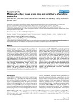
Human pancreatic cancer stem cells are sensitive to dual inhibition of IGF-IR and ErbB receptors
Bạn đang xem bản rút gọn của tài liệu. Xem và tải ngay bản đầy đủ của tài liệu tại đây (1 MB, 8 trang )
Urtasun et al. BMC Cancer (2015) 15:223
DOI 10.1186/s12885-015-1249-2
RESEARCH ARTICLE
Open Access
Human pancreatic cancer stem cells are sensitive
to dual inhibition of IGF-IR and ErbB receptors
Nerea Urtasun1,2*†, Anna Vidal-Pla1†, Sandra Pérez-Torras1,2,3 and Adela Mazo1,2,3
Abstract
Background: Pancreatic ductal adenocarcinoma is a particularly challenging malignancy characterized by poor
responsiveness to conventional chemotherapy. Although this tumor frequently overexpresses or possesses
constitutively activated variants of IGF-IR and EGFR/Her-2, clinical trials using inhibitors of these receptors have
failed. ErbB receptors have been proposed as one mechanism involved in the resistance to IGF-IR inhibitors. Therefore, combined treatment with inhibitors of both IGF-IR and ErbB receptors would appear to be a good strategy for
overcoming the emergence of resistance.
Methods: Sensitivity of cells to NVP-AEW541 and lapatinib in single or combination treatment was assessed by MTT
or WST-8 assays in a panel of human pancreatic cancer cell lines and cancer stem cells. Tumorspheres enriched in
cancer stem cells were obtained from cultures growing in non-adherent cell plates. The effects on cell signalling
pathways were analyzed by Western blot.
Results: We found that combined treatment with the IGF-IR and EGFR/Her-2 inhibitors NVP-AEW541 and lapatinib,
respectively, synergistically inhibited pancreatic cancer cell growth. Analysis at molecular level argued in favor of
cross-talk between IGF-IR and ErbBs pathways at IRS-1 level and indicated that the synergistic effect is associated
with the total abolishment of Akt, Erk and IRS-1 phosphorylation. Moreover, these inhibitors acted synergistically in
tumorsphere cultures to eliminate cancer stem cells, in contrast to their resistance to gemcitabine.
Conclusions: Taken together, these data indicate that simultaneous blockade of IGF-IR and EGFR/Her-2 using
NVP-AEW541 and lapatinib may overcome resistance in pancreatic cancer. Thus, the synergy observed with this
combined treatment indicates that it may be possible to maximize patient benefit with the appropriate combination of
currently known anticancer agents.
Keywords: Pancreatic ductal adenocarcinoma, Cancer stem cells, IGF-IR, EGFR, Her-2
Background
Pancreatic ductal adenocarcinoma (PDAC) is one of the
five most common causes of cancer death, owing to its late
diagnosis, high dissemination at early stages, and poor responsiveness to both radio- and chemotherapy [1]. Gemcitabine remains the current standard first-line treatment
[2]. However, chemotherapy in advanced disease confers
only modest survival advantage and symptoms palliation.
Recent clinical trials of gemcitabine combination therapies
* Correspondence:
†
Equal contributors
1
Departament de Bioquímica i Biologia Molecular, Universitat de Barcelona,
Barcelona, Spain
2
Institut de Biomedicina de la Universitat de Barcelona (IBUB), Barcelona,
Spain
Full list of author information is available at the end of the article
have produced significant, but low, response rates in advanced pancreatic cancer, underscoring the need for new
therapeutic approaches [3-5].
An important consideration in these strategies is the
heterogeneity of pancreatic tumors. In this context, several studies investigating pancreatic cancer biology have
identified a subpopulation of cells termed pancreatic
cancer stem cells (PCSCs) [6-8]. This subpopulation may
play a critical role in the resistance to chemotherapy and
radiation, suggesting that such cells may be the source
of some cases of pancreatic cancer relapse [9,10]. Therefore, therapeutic modalities that lead to the elimination
of CSCs could improve clinical outcome in patients with
pancreatic cancer.
© 2015 Urtasun et al.; licensee BioMed Central. This is an Open Access article distributed under the terms of the Creative
Commons Attribution License ( which permits unrestricted use, distribution, and
reproduction in any medium, provided the original work is properly credited. The Creative Commons Public Domain
Dedication waiver ( applies to the data made available in this article,
unless otherwise stated.
Urtasun et al. BMC Cancer (2015) 15:223
Receptor tyrosine kinases are currently among the
most promising therapeutic targets in a wide range of
tumors. Inhibition of receptor tyrosine kinases of the
ErbB family has been approved for the treatment of different tumors and is used extensively to treat breast cancer
[11]. There has also been growing research interest in
insulin-like growth factor-1 receptor (IGF-IR) as a target
for antitumor therapy [12-14], given the demonstrated ability of IGF-IR to potently contribute to a variety of oncogenic effects, including cell proliferation, cell survival, and
cell differentiation [15-17]. IGF-IR is frequently overexpressed or activated in pancreatic cancer, a factor that most
likely contributes to the aggressive growth characteristics
and poor prognosis of these tumors [18-20]. Moreover,
molecular mechanisms that lead to autocrine activation of
the IGF-IR and stimulation of downstream signaling
through phosphorylation (activation) of Akt have been
identified and could further substantially contribute to
tumor progression and invasion [21,22].
On the basis of these findings, IGF-IR has come to be
viewed as a rational therapeutic target in pancreatic cancer, prompting clinical investigations of IGF-IR inhibitors.
However, recent clinical trials of anti-IGF-IR compounds
in combination with gemcitabine have failed to demonstrate improved patient survival [23,24], a failure attributable, at least in part, to the development of resistance.
One mechanism proposed to account for resistance is activation of alternative survival pathways [25]. Among these
candidate alternative pathways are those activated by
members of the ErbB receptor family, which are important in regulating cell survival [26-28] and are frequently
overexpressed in pancreatic carcinomas [29,30]. Importantly, activation of mitogen-activated protein kinases
(MAPKs) by ErbB receptors signaling may counterbalance
the decrease in phosphorylated Akt induced by IGF-IR inhibitors. This compensatory mechanism could explain the
failure of treatments based on individual inhibition of
IGF-IR or ErbB [14,28,31], and suggests that therapeutic
strategies based on combined inhibition of IGF-IR and
ErbB receptors could overcome this resistance.
In the current study, we tested this hypothesis, investigating the impact of concurrent inhibition of IGF-IRs
and epidermal growth factor receptors (EGFR/Her-2) by
NVP-AEW541 and lapatinib tyrosine kinase inhibitors,
respectively, on pancreatic cancer cell lines and particularly on PCSCs.
Methods
Reagents and immunochemicals
Lapatinib was kindly provided by GlaxoSmithKline
(Brentford, UK) and NVP-AEW541 was a kind gift of
Novartis Pharma (Basel, Switzerland). Stock solutions of
drugs were prepared in dimethyl sulfoxide and stored
at −20°C, and diluted in fresh media before each
Page 2 of 8
experiment. Insulin-like growth factor (IGF-I) and Epidermal growth factor (EGF) (Peprotech, Rocky Hill, NJ, USA)
were dissolved in phosphate-buffered saline containing
0.1% bovine serum albumin (BSA). Cells were immunostained using antibodies against EGFR (1005), Her-2 (C18), Her-3 (C-17), IGF-IRβ (C-20) and Akt-1 (C-20) (Santa
Cruz Biotechnology, Santa Cruz, CA, USA); phosphoAkt (Ser473), phospho-p44/42 (Thr202/Tyr204) and
p44/42 (137 F5) (Cell Signaling Technology, Danvers,
MA, USA); phospho-IRS-1 (Tyr612) and phospho-IRS1 (Tyr896) (Invitrogen, Camarillo, CA, USA); and βactin (Sigma-Aldrich, St. Louis, MO, USA).
Cell culture
NP-9, NP-18, and NP-29 cell lines (kindly provided by
Dr Capella from Hospital de la Santa Creu i Sant Pau,
Barcelona, Spain) were derived from human pancreatic
adenocarcinomas xenografted in nude mice [32]. The
BxPC3 cell line was obtained from the American Type
Culture Collection (Manassas, VA, USA). CP15T and
CP15A cell lines were also derived from a human pancreatic adenocarcinoma xenografted in nude mice by
our group [33]. The research protocol complied with the
ethical guidelines of the 1975 Declaration of Helsinki
and was approved by the ethics committee of Universitat
de Barcelona. All participants provided written informed
consent. NP-9, NP-29, CP15T and CP15A cells were
grown in a 1:1 mixture of Dulbecco’s modified Eagle’s
medium (DMEM) and F12 medium; BxPC3 cells were
grown in DMEM; and NP-18 cell were grown in RPMI1640 medium (Gibco, Grand Island, NY, USA). All media
were supplemented with 5% fetal bovine serum and antibiotics (penicillin/streptomycin). Cells were maintained in
a humidified atmosphere of 5% CO2 at 37°C and subcultured every 3–4 days.
For tumorsphere cultures, cells were grown in ultralow attachment plates (Corning, Gendale, AZ, USA) using
serum-free DMEM:F12 (1:1) supplemented with B-27, N2,
antibiotic-antimycotic (Invitrogen), 20 ng/ml human EGF,
and 20 ng/ml human basic fibroblast growth factor (bFGF;
Peprotech). Tumorspheres were dissociated weekly using
trypsin and maintained for several passages. Experiments
were performed between the fourth and seventh passage
[34].
Dose–response assays
Dose–response assays were performed by seeding 2–
5 × 10³ cells/well in 96-well culture plates. Cultures
were exposed to increasing concentrations of lapatinib
and/or NVP-AEW541 for 72 h, at which time cell viability was determined by MTT (3-[4,5-dimethylthiazol2-yl]-2,5 diphenyl tetrazolium bromide) assay.
Assays comparing monolayers and tumorspheres were
performed by seeding single-cell suspensions at a density
Urtasun et al. BMC Cancer (2015) 15:223
of 1.5 × 10³ cells/well in standard or ultra-low-adhesion
96-well culture plates, respectively, with increasing concentrations of lapatinib and/or NVP-AEW541. Cell viability was determined 72 h post-treatment using a WST-8
assay (Sigma-Aldrich), as described by the manufacturer.
Data were fitted to a dose–response curve using standard nonlinear regression, adapting a Hill equation with
Grafit software (Erithacus Software, Ltd., Horley, UK) to
obtain 50% inhibitory concentration (IC50) values. Cell
survival for all experiments is expressed as the percentage of viable cells relative to that in untreated cells (defined as 100%).
The coefficient of drug interaction (CDI) was used to
analyze the effect of drug combination. CDI was calculated based on the absorbance in each group, as CDI =
AB/(A × B), where AB is the ratio for the combination
group relative to the control group, and A and B are the
ratios of each single agent group relative to the control
group. Thus, a CDI value < 1 indicates synergy, a CDI
value = 1 indicates additive effects, and a CDI value > 1
indicates antagonism. CDIs less than 0.7 indicate a significant synergistic effect.
Protein extraction and Western blot
Cells were lysed in ice-cold lysis buffer containing
20 mM Tris (pH 8), 150 mM NaCl, 10 mM EDTA,
10 mM Na4P2O7, 2 mM VO3−
4 , 100 mM NaF, 1 mM βglycerophosphate, 1% NP40, and protease and phosphatase inhibitor cocktails (Roche Applied Sciences,
Penzberg, Germany). Lysates containing equal amounts
Page 3 of 8
of protein (20 μg for monolayer experiments and 30 μg for
experiments comparing monolayers and tumorspheres),
assessed by Bradford assay (Bio-Rad, Hercules, CA, USA),
were electrophoretically separated on 8% polyacrylamidesodium dodecyl sulfate gels and transferred to nitrocellulose membranes (Schleicher and Schuell, Dassel, Germany).
Membranes were immunoblotted with the indicated primary antibodies. Antibody labeling was detected using an
enhanced chemiluminescence detection kit (Biological Industries, Kibbutz Beit Haemek, Israel).
Results
Sensitivity of human pancreatic cancer cell lines to
NVP-AEW541 and lapatinib
Expression levels of IGF-IR and ErbB family receptors
were examined in a panel of human pancreatic cancer
cell lines. IGF-IR expression levels varied, with high
levels detected in NP-29 and CP15A cell lines. Notably,
the highest levels of EGFR expression were also found in
NP-29 cells, whereas EGFR expression was negligible in
CP15T and CP15A cells. In contrast, Her-2, which was
observed in all cell lines, showed marked expression in
CP15T and CP15A cells. Her-3 expression was only
clearly detectable in NP-29, CP15T, and CP15A cells.
Intracellular signaling pathways were assessed by evaluating Akt and Erk (extracellular signal-regulated kinase)
phosphorylation. These experiments revealed a range of
activation levels, with NP-9 cells showing the highest
levels of Akt phosphorylation and CP15A cells showing
the lowest levels of Erk phosphorylation (Figure 1A).
Figure 1 Inhibition of IGF-I and ErbB receptors with NVP-AEW541 and lapatinib in pancreatic cancer cell lines. (A) Basal levels of IGF-I
and ErbB receptors and their signaling pathway components. Cells cultured to approximately 90% confluence were lysed and proteins in lysates were
analyzed by Western blot. (B) Dose–response curves and IC50 values for NVP-AEW541 and lapatinib in the panel of cell lines. Cells were treated 24 h
after seeding with increasing concentrations of NVP-AEW541 or lapatinib, and cell viability was measured by MTT assay 72 h after the start of treatment.
Data are presented as means ± standard deviation of a representative experiment (n = 3). ● NP-9, ♦ NP-18, ■ NP-29, CP15T, ▲ CP15A.
Urtasun et al. BMC Cancer (2015) 15:223
The effects of the IGF-IR inhibitor, NVP-AEW541,
and the EGFR and Her-2 inhibitor, lapatinib were then
examined in all five cell lines. NVP-AEW541 induced a
concentration-dependent inhibition of growth in all cell
lines. IC50 values ranged from 4.4 to 17.6 μM, with the
most potent effect observed in NP-18 cells. Lapatinib
also induced concentration-dependent growth inhibition
in all cell lines. Again, NP-18 cells showed the highest
sensitivity, and IC50 values ranged from 8.0 to 41.2 μM
(Figure 1B).
Response of pancreatic cancer cells to combined IGF-IR
and EGFR/Her-2 inhibition
Resistance to individual treatment with the IGF-IR and
EGFR/Her-2 inhibitors NVP-AEW541 and lapatinib, respectively, has been reported, reflecting the operation of
compensatory mechanisms between the two pathways. To
evaluate whether the individual effects of these drugs are
potentiated by concurrent inhibition of both pathways, we
assayed these two drugs in combination in the five cell
lines. Increasing concentrations of lapatinib were combined with a fixed (IC20) concentration of NVP-AEW541.
When used in combination, these drugs exhibited very potent synergy in all cell lines, with coefficients of drug interaction (CDIs) clearly < 0.7; remarkably, in some cases, CDI
values were < 0.1 (Figure 2A).
To evaluate the effects of these drugs on the intracellular signaling activity of both pathways, we selected
the NP-29 cell line, which exhibited the lowest CDI. In
control cells, an IGF-I stimulus promoted substantial
Akt and IRS-1 (Y612) phosphorylation and a small increase in IRS-1 (Y896) phosphorylation, but did not
affect Erk1/2 phosphorylation. This suggests that the
activity of the Ras-MAPK pathway is independent of
IGF-I in these cells. Conversely, EGF stimulation resulted in elevated phosphorylation of Erk1/2, IRS-1
(Y612), and IRS-1 Y896 (Figure 2B). Inhibition of IGF-IR
by NVP-AEW541 decreased IGF-I-induced phosphorylation of Akt and IRS-1 (Y612). In cells stimulated with
EGF or IGF-I + EGF, NVP-AEW541 treatment increased
EGFR pathway activation to a greater degree than in control cells, enhancing phosphorylation of Erk1/2, IRS1
(Y612), and IRS1 Y896 (Figure 2B). Whereas treatment
with lapatinib diminished EGF-stimulated activation of
Erk1/2 and IRS-1 (Y896), it did not significantly attenuate
IGF-I- or IGF-I + EGF-induced activation of Akt and IRS1 (Y612) (Figure 2B). Interestingly, simultaneous inhibition of both IGF-IR and EGFR/Her-2 by NVP-AEW541
and lapatinib completely abrogated IGF-I-, EGF-, and
IGF-I + EGF-stimulated phosphorylation of Akt, Erk1/2,
IRS-1 (Y612) and IRS-1 (Y896), confirming at the molecular level the strong synergy observed in cytotoxicity experiments (Figure 2B,C).
Page 4 of 8
Effect of IGF-IR and/or EGFR/Her-2 inhibition on
tumorspheres viability
The role of CSCs in the resistance to different drugs has
been extensively reported in recent years. Thus, the potent synergy obtained in tumor cells prompted us to
examine the effects of NVP-AEW541 and lapatinib on
cell viability in tumorspheres. These experiments were
performed using the two cell lines that exhibited the
highest synergy and in BxPC3 cells, a commercially
available cell line previously reported to be capable of
forming tumorspheres [35,36] that also exhibited a potent synergy (Additional file 1: Figure S1). An analysis of
morphology and cell cycle profile in tumorspheres obtained from CP15T and BxPC3 cells revealed PCSC
characteristics, but PCSC enrichment in NP-29 cells was
questionable (Additional file 2: Figure S2A,B, Additional
file 3: Supplemental methods).
Expression levels of receptors and the activity of their
pathways were then determined. These analyses showed
significant decreases in receptor expression and Akt
phosphorylation in the PCSC population (Figure 3A).
Despite this, both inhibitors were able to kill 100% of
cells, showing IC50 values in the same range as were obtained with the corresponding monolayers (Figure 3B,
Additional file 1: Figure S1A). These results contrast with
the resistance observed with gemcitabine (Additional file 2:
Figure S2C). Interestingly, combining these two drugs improved their inhibitory effect on cell viability, yielding CDI
values near 0.7, indicative of a potent synergistic effect, at
all concentrations (Figure 3C).
Discussion
Despite rapid advances on many fronts, PDAC remains
one of the most difficult human malignancies to treat.
The clinical outcome of patients with this disease has
not improved since the approval of gemcitabine, indicating the need for novel therapeutic strategies based on a
better understanding of the molecular basis of this disease [1]. In this context, several drugs designed to inhibit
IGF-IR have been developed, reflecting the fact that this
receptor is frequently overexpressed in PDAC and is associated with tumor progression and poor prognosis
[13,17,19,37]. However, several clinical trials of IGF-IR
inhibitors have failed, probably in part because of the activation of compensatory pathways [23,24]. ErbB receptors have been proposed as one mechanism involved in
the resistance to these inhibitors [28,38]. Therefore,
combined treatment with inhibitors of both IGF-IR and
ErbB receptors would appear to be a good strategy for
overcoming the emergence of resistance.
Inhibition of IGF-IR and EGFR/Her-2 by NVP-AEW541
and lapatinib caused a concentration-dependent reduction
of cell viability in all cell lines assayed. This cytotoxic effect has been previously described in other models, and,
Urtasun et al. BMC Cancer (2015) 15:223
Figure 2 (See legend on next page.)
Page 5 of 8
Urtasun et al. BMC Cancer (2015) 15:223
Page 6 of 8
(See figure on previous page.)
Figure 2 Effect of NVP-AEW541 and lapatinib combined treatment on the growth of pancreatic cancer cell lines. (A) Dose–response
curves and CDI values for NVP-AEW541 and lapatinib combinations. Twenty-four hours after seeding, cells were treated with increasing concentrations
of lapatinib alone (●) or combined with a fixed concentration of NVP-AEW541 (▲) equivalent to its IC20. Data are presented as means ± standard
deviation of a representative experiment (n = 3). (B) Molecular effects of NVP-AEW541 and lapatinib treatments. Cells were treated 24 h after seeding
with a concentration equivalent to the IC20 of NVP-AEW541, lapatinib, or their combination. After 72 h, 50 ng/ml of IGF-I, EGF or both were added for
20 min, and expression of IGF-IR and EGFR pathway components was analyzed by Western blot. (C) Schematic representation of the molecular
mechanism involved in NVP-AEW541 and lapatinib synergistic effect.
interestingly, it is tumor-selective, as it is higher in tumoral cells than in normal cells [39,40]. An evaluation of
the basal expression of IGF-IR and ErbB family receptors
and signaling pathway proteins showed no correlation between the levels of these receptors and sensitivity to their
inhibition, in good agreement with previous results in several types of cancer [38,41,42]. Moreover, when used in
combination, NVP-AEW541 and lapatinib strongly synergized in all cell lines at all concentrations assayed. Using
other inhibitors, this potentiation has been reported in
PDAC [38] and other tumors [43-45].
An analysis of the changes in signaling produced by
single and combined treatments argue in favor of crosstalk between IGF-IR and ErbBs pathways upstream of
their confluence at the MAPK and Akt level. IRS-1 is
generally considered to be a unique substrate of IGF-IR,
which phosphorylates IRS-1 at Y612. Notwithstanding this
presumption, a more recent study on breast cancer suggests that EGFR has the ability to recruit and phosphorylate
IRS-1 at Y896 [46]. This competence for the same substrate
is supported by our results and could contribute to the resistance caused by activation of mutual compensatory
Figure 3 IGF-IR and EGFR/Her-2 inhibition decreases the viability of pancreatic tumorspheres. (A) Basal levels of IGF-I and ErbB receptors
and their signaling pathway components in BxPC3 and CP15T tumorspheres were determined by Western blot. M, monolayer; T, tumorspheres.
(B) Dose–response curves and IC50 values for NVP-AEW541 and lapatinib. Cells were seeded with increasing concentrations of NVP-AEW541 or
lapatinib, and cell viability was measured by WST-8 assay 72 h after initiating treatment. ● BxPC3, ■ CP15T (C) Dose–response curves and CDI
values for NVP-AEW541 and lapatinib combinations. Cells were seeded with increasing concentrations of lapatinib alone (●) or combined with a
fixed concentration of NVP-AEW541 (▲) equivalent to its IC20. Data are presented as means ± standard deviation of three experiments.
Urtasun et al. BMC Cancer (2015) 15:223
pathways. The IRS-1 phosphorylation pattern clearly indicated that blocking IGF-IR signaling strongly induced phosphorylation of IRS-1 at Y896. This increase in IRS-1
phosphorylation highlights the crucial influence of this new
mechanism—activation of MAPK and especially Akt phosphorylation—in the resistance to IGF-IR inhibitors, and
points to preferential channeling of ErbB receptor signaling
to IRS-I (Y896) phosphorylation via phosphorylated Akt.
Interestingly, when both receptors were inhibited, IRS-1,
Akt and MAPK phosphorylation were completely abolished, reinforcing the utility of combined inhibition of both
pathways in averting the resistance induced by individual
treatments.
Despite these good in vitro results, the outcome in patients has been disappointing. One possible reason for the
failure of these targeted drugs could be the role of PCSCs
in resistance [47,48]. The importance of the IGF-IR pathway in treatments targeting PCSCs has not been previously described, although several recent reports have
demonstrated an association of this receptor with cell
stemness in some tumors [49,50]. Our results showed that
pancreatic cancer tumorspheres were sensitive to treatment with either NVP-AEW541 or lapatinib, in contrast
to their high resistance to gemcitabine. Remarkably, combining both drugs again produced a synergistic effect similar to that observed in monolayers. This synergy in
tumorspheres, which has not been previously described,
indicates that inhibition of both pathways in PCSCs can
also overcome the resistance caused by these compensatory pathways in this subpopulation.
Conclusions
Simultaneous inhibition of IGF-IR and ErbB receptors by
NVP-AEW541 and lapatinib circumvented the resistance
observed at the molecular level with individual treatments.
Interestingly, these inhibitors were also able to eliminate
PCSCs, overcoming their resistance to conventional
chemotherapy. Thus, the synergy observed with this combined treatment indicates that it may be possible to
maximize patient benefit with the appropriate combination of currently known anticancer agents.
Additional files
Additional file 1: Figure S1. Effect of NVP-AEW541 and lapatinib in the
BxPC3 monolayers. (A) Dose–response curves and IC50 values for NVPAEW541 and lapatinib. Cells were seeded with increasing concentrations of
NVP-AEW541 or lapatinib, and cell viability was measured by WST-8 assay
72 h after starting treatment. Data are presented as means ± standard
deviation of three experiments. (B) Dose–response curve and CDI values for
NVP-AEW541 and lapatinib combination. Twenty-four hours after seeding,
cells were treated with increasing concentrations of lapatinib alone (●) or
combined with a fixed concentration of NVP-AEW541 (▲) equivalent to its
IC20. Data are presented as means ± standard deviation of three experiments.
Additional file 2: Figure S2. Characterization of tumorspheres
obtained from different human pancreatic cancer cell lines. (A)
Page 7 of 8
Morphology of BxPC3, CP15T, and NP-29 tumorspheres. Cells were
maintained under standard culture conditions (monolayers) or in stem
cell medium on ultra-low-adhesion plates (tumorspheres). Scale bar =
5 μm. (B) Cell cycle profiles of monolayers and tumorspheres. S-phase
represented in light grey, G2/M-phase in dark grey, and G0/G1-phase in
black. (C) Dose–response curve and IC50 values of gemcitabine for monolayers and tumorspheres. Cells were seeded with increasing
concentrations of gemcitabine, and cell viability was measured by WST-8
assay 72 h after starting treatment. Data are presented as means ±
standard deviation of three experiments. ■BxPC3 monolayer, □BxPC3
tumorspheres, ●CP15T monolayer, ○CP15T tumorspheres.
Additional file 3: Analysis of cell cycle by flow cytometry.
Abbreviations
CDI: Coefficient of drug interaction; CSC: Cancer stem cells; EGF: Epidermal
growth factor; EGFR: Epidermal growth factor receptor; Erk: Extracellular signalregulated kinase; IC50: 50% inhibitory concentration; IGF: Insulin-like growth
factor; IGF-IR: Insulin-like growth factor-1 receptor; IRS-1: Insulin receptor substrate
1; MAPKs: Mitogen-activated protein kinases; pAkt: Phosphorylated Akt;
PCSC: Pancreatic cancer stem cells; PDAC: Pancreatic ductal adenocarcinoma.
Competing interests
The authors declare that they have no competing interests.
Authors’ contributions
NU carried out the experiments related to tumorspheres and helped to draft
the manuscript. AVP carried out the experiments related to monolayers and
helped to draft the manuscript. SPT participated in the design of the study
and helped to draft the manuscript. AM participated in the design of the
study and helped to draft the manuscript. All authors read and approved the
final manuscript.
Acknowledgements
This work has been supported by grants BIO2008-04692-C03-03 and SAF201123660 (Ministerio de Economia y Competitividad) and receives partial support
of the Generalitat de Catalunya (2009SGR624). The group belongs to the
National Biomedical Research Institute on Liver and Gastrointestinal Diseases
(CIBERehd) and SPT is a CIBER researcher. CIBER is an initiative of the Instituto
de Salud Carlos III (ISCIII, Ministerio de Economia y Competitividad). AVP has
been the recipient of a FI fellow from the Generalitat de Catalunya. We are
grateful to GlaxoSmithKline and Novartis Pharma for kindly provided lapatinib
and NVP-AEW541, respectively.
In memoriam of Dr. Adela Mazo, who passed away on March 24th 2015.
Author details
1
Departament de Bioquímica i Biologia Molecular, Universitat de Barcelona,
Barcelona, Spain. 2Institut de Biomedicina de la Universitat de Barcelona
(IBUB), Barcelona, Spain. 3CIBERehd, Madrid, Spain.
Received: 28 August 2014 Accepted: 24 March 2015
References
1. Siegel R, Naishadham D, Jemal A. Cancer statistics, 2012. CA Cancer J Clin.
2012;62(1):10–29.
2. Burris 3rd HA, Moore MJ, Andersen J, Green MR, Rothenberg ML, Modiano
MR, et al. Improvements in survival and clinical benefit with gemcitabine as
first-line therapy for patients with advanced pancreas cancer: a randomized
trial. J Clin Oncol. 1997;15(6):2403–13.
3. Di Marco M, Di Cicilia R, Macchini M, Nobili E, Vecchiarelli S, Brandi G, et al.
Metastatic pancreatic cancer: is gemcitabine still the best standard
treatment? (Review). Oncol Rep. 2010;23(5):1183–92.
4. Saif MW, Lee Y, Kim R. Harnessing gemcitabine metabolism: a step towards
personalized medicine for pancreatic cancer. Ther Adv Med Oncol.
2012;4(6):341–6.
5. Yang ZY, Yuan JQ, Di MY, Zheng DY, Chen JZ, Ding H, et al. Gemcitabine
plus erlotinib for advanced pancreatic cancer: a systematic review with
meta-analysis. PLoS One. 2013;8(3):e57528.
6. Hermann PC, Huber SL, Herrler T, Aicher A, Ellwart JW, Guba M, et al.
Distinct populations of cancer stem cells determine tumor growth and
Urtasun et al. BMC Cancer (2015) 15:223
7.
8.
9.
10.
11.
12.
13.
14.
15.
16.
17.
18.
19.
20.
21.
22.
23.
24.
25.
26.
27.
28.
29.
30.
metastatic activity in human pancreatic cancer. Cell Stem Cell.
2007;1(3):313–23.
Li C, Heidt DG, Dalerba P, Burant CF, Zhang L, Adsay V, et al. Identification
of pancreatic cancer stem cells. Cancer Res. 2007;67(3):1030–7.
Li C, Wu JJ, Hynes M, Dosch J, Sarkar B, Welling TH, et al. c-Met is a marker
of pancreatic cancer stem cells and therapeutic target. Gastroenterology.
2011;141(6):2218–27. e2215.
Balic A, Dorado J, Alonso-Gomez M, Heeschen C. Stem cells as the root of
pancreatic ductal adenocarcinoma. Exp Cell Res. 2012;318(6):691–704.
Bednar F, Simeone DM. Pancreatic cancer stem cell biology and its
therapeutic implications. J Gastroenterol. 2011;46(12):1345–52.
Higgins MJ, Baselga J. Targeted therapies for breast cancer. J Clin Invest.
2011;121(10):3797–803.
Sachdev D, Yee D. Disrupting insulin-like growth factor signaling as a potential
cancer therapy. Mol Cancer Ther. 2007;6(1):1–12.
Pollak M. Insulin and insulin-like growth factor signalling in neoplasia. Nat
Rev Cancer. 2008;8(12):915–28.
Ioannou N, Dalgleish AG, Seddon AM, Mackintosh D, Guertler U, Solca F,
et al. Anti-tumour activity of afatinib, an irreversible ErbB family blocker, in
human pancreatic tumour cells. Br J Cancer. 2011;105(10):1554–62.
Samani AA, Yakar S, LeRoith D, Brodt P. The role of the IGF system in cancer
growth and metastasis: overview and recent insights. Endocr Rev.
2007;28(1):20–47.
Werner H, Bruchim I. The insulin-like growth factor-I receptor as an
oncogene. Arch Physiol Biochem. 2009;115(2):58–71.
Subramani R, Lopez-Valdez R, Arumugam A, Nandy S, Boopalan T,
Lakshmanaswamy R. Targeting insulin-like growth factor 1 receptor inhibits
pancreatic cancer growth and metastasis. PLoS One. 2014;9(5):e97016.
Karna E, Surazynski A, Orlowski K, Laszkiewicz J, Puchalski Z, Nawrat P, et al.
Serum and tissue level of insulin-like growth factor-I (IGF-I) and IGF-I binding
proteins as an index of pancreatitis and pancreatic cancer. Int J Exp Pathol.
2002;83(5):239–45.
Bardeesy N, DePinho RA. Pancreatic cancer biology and genetics. Nat Rev
Cancer. 2002;2(12):897–909.
Ioannou N, Seddon AM, Dalgleish A, Mackintosh D, Modjtahedi H.
Expression pattern and targeting of HER family members and IGF-IR in
pancreatic cancer. Front Biosci (Landmark Ed). 2012;17:2698–724.
Bergmann U, Funatomi H, Yokoyama M, Beger HG, Korc M. Insulin-like
growth factor I overexpression in human pancreatic cancer: evidence for
autocrine and paracrine roles. Cancer Res. 1995;55(10):2007–11.
Zhang D, Brodt P. Type 1 insulin-like growth factor regulates MT1-MMP
synthesis and tumor invasion via PI 3-kinase/Akt signaling. Oncogene.
2003;22(7):974–82.
Kindler HL, Richards DA, Garbo LE, Garon EB, Stephenson Jr JJ, Rocha-Lima CM,
et al. A randomized, placebo-controlled phase 2 study of ganitumab (AMG
479) or conatumumab (AMG 655) in combination with gemcitabine in patients
with metastatic pancreatic cancer. Ann Oncol. 2012;23(11):2834–42.
von Mehren M, Britten CD, Pieslor P, Saville W, Vassos A, Harris S, et al.
Phase I, dose-escalation study of BIIB022 (anti-IGF-1R antibody) in advanced
solid tumors. J Clin Oncol. 2010;28(Suppl.). Abstract 2612.
Huang F, Hurlburt W, Greer A, Reeves KA, Hillerman S, Chang H, et al.
Differential mechanisms of acquired resistance to insulin-like growth factor-i
receptor antibody therapy or to a small-molecule inhibitor, BMS-754807, in
a human rhabdomyosarcoma model. Cancer Res. 2010;70(18):7221–31.
Scaltriti M, Baselga J. The epidermal growth factor receptor pathway: a
model for targeted therapy. Clin Cancer Res. 2006;12(18):5268–72.
Buck E, Eyzaguirre A, Rosenfeld-Franklin M, Thomson S, Mulvihill M, Barr S,
et al. Feedback mechanisms promote cooperativity for small molecule
inhibitors of epidermal and insulin-like growth factor receptors. Cancer Res.
2008;68(20):8322–32.
Haluska P, Carboni JM, TenEyck C, Attar RM, Hou X, Yu C, et al. HER receptor
signaling confers resistance to the insulin-like growth factor-I receptor
inhibitor, BMS-536924. Mol Cancer Ther. 2008;7(9):2589–98.
Ueda S, Hatsuse K, Tsuda H, Ogata S, Kawarabayashi N, Takigawa T, et al.
Potential crosstalk between insulin-like growth factor receptor type 1 and
epidermal growth factor receptor in progression and metastasis of pancreatic
cancer. Mod Pathol. 2006;19(6):788–96.
Papageorgio C, Perry MC. Epidermal growth factor receptor-targeted
therapy for pancreatic cancer. Cancer Invest. 2007;25(7):647–57.
Page 8 of 8
31. Lu Y, Zi X, Zhao Y, Pollak M. Overexpression of ErbB2 receptor inhibits IGF-Iinduced Shc-MAPK signaling pathway in breast cancer cells. Biochem
Biophys Res Commun. 2004;313(3):709–15.
32. Capella G, Farre L, Villanueva A, Reyes G, Garcia C, Tarafa G, et al. Orthotopic
models of human pancreatic cancer. Ann N Y Acad Sci. 1999;880:103–9.
33. Perez-Torras S, Vidal-Pla A, Miquel R, Almendro V, Fernandez-Cruz L, Navarro
S, et al. Characterization of human pancreatic orthotopic tumor xenografts
suitable for drug screening. Cell Oncol (Dordr). 2011;34(6):511–21.
34. Cao L, Zhou Y, Zhai B, Liao J, Xu W, Zhang R, et al. Sphere-forming cell
subpopulations with cancer stem cell properties in human hepatoma cell
lines. BMC Gastroenterol. 2011;11:71.
35. Ji Q, Hao X, Zhang M, Tang W, Yang M, Li L, et al. MicroRNA miR-34 inhibits
human pancreatic cancer tumor-initiating cells. PLoS One. 2009;4(8):e6816.
36. Wei HJ, Yin T, Zhu Z, Shi PF, Tian Y, Wang CY. Expression of CD44, CD24
and ESA in pancreatic adenocarcinoma cell lines varies with local
microenvironment. Hepatobiliary Pancreat Dis Int. 2011;10(4):428–34.
37. Tognon CE, Sorensen PH. Targeting the insulin-like growth factor 1 receptor
(IGF1R) signaling pathway for cancer therapy. Expert Opin Ther Targets.
2012;16(1):33–48.
38. Ioannou N, Seddon AM, Dalgleish A, Mackintosh D, Modjtahedi H.
Treatment with a combination of the ErbB (HER) family blocker afatinib and
the IGF-IR inhibitor, NVP-AEW541 induces synergistic growth inhibition of
human pancreatic cancer cells. BMC Cancer. 2013;13:41.
39. Howes AL, Richardson RD, Finlay D, Vuori K. 3-Dimensional culture systems
for anti-cancer compound profiling and high-throughput screening reveal
increases in EGFR inhibitor-mediated cytotoxicity compared to monolayer
culture systems. PLoS One. 2014;9(9):e108283.
40. Premkumar DR, Jane EP, Pollack IF. Co-administration of NVP-AEW541 and
dasatinib induces mitochondrial-mediated apoptosis through Bax activation
in malignant human glioma cell lines. Int J Oncol. 2010;37(3):633–43.
41. Cunningham MP, Thomas H, Marks C, Green M, Fan Z, Modjtahedi H. Cotargeting the EGFR and IGF-IR with anti-EGFR monoclonal antibody ICR62
and the IGF-IR tyrosine kinase inhibitor NVP-AEW541 in colorectal cancer
cells. Int J Oncol. 2008;33(5):1107–13.
42. Mukohara T, Shimada H, Ogasawara N, Wanikawa R, Shimomura M,
Nakatsura T, et al. Sensitivity of breast cancer cell lines to the novel insulinlike growth factor-1 receptor (IGF-1R) inhibitor NVP-AEW541 is dependent
on the level of IRS-1 expression. Cancer Lett. 2009;282(1):14–24.
43. Browne BC, Crown J, Venkatesan N, Duffy MJ, Clynes M, Slamon D, et al.
Inhibition of IGF1R activity enhances response to trastuzumab in HER-2positive breast cancer cells. Ann Oncol. 2011;22(1):68–73.
44. Esparis-Ogando A, Ocana A, Rodriguez-Barrueco R, Ferreira L, Borges J,
Pandiella A. Synergic antitumoral effect of an IGF-IR inhibitor and
trastuzumab on HER2-overexpressing breast cancer cells. Ann Oncol.
2008;19(11):1860–9.
45. Kaulfuss S, Burfeind P, Gaedcke J, Scharf JG. Dual silencing of insulin-like
growth factor-I receptor and epidermal growth factor receptor in colorectal
cancer cells is associated with decreased proliferation and enhanced
apoptosis. Mol Cancer Ther. 2009;8(4):821–33.
46. Knowlden JM, Jones HE, Barrow D, Gee JM, Nicholson RI, Hutcheson IR.
Insulin receptor substrate-1 involvement in epidermal growth factor
receptor and insulin-like growth factor receptor signalling: implication for
Gefitinib (‘Iressa’) response and resistance. Breast Cancer Res Treat.
2008;111(1):79–91.
47. Abel EV, Simeone DM. Biology and clinical applications of pancreatic cancer
stem cells. Gastroenterology. 2013;144(6):1241–8.
48. Clarke MF, Dick JE, Dirks PB, Eaves CJ, Jamieson CH, Jones DL, et al. Cancer
stem cells–perspectives on current status and future directions: AACR
Workshop on cancer stem cells. Cancer Res. 2006;66(19):9339–44.
49. Dallas NA, Xia L, Fan F, Gray MJ, Gaur P, Van Buren 2nd G, et al.
Chemoresistant colorectal cancer cells, the cancer stem cell phenotype, and
increased sensitivity to insulin-like growth factor-I receptor inhibition. Cancer
Res. 2009;69(5):1951–7.
50. Xu C, Xie D, Yu SC, Yang XJ, He LR, Yang J, et al. beta-Catenin/POU5F1/
SOX2 transcription factor complex mediates IGF-I receptor signaling and
predicts poor prognosis in lung adenocarcinoma. Cancer Res.
2013;73(10):3181–9.