High expression of NQO1 is associated with poor prognosis in serous ovarian carcinoma
Bạn đang xem bản rút gọn của tài liệu. Xem và tải ngay bản đầy đủ của tài liệu tại đây (1.09 MB, 8 trang )
Cui et al. BMC Cancer (2015) 15:244
DOI 10.1186/s12885-015-1271-4
RESEARCH ARTICLE
Open Access
High expression of NQO1 is associated with poor
prognosis in serous ovarian carcinoma
Xuelian Cui1,2†, Lianhua Li3†, Guanghai Yan2, Kai Meng2, Zhenhua Lin1,2, Yunze Nan3*, Guang Jin1* and Chunyu Li2*
Abstract
Background: NAD(P)H:quinone oxidoreductase (NQO1) is a flavoprotein that catalyzes two-electron reduction and
detoxification of quinones and its derivatives. NQO1 catalyzes reactions that have a protective effect against redox
cycling, oxidative stress and neoplasia. High expression of NQO1 is associated with many solid tumors including
those affecting the colon, breast and pancreas; however, its role in the progression of ovarian carcinoma is largely
undefined. This study aimed to investigate the clinicopathological significance of high NQO1 expression in serous
ovarian carcinoma.
Methods: NQO1 protein expression was assessed using immunohistochemical (IHC) staining in 160 patients with
serous ovarian carcinoma, 62 patients with ovarian borderline tumors and 53 patients with benign ovarian tumors.
Quantitative real-time polymerase chain reaction (qRT-PCR) was performed to detect NQO1 mRNA expression levels.
The correlation between high NQO1 expression and clinicopathological features of ovarian carcinoma was evaluated
by Chi-square and Fisher’s exact test. Overall survival (OS) rates of all of ovarian carcinoma patients were calculated
using the Kaplan-Meier method, and univariate and multivariate analyses were performed using the Cox proportional
hazards regression model.
Results: NQO1 protein expression in ovarian carcinoma cells was predominantly cytoplasmic. Strong, positive expression
of NQO1 protein was observed in 63.8% (102/160) of ovarian carcinomas, which was significantly higher than in
borderline serous tumors (32.3%, 20/62) or benign serous tumors (11.3%, 6/53). Importantly, the rate of strong,
positive NQO1 expression in borderline serous tumors was also higher than in benign serous tumors. High expression
of NQO1 protein was closely associated with higher histological grade, advanced clinical stage and lower OS rates in
ovarian carcinomas. Moreover, multivariate analysis indicated that NQO1 was a significant independent prognostic
factor, in addition to clinical stage, in patients with ovarian carcinoma.
Conclusions: NQO1 is frequently upregulated in ovarian carcinoma. High expressin of NQO1 protein may be an effective
biomarker for poor prognostic evaluation of patients with serous ovarian carcinomas.
Keywords: Ovarian carcinoma, NQO1, Immunohistochemistry, Survival analysis
Background
Ovarian carcinoma is one of the most lethal gynecological
carcinomas, and the second leading cause of carcinomarelated deaths among women [1]. The majority of ovarian
carcinoma cases are of epithelial origin (> 90%), and of
these, serous ovarian carcinoma is the most common
* Correspondence: ; ;
†
Equal contributors
3
Department of Gynecology & Obstetrics, Yanbian University Hospital, Yanji
133000, China
1
Department of Pathology, Yanbian University Medical College, Yanji 133002,
China
2
Cancer Research Center, Yanbian University, Yanji 133002, China
subtype [2]. As ovarian carcinoma is often asymptomatic in the early stages, or presents with vague symptoms mimicking extra-ovarian disease, the majority of
patients (70–75%) present with widespread disease at
diagnosis, and as such the mortality rate is high [3].
Therefore, understanding the molecular pathogenesis
and mechanisms underlying ovarian cancer initiation
and progression is critical for the prevention and treatment of this disease.
The NAD(P)H:quinone oxidoreductase 1 (NQO1) is a
predominantly cytosolic enzyme, which uses NADH or
NADPH as substrates to directly reduce quinones to
© 2015 Cui et al.; licensee BioMed Central. This is an Open Access article distributed under the terms of the Creative
Commons Attribution License ( which permits unrestricted use, distribution, and
reproduction in any medium, provided the original work is properly credited. The Creative Commons Public Domain
Dedication waiver ( applies to the data made available in this article,
unless otherwise stated.
Cui et al. BMC Cancer (2015) 15:244
hydroquinones [4]. The direct two-electron reduction of
quinones to hydroquinone by NQO1 is historically considered a detoxification mechanism, since this reaction
bypasses the formation of the highly reactive semiquinone [5]. NQO1 provides cells with multiple layers of
protection against oxidative stress, including the direct
detoxification of highly reactive quinones, the maintenance of lipid-soluble antioxidants in reduced forms, and
stabilization of the tumor suppressor p53 [6]. Chronic
inflammation suppresses NQO1 expression and may increase susceptibility to cell injury. Paradoxically, increasing evidence suggests that high expression of NQO1 at
the early stages of carcinogenesis may provide cancer cells
with a growth advantage [7-9]. Moreover, certain quinones, such as mitomycin C, streptonigrin, E09 and RH1,
are bioactivated by NQO1 [10-14]. The bioactivation
property of NQO1 renders it an ideal target for developing
anti-tumor drugs, since NQO1 activities are elevated in
various human tumors [15]. NQO1 was shown to be
expressed at high levels in many solid tumors, including
uterine cervix [16], lung [17] and pancreas [18], and was
also detected following the induction of cell cycle progression and proliferation of melanoma cells [19]. However, to
date, the role of NQO1 in ovarian carcinoma progression
remains unclear.
In the present study, we investigated the correlation between high expression of NQO1 and clinicopathological
features of serous ovarian carcinomas. We also assessed
the prognostic value of high NQO1 expression in patients
with serous ovarian carcinoma.
Methods
Ethics statement
This study complied with the Helsinki Declaration and
was approved by the Human Ethics and Research Ethics
committees of Yanbian University Medical College of
China. Through the surgery consent form, patients were
informed that the resected specimens were stored by
our hospital and potentially used for scientific research,
and that their privacy would be maintained. Follow-up
survival data were collected retrospectively through
medical-record analyses.
Patients and tissue specimens
A total of 275 human ovarian tumor specimens, including
160 serous carcinomas, 62 borderline serous tumors, 53
benign serous tumors were used for this study. These tumors were selected randomly from patients undergoing
surgery between 2005 and 2010 and stored in the Tumor
Tissue Bank of Yanbian University Medical College. Pathological parameters, including age, menopausal status, grade
and survival data, were carefully reviewed in all 160 serous
ovarian carcinomas.
Page 2 of 8
In 160 cases, patients’ ages ≥ 48 years to <48 years was
97:63. The hematoxylin and eosin-stained slides of the
different biopsies were reviewed by two experienced pathologists and one appropriate paraffin block was selected for this study. Histopathological grades were
made using the World Health Organization (Pathology
& Genetics Tumors of gynecological system) criteria. All
of the ovarian carcinoma patients were clinically staged
according to the FIGO staging system [with 92 early- stage
tumors (FIGO stages I and II) and 68 late-stage tumors
(FIGO stages III and IV)]. None of the ovarian carcinoma
patients received preoperative radiation or chemotherapy
before surgery. All ovarian carcinoma patients had followup records for more than 5 years. By February 2014, 111
patients had died while 49 patients remained alive. The
median survival time was 44.5 months.
Immunohistochemical (IHC) analysis
IHC analysis was performed using the DAKO LSAB kit
(DAKO A/S, Glostrup, Denmark). Briefly, to eliminate
endogenous peroxidase activity, 4 μm thick tissue sections were deparaffinized, rehydrated and incubated with
3% H2O2 in methanol for 15 min at room temperature
(RT). The antigen was retrieved at 95°C for 20 min by placing the slides in 0.01 M sodium citrate buffer (pH 6.0).
The slides were then incubated with the NQO1 antibody
(1:200, Santa Cruz Biotechnology, Dallas, TX, USA) at 4°C
overnight. After incubation with the biotinylated secondary
antibody at RT for 30 min, the slides were incubated with
streptavidin-peroxidase complex at RT for 30 min. Immunostaining was developed by using 3,3′-diaminobenzidine
and Mayer’s hematoxylin was used for counterstaining [16].
Mouse IgG was used as an isotype control. In addition,
positive tissue sections were processed while omitting the
primary antibody (mouse anti-NQO1) as negative controls.
Evaluation of the IHC staining
As described previously [20], the expressions were
scored by two pathologists (Lin Z & Liu S) who did
not possess knowledge of the clinical data. In case of
discrepancies, a final score was established by reassessment on a double-headed microscope. Briefly, the immunostaining for NQO1 was semi-quantitatively scored as ‘-’
(negative, no or less than 5% positive cells), ‘+’ (5–25%
positive cells), ‘++’ (26–50% positive cells) and ‘+++’ (more
than 50% positive cells). Only the cytoplasmic expression
pattern was considered as positive staining. Tissue sections scored as ‘++’ and ‘+++’ were considered as strong
positives (high expression) of NQO1. For survival data
analysis, ‘++’ or ‘+++’ scored samples were considered as
high NQO1 expression and ‘-’or ‘+’ scored samples were
considered as low NQO1 expression.
Cui et al. BMC Cancer (2015) 15:244
qRT-PCR
Total RNA was extracted using TRIzol Reagent (Invitrogen, Carlsbad, CA, USA) from fresh tissue samples of 19
serous ovarian carcinomas and 15 benign serous ovarian
tumors. First-strand cDNA was synthesized by PrimeScript reverse transcriptase (TaKaRa Biotechnology, Dalian, China) and oligo (dT) following the manufacturer’s
instructions. Real-time PCR was performed using doublestranded DNA-specific SYBR Premix Ex Taq™ II Kit
(TaKaRa Biotechnology) on a Bio-Rad sequence detection
system according to the manufacturer’s instructions.
Double-stranded DNA specific expression was tested
by the comparative Ct method using 2-ΔΔCt. NQO1
primers were as follows: 5′-GGCAGAAGAGCACTGATCGTA-3′, and 5′-TGATGGGATTGAAGTTCATG
GC-3′. Primers for GAPDH, which was used as an internal control, were: 5′-GGTCTCCTCTGACTTCAACA3′ and 5′-ATACCAGGAAATGAGCTTGA-3′. All assays
were performed at least three times.
Statistical analysis
Statistical analyses included descriptive statistics with
determination of minimal and maximal values, means
and medians, with 95% confidence interval (CI) for particular variables. Chi-square test and Fisher’s exact test
were used to assess correlation between clinicopathological characteristics and the expression of studied protein.
Page 3 of 8
Kaplan-Meier method was used to calculate the survival
rates after tumor removal and Log-rank was used to
analyze the differences in survival curves. Multivariate
survival analysis was performed on all the significant
characteristics measured by univariate survival analysis
(age, menopausal status, histological grade, FIGO stage
and NQO1 expression) through the Cox proportional
hazard regression model. Statistical calculation was performed using the SPSS 17.0. P < 0.05 was considered statistically significant.
Results
High expression of NQO1 protein in serous ovarian
carcinoma
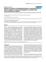
IHC analysis of NQO1 in ovarian carcinoma cells from
160 patients revealed predominantly cytoplasmic expression (Figure 1). The rate of positive NQO1 protein expression was significantly higher in serous carcinomas
(85.6%, 137/160) than in borderline serous tumors
(56.5%, 35/62) or benign serous tumors (34.0%, 18/53)
(P < 0.01, respectively). Similarly, the rate of strong, positive NQO1 protein expression was significantly higher in
serous carcinomas (63.8%, 102/160) than in either borderline serous tumors (32.3%, 20/62) or benign serous
tumors (11.3%, 6/53) (P < 0.01, respectively). More importantly, the rates of positive and strongly positive
NQO1 protein expression in borderline serous tumors
Figure 1 IHC staining of NQO1 protein in ovarian tumor samples. (A) Negative expression of NQO1 protein in a benign serous tumor.
(B–C) Weak positive expression of NQO1 protein (B) and positive expression (C) in atypical cells of borderline serous tumors. (D) Strong
positive expression of NQO1 protein in serous carcinoma cells, in a patient with metastasis. (E) Positive expression of NQO1 protein in a
serous carcinoma patient without metastasis. Scattered, strongly positive-staining cancer cells are seen (arrows). (F) Negative expression
of NQO1 protein in a serous carcinoma patient without metastasis. Original magnification, A–F: ×200.
Cui et al. BMC Cancer (2015) 15:244
Page 4 of 8
were significantly higher than in benign serous tumors
(P < 0.05) (Table 1).
In keeping with these results, analysis of NQO1
mRNA levels by qRT-PCR confirmed elevated levels of
NQO1 transcript in serous ovarian carcinoma samples
compared with benign ovarian tumors in fresh tissues
(Figure 2).
Correlation between NQO1 expression status and
clinicopathological features of serous ovarian carcinoma
To evaluate the relationship between NQO1 protein and
ovarian carcinoma progression, we analyzed the correlation between high NQO1 expression and clinicopathological features of ovarian carcinomas. The strongly
positive rates of NQO1 protein were significantly higher
in Grade 2 (G2) (61.7%, 29/47) and Grade 3 (G3) (69.7%,
53/76) ovarian carcinomas than those in Grade 1 (G1)
(27.0%, 10/37) cases (P = 0.000). For the FIGO clinical
stages, the strongly positive rate of NQO1 protein was
80.9% (55/68) in the late-stage (IIB–IIIC) ovarian carcinomas, but only 40.2% (37/92) in early-stage (I–IIA) cases
(P = 0.000). However, high expression of NQO1 protein
was not related with age, menopausal status of patients
with ovarian carcinoma (Table 2).
High NQO1 expression is an independent biomarker of
poor prognosis in patients with serous ovarian carcinoma
To further substantiate the importance of high NQO1
expression in ovarian carcinoma progression, we analyzed the OS of 160 ovarian carcinoma patients using
the Kaplan–Meier method. Patients with high NQO1 expression exhibited a lower rate of OS than those with
low NQO1 expression (Log-rank = 21.699, P = 0.000)
(Figure 3A). Similarly, ovarian carcinoma patients with
high NQO1 expression had decreased OS compared
with those with low NQO1 expression in either earlystage cases (Log-rank = 6.527, P = 0.011) or late-stage
cases (Log-rank = 4.806, P = 0.028) (Figure 3B–C). Moreover, survival of patients with G1 (Log-rank = 4.359, P =
0.037), G2 (Log-rank = 7.020, P = 0.008) and G3 (Logrank = 5.978, P = 0.015) ovarian carcinoma was significantly lower in patients with tumors exhibiting high versus low NQO1 expression (Figure 3D–F).
Figure 2 qRT-PCR analysis of NQO1 mRNA. Serous carcinoma
specimens (n = 19) and benign serous tumors (n = 15) were
collected, and NQO1 mRNA levels were assessed by qRT-PCR. Error
bars represent the standard deviation of the mean (SD) calculated
from three parallel experiments. **P < 0.01.
Univariate analysis demonstrated that histological grade
(P = 0.020), FIGO stage (P = 0.000) and NQO1 expression
status (P = 0.000) were all significantly associated with OS
in patients with ovarian carcinoma. These data suggest
that NQO1 may be a valuable prognostic factor in ovarian
carcinoma. Multivariate analysis was subsequently performed using the Cox proportional hazards model for all
significant variables examined in the univariate analysis.
We found that high expression of NQO1 (HR: 1.796, 95%
CI: 1.250–2.580, P = 0.002) and FIGO stage (HR: 1.736,
95% CI: 1.228–2.453, P = 0.002) were significant independent prognostic factors for survival in ovarian carcinoma
(Table 3).
Discussion
The catalytic properties of NQO1 were first reported by
Ernster and Navazio in 1958 [21]. NQO1 is predominantly located in the cytoplasm, but low levels of NQO1
have also been identified in the nucleus under normal
conditions [22]. Several studies have indicated that the
phase II enzyme, NQO1, catalyzes the metabolic detoxification of quinones and protects cells against chemicalinduced oxidative stress and cancer [23,24]. The importance of NQO1 in cancer prevention was supported by
Table 1 NQO1 expression in ovarian carcinomas
Diagnosis
No. of cases
-
+
++
+++
Serous carcinoma
160
23
35
67
35
Borderline serous tumor
62
27
15
12
8
56.5%*
32.3%*
Benign serous tumor
53
35
12
4
2
34.0%
11.3%
*P < 0.05, **P < 0.01, compared with benign serous tumor.
#P < 0.01, compared with borderline serous tumor.
Positive cases
Positive cases
rates
Strongly positive
rates
85.6%**#
63.8%**#
Cui et al. BMC Cancer (2015) 15:244
Page 5 of 8
Table 2 Correlation between NQO1 protein expression and the clinicopathological parameters of ovarian carcinoma
Variables
No. of cases
NQO1 strongly positive cases (%)
Age
≥48
97
57 (58.8%)
<48
63
35 (55.6%)
Premenopausal
76
43 (56.6%)
Postmenopausal
84
49 (58.3%)
Menopausal status
Histological grade
Grade-1
37
10 (27.0%)
Grade-2
47
29 (61.7%)
Grade-3
76
53 (69.7%)
FIGO stage
I-II
92
37 (40.2%)
III-IV
68
55 (80.9%)
χ2
P value
0.161
0.689
0.050
0.823
19.056
0.000**
26.458
0.000**
**P < 0.01.
the finding that NQO1-null mice are more susceptible to
7,12-dimethylbenz(a)anthracene- and benzo(a)pyrene-induced skin cancer [25,26]. In contrast, Choi et al. demonstrated that inhibition of NQO1 activity decreases
melanogenesis, whereas overexpression of NQO1 enhances
pigmentation [27]. Studies using an NQO1 inhibitor suggest that this oxidoreductase plays a role in inducing the
growth of pancreatic cells [28]. Beyond these reports, however, the function of NQO1 in tumor progression remains
controversial.
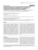
Figure 3 Kaplan-Meier survival curves illustrating the significance of NQO1 expression in ovarian carcinomas. (A) OS rates of patients
with high (solid, n = 92) and low (dashed, n = 68) NQO1 expression. A: Log-rank = 21.699, P = 0.000. (B-C) High NQO1 expression was strongly associated
with poor OS in early-stage (solid, n = 37) and late-stage (solid, n = 55). B: Log-rank = 6.527, P = 0.011; C: Log-rank = 4.806, P = 0.028. (D-F) High
NQO1 expression was strongly associated with poor OS in G1 (solid, n = 10), G2 (solid, n = 29) and G3 (solid, n = 53). D: Log-rank = 4.359, P = 0.037;
E: Log-rank = 7.020, P = 0.008; F: Log-rank = 5.978, P = 0.015).
Cui et al. BMC Cancer (2015) 15:244
Page 6 of 8
Table 3 Univariate and multivariate analysis of clinicopathological factors for the overall survival rate of 160 patients
with ovarian carcinoma
Characteristics
Univariate analysis HR (95%CI)
P value
Multivariate analysis HR (95%CI)
P value
Age
1.339(0.964-1.860)
0.082
1.391(0.992-1.951)
0.056
Menopausal status
1.115(0.815-1.525)
0.496
1.177(0.842-1.646)
0.340
Histological grade
1.268(1.038-1.548)
0.020*
1.099(0.881-1.371)
0.404
FIGO stage
1.944(1.414-2.672)
0.000**
1.736(1.228-2.453)
0.002**
NQO1
2.167(1.555-3.021)
0.000**
1.796(1.250-2.580)
0.002**
HR: hazard ratio; CI: confidence interval.
*P < 0.05, **P < 0.01.
To date, many studies have shown that polymorphisms
in the NQO1 gene affect the translation of the NQO1
protein. The NQO1 C609T polymorphism has been associated with an increased risk of various malignancies,
including lung [29], esophageal [30], gastric [31] and
uterine cervix [16] cancers. Goode et al. reported that
an NQO1 single-nucleotide polymorphism (SNP) is associated with an increased risk of ovarian cancer [32].
Moreover, high NQO1 expression has been observed in
many cancers of the liver, thyroid, breast, colon and
pancreas. Siegel et al. also found that NQO1 was overexpressed in ovarian carcinoma compared with normal
tissue [33]. However, to date, the role of NQO1 as a
biomarker in ovarian carcinoma progression has not
been elucidated.
Here, we performed IHC staining of NQO1 protein
using 160 serous ovarian carcinomas, 62 borderline serous tumors and 53 benign serous tumors. We observed
that expression of NQO1 protein (positive and strongly
positive) was significantly higher in serous carcinomas
compared with either borderline or benign serous tumors (P < 0.01). More importantly, we observed a significant difference in the rates of positive and strongly
positive NQO1 expression between borderline serous
tumors and benign serous tumors (P < 0.05), indicating
that NQO1 may play an important role in the progression
of ovarian carcinoma. Compatible with these findings, we
also observed that the rate of strongly positive NQO1 protein expression was significantly higher in patients with
late-stage serous ovarian carcinoma, compared with earlystage cases. Similarly, the rate of strongly positive NQO1
protein expression was higher in patients with G2 or G3,
compared with G1 ovarian carcinomas. High-grade serous
ovarian carcinoma is the most lethal form of gynecological
malignant carcinoma, and the majority of patients present
with late clinical stages (FIGO stages III and IV) of disease
at the time of diagnosis. Our data also demonstrate that
positive NQO1 expression is significantly correlated with
high-grade and late-stage ovarian carcinoma. qRT-PCR
analysis also confirmed increased levels of NQO1 mRNA
in serous ovarian carcinoma samples compared with
benign ovarian tumors in fresh tissues. These results indicate that NQO1 may be a useful biomarker for poor prognosis in patients with ovarian carcinoma.
Previously, we have shown that high expression of NQO1
protein was strongly associated with advanced stage, lymph
node metastasis, Her2 overexpression and shortened
survival of patients with breast cancer [34]. Moreover,
we demonstrated that high expression of NQO1 in cervical
squamous cell carcinoma patients was associated with
lower disease-free survival (DFS) and 5-year OS rates compared with patients with low-level NQO1 expression [16].
Buranrat et al. also reported a significant association between high NQO1 expression and short overall survival in
cholangiocarcinoma patients, raising the exciting possibility
of using NQO1 as a tumor marker [35]. With respect to
survival, we found that ovarian carcinoma patients exhibiting high NQO1 expression had lower OS rates compared
with patients with low NQO1 expression (P < 0.01).
Univariate survival analysis revealed that tumor histological grade, FIGO stage and NQO1 expression status
were all significantly related to OS of patients with serous ovarian carcinoma (P < 0.05). Further multivariate
survival analysis revealed that NQO1 expression was an
independent prognostic factor, as was FIGO stage. Our
clinical and experimental data indicate that NQO1 is a
prognostic factor and a potential therapeutic target in
patients with serous ovarian carcinoma.
Recently, NQO1 has been targeted in tumor cells,
exemplifying an ‘enzyme directed’ approach to anticancer drug development [36]. Kung et al. demonstrated
that β-Lapachone-induced cytotoxicity of three different
lung cancer cell lines was positively correlated with NQO1
expression and enzyme activity [37]. Hadley et al. suggested that stratification of patients on the basis of NQO1
protein levels could identify a subset of esophageal squamous cell carcinomas patients that may potentially benefit
from administration of low doses of 17-AAG, possibly in
combination with other chemotherapeutics [38]. Huang
et al. reported that the potency and NQO1-dependent
therapeutic window of deoxynyboquinone and its apparent
reduced metabolism by one-electron oxidoreductases
Cui et al. BMC Cancer (2015) 15:244
make this drug (or derivatives) very promising [39]. Further
studies are therefore necessary to verify whether NQO1 inhibitors may be of clinical benefit to patients with ovarian
carcinoma.
Conclusions
NQO1 is frequently upregulated in ovarian carcinoma,
and its high expression predicts poor prognosis of patients
with ovarian carcinoma. NQO1 may serve as a new prognostic factor and potential therapeutic target for patients
with serous ovarian carcinoma.
Competing interests
The authors declare that they have no competing interests.
Authors’ contributions
CX, LL, and YG participated in the study conception, design, case selection
and experiments. YG, MK, NY and JG carried out data collection. JG, LZ and
LC performed the data analysis and wrote the manuscript. All authors read
and approved the final manuscript.
Acknowledgments
This study was supported by grants from National Natural Science Funds of
China (31360269), and The Projects of Research & Innovation of Jilin Youth
Leader and Team in China (20130521017JH).
Received: 30 July 2014 Accepted: 26 March 2015
References
1. Siegel R, Naishadham D, Jemal A. Cancer statistics. CA Cancer J Clin.
2012;62(1):10–29.
2. Smolle E, Taucher V, Pichler M, Petru E, Lax S, Haybaeck J. Targeting signaling
pathways in epithelial ovarian cancer. Int J Mol Sci. 2013;14:9536–55.
3. Marsden DE, Friedlander M, Hacker NF. Current management of epithelial
ovarian carcinoma. Semin Surg Oncol. 2000;19:11–9.
4. Ross D, Kepa JK, Winski SL, Beall HD, Anwar A, Siegel D. NAD(P)H:quinone
oxidoreductase 1 (NQO1): chemoprotection, bioactivation, gene regulation
and genetic polymorphisms. Chem Biol Interact. 2000;129:77–97.
5. Siegel D, Yan C, Ross D. NAD (P) H:quinone oxidoreductase 1 (NQO1) in
the sensitivity and resistance to antitumor quinones. Biochem Pharmacol.
2012;83(8):1033–40.
6. Nioi P, Hayes JD. Contribution of NAD(P)H:quinone oxidoreductase 1 to
protection against carcinogenesis, and regulation of its gene by the Nrf2
basic-region leucine zipper and the arylhydrocarbon receptor basic helix-loophelix transcription factors. Mutat Res. 2004;555:149–71.
7. Prawan A, Buranrat B, Kukongviriyapan U, Sripa B, Kukongviriyapan V.
Inflammatory cytokines suppress NAD(P)H:quinone oxidoreductase-1 and
induce oxidative stress in cholangiocarcinoma cells. J Cancer Res Clin Oncol.
2009;135:515–22.
8. Kolesar JM, Pritchard SC, Kerr KM, Kim K, Nicolson MC, McLeod H. Evaluation
of NQO1 gene expression and variant allele in human NSCLC tumors and
matched normal lung tissue. Int J Oncol. 2002;21:1119–24.
9. Cresteil T, Jaiswal AK. High levels of expression of the NAD(P)H: quinone
oxidoreductase (NQO1) gene in tumor cells compared to normal cells of
the same origan. Biochem Pharmacol. 1991;42:1021–7.
10. Ross D, Siegel D, Beall H, Prakash AS, Mulcahy RT. DTdiaphorase in
activation and detoxification of quinones. Bioreductive activation of
mitomycin C. Cancer Metastasis Rev. 1993;12:83–101.
11. Tedeschi G, Chen S, Massey V. DT-diaphorase. Redox potential, steadystate,
and rapid reaction studies. J Biol Chem. 1995;270:1198–204.
12. Smitskamp-Wilms E, Hendriks HR, Peters GJ. Development, pharmacology,
role of DT-diaphorase and prospects of the indoloquinone EO9. Gen Pharmacol.
1996;27:421–9.
13. Ross D, Beall H, Traver RD, Siegel D, Phillips RM. Bioactivation of quinones
by DT-diaphorase, molecular, biochemical, and chemical studies. Oncol Res.
1994;6:493–500.
Page 7 of 8
14. Cadenas E. Antioxidant and prooxidant functions of DT-diaphorase in quinone
metabolism. Biochem Pharmacol. 1995;49:127–40.
15. Ross D, Siegel D. NAD(P)H:quinone oxidoreductase 1 (NQO1, DTdiaphorase),
functions and pharmacogenetics. Methods Enzymol. 2004;382:115–44.
16. Ma Y, Kong J, Yan G, Ren S, Jin D, Jin T, et al. NQO1 overexpression is
associated with poor prognosis in squamous cell carcinoma of the uterine
cervix. BMC Cancer. 2014;14:414.
17. Siegel D, Franklin WA, Ross D. Immunohistochemical detection of NAD(P)H:
quinone oxidoreductase in human lung and lung tumors. Clin Cancer Res.
1998;4(9):2065–70.
18. Awadallah NS, Dehn D, Shah RJ, Russell Nash S, Chen YK, Ross D, et al.
NQO1 expression in pancreatic cancer and its potential use as a biomarker.
Appl Immunohistochem Mol Morphol. 2008;16(1):24–31.
19. Garate M, Wani AA, Li G. The NAD(P)H:Quinone Oxidoreductase 1 induces
cell cycle progression and proliferation of melanoma cells. Free Radic Biol
Med. 2010;48(12):1601–9.
20. Lin L, Piao J, Gao W, Piao Y, Jin G, Ma Y, et al. DEK over expression as an
independent biomarker for poor prognosis in colorectal cancer. BMC
Cancer. 2013;13:366.
21. Ernster L, Navazio F. Soluble diaphorase in animal tissues. Acta Chem Scand.
1958;12:595–602.
22. Winski SL, Koutalos Y, Bentley DL, Ross D. Subcellular localization of
NAD(P)H:quinone oxidoreductase 1 in human cancer cells. Cancer Res.
2002;62:1420–4.
23. Lee H, Oh ET, Choi BH, Park MT, Lee JK, Lee JS, et al. NQO1-induced activation
of AMPK contributes to cancer cell death by oxygen-glucose deprivation. Sci
Rep. 2015;5:7769.
24. Girolami F, Abbadessa G, Racca S, Spaccamiglio A, Piccione F, Dacasto M,
et al. Time-dependent acetylsalicylic acid effects on liver CYP1A and antioxidant
enzymes in a rat model of 7,12-dimethylbenzanthracene(DMBA)-induced
mammary carcinogenesis. Toxicol Lett. 2008;181:87–92.
25. Iskander K, Gaikwad A, Paquet M, Long II DJ, Brayton C, Barrios R, et al.
Lower induction of p53 and decreased apoptosis in NQO1-null mice lead to
increased sensitivity to chemical-induced skin carcinogenesis. Cancer Res.
2005;65:2054–8.
26. Long DJ, Waikel RL, Wang XJ, Roop DR, Jaiswal AK. NAD(P)H:quinone
oxidoreductase 1 deficiency and increased susceptibility to 7,12dimethylbenz[a]-anthracene-induced carcinogenesis in mouse skin.
J Natl Cancer Inst. 2001;93:1166–70.
27. Choi TY, Sohn KC, Kim JH, Kim SM, Kim CH, Hwang JS, et al. Impact of NAD
(P)H:quinone oxidoreductase-1 on pigmentation. J Invest Dermatol.
2010;130(3):784–92.
28. Reigan P, Colucci MA, Siegel D, Chilloux A, Moody CJ, Ross D. Development of
indolequinone mechanism-based inhibitors of NAD(P)H:quinone oxidoreductase
1 (NQO1): NQO1 inhibition and growth inhibitory activity in human pancreatic
MIA PaCa-2 cancer cells. Biochemistry. 2007;46:5941–50.
29. Nagata M, Kimura T, Suzumura T, Kira Y, Nakai T, Umekawa K, et al. C609T
polymorphism of NADPH quinone oxidoreductase 1 correlates clinical
hematological toxicities in lung cancer patients treated with amrubicin.
Clin Med Insights Oncol. 2013;7:31–9.
30. Malik MA, Zargar SA, Mittal B. Role of NQO1 609C > T and NQO2–3423G > A
gene polymorphisms in esophageal cancer risk in Kashmir valley and meta
analysis. Mol Biol Rep. 2012;39(9):9095–104.
31. Lin L, Qin Y, Jin T, Liu S, Zhang S, Shen X, et al. Significance of NQO1
overexpression for prognostic evaluation of gastric adenocarcinoma. Exp
Mol Pathol. 2014;96(2):200–5.
32. Goode EL, White KL, Vierkant RA, Phelan CM, Cunningham JM, Schildkraut
JM, et al. Xenobiotic-metabolizing gene polymorphisms and Ovarian cancer
risk. Mol Carcinog. 2011;50(5):397–402.
33. Siegel D, Ross D. Immunodetection of NAD(P)H:quinone oxidoreductase 1
(NQO1) in human tissues. Free Radic Biol Med. 2000;29:246–53.
34. Yang Y, Zhang Y, Wu Q, Cui X, Lin Z, Liu S, et al. Clinical implications
of high NQO1 expression in breast cancers. J Exp Clin Cancer Res.
2014;33:14.
35. Buranrat B, Chau-In S, Prawan A, Puapairoj A, Zeekpudsa P, Kukongviriyapan
V. NQO1 Expression correlates with cholangiocarcinoma prognosis. Asian
Pac J Cancer Prev. 2012;13(Suppl):131–6.
36. Workman P. Enzyme-directed bioreductive drug development revisited: a
commentary on recent progress and future prospects with emphasis on
quinone anticancer agents and quinone metabolizing enzymes, particularly
DT-diaphorase. Oncol Res. 1994;6:461–75.
Cui et al. BMC Cancer (2015) 15:244
Page 8 of 8
37. Kung H, Weng T, Liu Y, Lu K, Chau Y. Sulindac compounds facilitate the
cytotoxicity of β-lapachone by up-regulation of NAD(P)H quinone
oxidoreductase in human lung cancer cells. PLoS One. 2014;9(2):e88122.
38. Hadley K, Hendricks D. Use of NQO1 status as a selective biomarker for
oesophageal squamous cell carcinomas with greater sensitivity to 17-AAG.
BMC Cancer. 2014;14:334.
39. Huang X, Dong Y, Bey E. An NQO1 substrate with potent antitumor activity
that selectively kills by PARP1-induced programmed necrosis. Cancer Res.
2012;72:3038–47.
Submit your next manuscript to BioMed Central
and take full advantage of:
• Convenient online submission
• Thorough peer review
• No space constraints or color figure charges
• Immediate publication on acceptance
• Inclusion in PubMed, CAS, Scopus and Google Scholar
• Research which is freely available for redistribution
Submit your manuscript at
www.biomedcentral.com/submit