Poor prognosis of single hormone receptorpositive breast cancer: Similar outcome as triple-negative breast cancer
Bạn đang xem bản rút gọn của tài liệu. Xem và tải ngay bản đầy đủ của tài liệu tại đây (627.2 KB, 9 trang )
Bae et al. BMC Cancer (2015) 15:138
DOI 10.1186/s12885-015-1121-4
RESEARCH ARTICLE
Open Access
Poor prognosis of single hormone receptorpositive breast cancer: similar outcome as
triple-negative breast cancer
Soo Youn Bae, Sangmin Kim, Jun Ho Lee, Hyun-chul Lee, Se Kyung Lee, Won Ho Kil, Seok Won Kim,
Jeong Eon Lee and Seok Jin Nam*
Abstract
Background: Response to endocrine therapy in breast cancer correlates with estrogen receptor (ER) and
progesterone receptor (PR) status. Generally, hormone receptor-positive (HR+) breast cancers have favorable
prognosis. In order to understand the exact clinical characteristics and prognosis of single HR-positive breast
cancer (ER + PR- tumors and ER-PR+ tumors), we compared these tumors to double HR+ tumors as well as
HR- negative tumors (ER-PR-).
Methods: We examined the clinical and biological features of 6,980 women with invasive ductal carcinoma,
and these patients were stratified according to ER and PR expression as double HR+ (ER + PR+), single HR+
(ER + PR- and ER-PR+) and double HR-negative (HR-, ER-PR-) tumors.
Results: In this study, 571 (8.2%) cases were single HR+ tumors, of which 90 (1.3%) were ER-PR+ tumors and 481
(6.9%) were ER + PR- tumors. Our multivariate analysis showed that in patients without HER2 overexpression
ER + PR- tumors were associated with an increased risk of recurrence and death compared with ER + PR+
tumors, with a hazard ratio of 2.12 for disease-free survival (DFS) and 4.79 for overall survival (OS). In patients
without HER2 overexpression ER-PR+ tumors had increased risk of recurrence and death compared with ER +
PR+ tumor, with a hazard ratio of 4.19 for DFS and 7.22 for OS. In contrast, in patients with HER2 overexpression,
the difference in survival between single HR+ tumors and double HR+ HR- tumors was not statistically
significant. In patients without HER2 overexpression the DFS and OS of ER + PR- and ER-PR+ tumors
were not significantly different from those of ER-PR- tumors.
Conclusion: We have identified clinically and biologically distinct features of single HR+ tumors (ER–PR+ and
ER + PR–) through comparison with both ER + PR+ and ER-PR- tumors. These differences were only significant in
HER2- tumors, not in HER2+ tumors. Single HR+ tumors without HER2 overexpression (ER + PR-HER2- or ER-PR +
HER2-) were associated with poorer survival than ER + PR + HER2- tumors, and had comparable poor survival to
ER-PR-HER2- tumors (triple-negative breast cancer).
Keywords: Breast cancer, Estrogen receptor, Progesterone receptor, Human epidermal growth factor receptor 2,
Prognosis
* Correspondence:
Department of Surgery, Samsung Medical Center, Sungkyunkwan University
School of Medicine, 50 Irwon-dong, Kangnam-gu, 135-710 Seoul, South
Korea
© 2015 Bae et al.; licensee BioMed Central. This is an Open Access article distributed under the terms of the Creative
Commons Attribution License ( which permits unrestricted use, distribution, and
reproduction in any medium, provided the original work is properly credited. The Creative Commons Public Domain
Dedication waiver ( applies to the data made available in this article,
unless otherwise stated.
Bae et al. BMC Cancer (2015) 15:138
Background
In breast cancer, steroid hormone receptors (HRs; i.e.,
estrogen receptor [ER] or progesterone receptor [PR])
have been shown to be important prognostic factors
and predictive markers for response to endocrine therapy in the treatment of breast cancer. About 70% of
breast cancers are hormone receptor-positive tumors
(HR+). HR+ breast cancers generally have a favorable
prognosis, but HR-negative (HR-) breast cancers have
a poor prognosis. PR is an estrogen-regulated gene;
ER-positive (ER+) tumors are usually also PR positive
(PR+), whereas ER-negative (ER-) tumors are usually
PR negative (PR-). Therefore, single HR+ (i.e., ER+/PRor ER-/PR+) tumors represent a minority of breast
cancers.
Clinical data have shown in both the metastatic and
adjuvant treatment settings that tamoxifen is less efficacious in ER + PR− tumors than in ER + PR+ tumors
[1-3], and single HR+ breast cancers, especially ER +
PR- breast cancers, have aggressive features and poorer
prognosis in comparison to double HR+ (ER + PR+)
breast cancer [4,5]. However, to our knowledge, comparative studies of HR- (ER-PR-) breast cancers are very
limited [6,7].
Previous studies have shown that ER + PR- tumors exhibit high expression of epidermal growth factor receptors [1,4,7-11], but in most studies, the prognosis of ER
+ PR- tumors was determined without considering human epidermal growth factor receptor 2 (HER2) expression. Moreover, these studies had a common limitation,
in that prognosis was evaluated without considering
trastuzumab treatment.
Previous studies have suggested that ER-PR+ tumors
have poorer prognosis than ER + PR+ tumors [10,12-16].
However, due to the rarity of ER-PR+ breast cancer
(a reported incidence of 1.5-3.4% [10,12-15]), the
characteristics and prognosis of this tumor are not
well known.
Therefore, in order to understand the exact clinical
characteristics and prognosis of single HR-positive breast
cancer (ER + PR- tumors and ER-PR+ tumors), we compared these tumors to double HR+ tumors as well as
HR- tumors (ER-PR-), and stratified these results according to HER2 overexpression.
Methods
Patients were selected from the clinical database of
the Breast Cancer Center at Samsung Medical Center,
Korea, between January 2003 and July 2013. A total
of 7,010 women with invasive ductal carcinoma were
identified. Of them, 6,980 patients were selected for
this study excluding patients who were diagnosed with bilateral tumors or with distant metastases at preoperative
work-up or underwent neoadjuvant chemotherapy.
Page 2 of 9
We reviewed the clinicopathologic characteristics of
patients, including biologic factors, such as ER, PR,
HER2, epidermal growth factor receptors (EGFR), and
Ki-67. The pathologic tumor stage was assessed according to the American Joint Committee on Cancer (AJCC)
6th Staging System. For ER and PR staging, nuclear (not
cytoplasmic) staining was scored using the Allred score
(AS) interpretation system, a method that provides
semi-quantitative measurement of the proportion of
positive cells (scored on a 0 to 5 scale) and staining intensity (scored on a 0 to 3 scale), with a maximum score
of 8; an AS > 2 considered positive.
HER2 positivity was defined as an intensity of 3+ by
IHC, a score of 2+ was interpreted as equivocal. A negative test was defined as staining with a score of 0/1+.
For equivocal stating, silver in situ hybridization (SISH)
or fluorescence in situ hybridization (FISH) were performed; the results were positive for HER2 amplification
when the ratio of HER2 to CEP17 was > 2.2.
For EGFR and/or Ki-67, results were considered positive based on identification of the following criteria in at
least one core. Immunostaining for EGFR was interpreted as positive when at least 10% of the tumor cells
showed moderate to strong membrane staining. Ki-67
was considered positive when ≥ 14.0% of cells showed
staining [17].
Differences in the frequencies of clinicopathological
factors and subtypes were statistically analyzed using the
chi-square test and Fisher’s exact test. Disease-free survival (DFS) was defined as the time from surgery to the
date of documentation of relapse, including locoregional
recurrence and/or distant metastasis. Overall survival
(OS) was defined as the number of months from surgery
to the date of death. Survival curves were constructed
using the Kaplan-Meier method. Hazard ratios were
estimated using a Cox regression for DFS/OS in a multivariate analysis. Statistical significance was defined as
P < 0.05. All statistical analyses were performed using
SPSS Statistics 21.0 (IBM).
Study data were collected using a protocol approved
by the Institutional Review Board of Samsung Medical
Center, Korea (IRB number 2014-09-111). Specific patient consent was not required because we used retrospective data from medical records of patients who had
previously signed information release documents.
Results and discussion
Clinicopathologic characteristics of single hormone
receptor- positive breast cancer
The median follow-up duration for the 6,980 patients included in this analysis was 45 months (range, 1-133
months). In this study, 4,651 (66.6%) cases were double
HR+ (ER + PR+) tumors, 1,758 (25.2%) were double
HR- (ER-PR-) tumors, and 571 (8.2%) cases were single
Bae et al. BMC Cancer (2015) 15:138
hormone-receptor positive tumors, of which 90 (1.3%)
cases were ER-PR+ tumors and 481 (6.9%) were ER +
PR- tumors. The clinicopathological characteristics of
the four subtypes are summarized in Table 1. Overall,
ER+/PR- tumors were found more frequently in
postmenopausal women (61.5%) than other subtypes
(P < 0.001). Compared with ER + PR+ tumor, ER +
PR- tumors were not significantly different in staging
(P = 0.083), but ER + PR- tumors exhibited higher nuclear
grade (NG,P <0.001), higher Ki-67 level (Ki-67 ≥ 14.0,
76.0% vs. 53.9%, P < 0.001), and higher EGFR and HER2
expression (p < 0.001). However, compared with ER-PRtumors, ER + PR- tumors showed lower stage (stage I,
ER + PR- 43.7% vs. ER-PR- 35.8%, P = 0.027), lower NG
(P < 0.001), lower Ki-67 level (P < 0.001), lower p53 expression (P <0.001) and lower EGFR expression (P < 0.001),
but there was no difference in HER2 overexpression
(P = 0.089).
ER-PR+ tumors had higher NG (P <0.001), higher Ki-67
level (P < 0.001), and higher expression of p53 and EGFR
(P < 0.001) than ER + PR+ tumors. However, compared
with ER-PR- tumors, there was no difference in stage
(P = 0.979) or NG (P = 0.0117). Also, there was no difference in expression of Ki-67 (P = 0.511), p53 (P = 0.531),
EGFR (P = 0.055) or HER2 (P = 0.419).
Both ER-PR+ and ER + PR- tumors were shown to
have higher HER2 overexpression (34.5%) than ER +
PR+ tumors (11.4%, P < 0.001), but had similar HER2
overexpression to ER-PR- tumors (38.8%, P = 0.192).
The characteristics of single hormone receptorpositive (ER + PR- and ER-PR+) tumors were more
distinct in HER2-negative (HER2-) tumors than in
HER2 overexpressing (HER2+) tumors. (Additional file 1:
Table S1 and Table S2).
Survival analysis of single hormone receptor- positive
breast cancer
Approximately 97% of patients with ER + PR- tumors
and 88% of patients with ER-PR+ tumors received endocrine therapy. More patients with ER + PR- (73.7%) and
ER-PR+ (89.7%) tumors received chemotherapy than the
group with ER + PR+ tumors (68.7%), but less than the
group with ER-PR- tumors (91.9%, Table 1). Approximately 72% of patients with ER + PR- tumors received
both endocrine therapy and chemotherapy, and 24.9% of
patients received only endocrine therapy. In ER-PR+
tumors, 80% of patients received both chemotherapy
and endocrine therapy, 8.2% of patients received only
endocrine therapy and 9.4% of patients received only
chemotherapy.
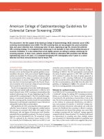
With univariate analysis by Kaplan-Meier method, the
survival graph of ER + PR- tumors was located between
that of ER + PR+ tumors and ER-PR- tumors. The 5-year
and 10-year DFS of ER + PR- tumors was 91.4% and
Page 3 of 9
79.6%, respectively, and the 5-year and 10-year OS was
95.9% and 93.9%, respectively. Patients with ER-PR+ tumors had worse DFS (5-year 81.0%; 10 year 73.1%) and
OS (5-year 95.3%; 10-year 88.7%, Figure 1) than those with
ER + PR-.
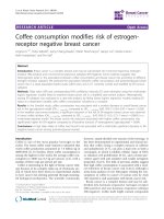
Among 1,376 patients with HER2 overexpression,
there was no significant difference in DFS between four
subgroups (P = 0.529), and patients with ER-PR-HER+
tumors had the worst OS (P = 0.010, Figure 2). However,
the 790 patients who received trastuzumab therapy had
similar OS (P = 0.113), as did the 586 patients who did
not receive trastuzumab therapy (P = 0.147).
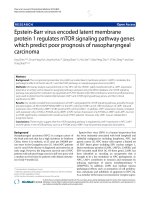
In 5,433 patients without HER2 overexpression, ER +
PR- tumors were associated with poorer OS than ER +
PR+ tumors (P < 0.001), but similar OS to ER-PR- tumors (P =0.338). ER-PR+ tumors also had poorer OS
than ER + PR+ tumors (P < 0.001), but there was no
significant difference from the OS of ER-PR- tumors
(P = 0.165, Figure 3).
With multivariate analysis, in patients with HER2
overexpression, the single HR+ (ER + PR- and ER-PR+)
tumors seem to increase risk of recurrence, but this difference was not significant (Table 2). In patients without
HER2 overexpression, ER + PR- tumors had increased
risk of recurrence and death compared with ER + PR+
tumors, with a hazard ratio of 2.12 (95% CI 1.20 -3.75)
for DFS and 4.79 (95% CI 1.84-12.18) for OS. ER-PR+
tumors were at increased risk of recurrence and death
compared with ER + PR+ tumors, with a hazard ratio of
4.19 (95% CI; 1.86-10.02) for DFS and 7.22 (95% CI
1.62-32.06) for OS (Table 3). ER + PR- tumors and ERPR+ tumors were not significantly different in terms of
DFS and OS compared ER-PR- tumors.
Discussion
We have evaluated in detail the biological characteristics
and prognosis of single HR+ tumors through comparison with ER + PR+ tumors as well as ER-PR- tumors. In
our series, 8.2% of cases were ER + PR- and 1.4% were
ER-PR+. These numbers are somewhat smaller than
those from previously published series where 10-15% of
cases were ER + PR- and 2-4% were ER-PR+. Most previous studies included patients with breast cancer
regardless of histologic type, but we analyzed patients
with invasive ductal carcinoma [10,12,13,18]. However,
the clinical and biological features of ER + PR- tumors
were consistent with those found in previous studies,
and there was a high incidence in postmenopausal
women. In terms of NG and IHC of Ki-67 level, p53 and
EGFR, ER + PR- tumors showed moderate characteristics between the levels of ER + PR+ and ER-PR- tumors,
while ER-PR+ tumors were more similar to ER-PR- tumors
than ER + PR+ tumors. In addition, on Kaplan-Meier
analysis, the survival graph of the ER + PR- tumors was
Bae et al. BMC Cancer (2015) 15:138
Page 4 of 9
Table 1 Clinicopathologic characteristics of patients with ER + PR+, ER + PR-, ER-PR+ and ER-PR- tumors
Age, median (range)
ER + PR+ (N = 4651)
ER-PR+ (N = 90)
ER-PR- (N = 1758)
ER + PR- (N = 481)
47 (20-90)
48 (22-72)
49 (21-85)
54 (27-84)
Menopause
Postmenopause
1489
(32.4%)
34
38.6%
836
(48.4%)
319
(67.3%)
Premenopause
3110
(67.6%)
54
61.4%
893
(51.6%)
155
(32.7%)
Uknown
52
2
29
7
Operation
MRM
1363
(29.3%)
40
(44.4%)
585
(33.3%)
171
(35.6%)
BCS
3288
(70.7%)
50
(55.6%)
1173
(66.7%)
310
(64.4%)
T1
2985
(64.2%)
49
(54.4%)
860
(48.9%)
286
(59.5%)
T2
1472
(31.6%)
38
(42.2%)
835
(47.5%)
184
(38.3%)
T3
184
(4.0%)
3
(3.3%)
62
(3.5%)
10
(2.1%)
T4
10
(0.2%)
0
(0.0%)
1
(0.1%)
1
(0.2%)
N0
2717
(58.4%)
48
(53.3%)
1105
(62.9%)
303
(63.0%)
N1
1368
(29.4%)
29
(32.2%)
445
(25.3%)
129
(26.8%)
N2
363
(7.8%)
8
(8.9%)
133
(7.6%)
32
(6.7%)
N3
203
(4.4%)
5
(5.6%)
75
(4.3%)
17
(3.5%)
I
2156
(46.4%)
32
(35.6%)
630
(35.8%)
210
(43.7%)
IIA
1266
(27.2%)
30
(33.3%)
635
(36.1%)
162
(33.7%)
IIB
605
(13.0%)
14
(15.6%)
266
(15.1%)
54
(11.2%)
IIIA
413
(8.9%)
9
(10.0%)
151
(8.6%)
37
(7.7%)
IIIB
8
(0.2%)
0
1
(0.1%)
1
(0.2%)
IIIC
203
(4.4%)
5
(5.6%)
75
(4.3%)
17
(3.5%)
I
974
(21.0%)
1
(1.1%)
12
(0.7%)
67
(14.0%)
II
2656
(57.3%)
26
(29.2%)
358
(20.4%)
206
(43.1%)
III
1006
(21.7%)
62
(69.7%)
1383
(78.9%)
205
(42.9%)
unknown
15
pT
pN
Stage
Nuclear Grade
1
5
3
HER2
Positive
518
(11.4%)
30
(34.5%)
671
(38.8%)
159
(34.5%)
Negative
4018
(88.6%)
57
(65.5%)
1058
(61.2%)
302
(65.5%)
Unknown
115
3
29
20
Ki-67
≥ 14.0%
2202
(53.9%)
56
(91.8%)
1334
(93.9%)
292
(76.0%)
< 14.0%
1887
(46.1%)
5
(8.2%)
87
(6.1%)
92
(24.0%)
Unknown
562
29
337
97
p53
Positive
1008
(21.8%)
49
(55.1%)
1021
(58.4%)
162
(34.2%)
Negative
3622
(78.2%)
40
(44.9%)
727
(41.6%)
311
(65.8%)
Unknown
21
1
10
8
Bae et al. BMC Cancer (2015) 15:138
Page 5 of 9
Table 1 Clinicopathologic characteristics of patients with ER + PR+, ER + PR-, ER-PR+ and ER-PR- tumors (Continued)
Chemotherapy
Yes
3127
(68.7%)
78
(89.7%)
1575
(91.9%)
345
(73.7%)
No
1424
(31.3%)
9
(10.3%)
138
(8.1%)
123
(26.3%)
Unknown
100
3
45
13
Radiotherapy
Yes
3543
(78.0%)
55
(64.0%)
1258
(73.8%)
334
(70.9%)
No
1001
(22.0%)
31
(36.0%)
446
(26.2%)
137
(29.1%)
Unknown
107
4
54
10
Endocrine Therapy
Yes
4490
(99.2%)
75
(88.2%)
2
(0.1%)
454
(97.0%)
No
34
(0.8%)
10
(11.8%)
1704
(99.9%)
14
(3.0%)
Unknown
127
5
located in between those of ER + PR+ tumors and ERPR- tumors, and ER-PR+ tumors were shown to have
worse survival than ER-PR- tumors.
In previous studies, loss of PR has been suggested to
be a marker of aberrant growth factor signaling and has
been associated with one mechanism for endocrine resistance[11], and several studies have shown that ER +
PR- tumors exhibit high expression of epidermal growth
factor receptors [1,4,7-11]. In our cases, HER2 overexpression was 34.5% in single HR+ tumors, and as high
as 38.8% in ER-PR- tumors, but the rate of HER2 overexpression in ER + PR+ tumors was 11.4%. Therefore,
we stratified our cases according to HER2 overexpression and we found that differences in clinicopathologic
characteristics were not significantly different between
the four subgroups (ER + PR+, ER-PR+, ER-PR- and
52
13
ER + PR-) in patients with HER2 overexpression. In
addition, there was no difference in survival between
these four subgroups. However, in patients without
HER2 overexpression, significant differences in biological
characteristics were shown more distinctly; ER + PR-, ERPR+ tumors and ER-PR-HER2- tumors (triple-negative
breast cancer, TNBC) were both associated with poor
survival.
As demonstrated in previous studies, PR negativity
may be association with cross talk with epidermal
growth factor receptor- i.e., HER2 or EGFR. In our
study, ER + PR- tumors showed high HER2 overexpression. Nevertheless, PR negativity was not a significant
prognostic factor in tumors with HER2 overexpression.
This suggests that HER2 expression may be a more significant prognostic factor than PR loss in tumors with
Figure 1 (a) Disease-free survival (DFS) and (b) overall survival (OS) of all patients.
Bae et al. BMC Cancer (2015) 15:138
Page 6 of 9
Figure 2 (a) Disease-free survival (DFS) and (b) overall survival (OS) of patients with HER2-positive tumors.
HER2 overexpression (HER2+) or may be associated with
the results of trastuzumab treatment.
However, in tumors without HER2 overexpression,
single HR positivity is a significant prognostic factor.
The survival graph of ER + PR-tumors is between that
of ER + PR+ tumors and ER-PR- tumors initially, but
falls to as poor as TNBC at about the 10-year follow-up.
Therefore, ER + PR-HER2- and TNBC tumors show no
difference in terms of long-term survival. ER-PR +
HER2- tumors show similar biological features to
TNBC, including high Ki-67 level and high expression
of EGFR (about 90%) and p53 (50%). Previous studies
have shown incidence rate and clinicopathologic features, and ER-PR+ tumors have increased an incidence
in premenopausal women and of an aggressive phenotype with higher tumor grade and HER2 overexpression [10,19,20]. Our results are consistent with those
of previous studies. In our series, although there were
only a few ER-PR + HER2- tumors, approximately 80%
(43/53) of patients with ER-PR + HER2- tumors received
chemotherapy and endocrine therapy. Nevertheless, the
aggressive behavior of these tumors suggests that ER-PR+
tumors are very rare and represent a distinct biological
subtype.
Figure 3 (a) Disease-free survival (DFS) and (b) overall survival (OS) of patients with HER2-negative tumors.
Bae et al. BMC Cancer (2015) 15:138
Page 7 of 9
Table 2 Multivariate analysis of disease-free survival (DFS) and overall survival (OS) in 1.376 women with HER2-positive
breast cancer
DFS
OS
ER + PR+ vs. ER-PR+
B coefficients
Standard
error
Wald
P
Hazard
ratio
95.0% confidence interval
Lower
Upper
.391
.619
.399
0.528
1.478
.439
4.973
ER + PR+ vs. ER-PR-
.394
.261
2.284
0.131
1.483
.890
2.471
ER + PR+ vs. ER + PR-
.457
.388
1.389
0.239
1.580
.738
3.382
ER-PR- vs. ER-PR+
-.003
.619
.000
0.996
.997
.296
3.357
ER-PR- vs. ER + PR-
.064
.373
.029
0.864
1.066
.513
2.214
ER + PR+ vs. ER-PR+
−7.587
82.200
.009
0.926
.001
.000
4.71E + 66
ER + PR+ vs. ER-PR-
1.376
.583
5.576
0.018
3.958
1.263
12.398
ER + PR+ vs. ER + PR-
.241
1.125
.046
0.830
1.273
.140
11.549
ER-PR- vs. ER-PR+
−8.962
82.199
.012
0.913
.000
.000
1.18E + 66
ER-PR- vs. ER + PR-
−1.135
1.036
1.200
0.273
.322
.042
2.449
(Adjusted for age, stage, nuclear grade, Ki-67and trastuzumab treatment).
Our study is consistent with previous large studies
demonstrating that single HR+ tumors have high expression of EGFR/HER2 and more aggressive features than
ER + PR+ tumors. However, our results show that single
HR positivity was not a significant prognostic factor in
HER2+ breast cancer. Therefore, the aggressiveness of
single HR+ tumors is not simply due to hyperactive
growth factor signaling pathways. As in recent studies
[7-9], our cases have shown that single HR+ tumors are
associated with a high level of Ki-67 (≥14.0%), and, in
multivariate analysis, Ki-67 was shown to have borderline significance (P = 0.068). When we additionally analyzed according to Ki-67 level, in cases with a high level
of Ki-67 (≥14.0%), the differences among the four subtypes were still shown consistently. However, in patients
with a low Ki-67 level (<14.0%), the prognosis of ER +
PR- tumors was not different from that of ER + PR+ tumors. These results may suggest that PR is a significant
prognostic factor in HR + HER2- tumors with a high
level of Ki-67 expression, but not in HR + HER2+ tumors. These suggest that proliferation-related genes may
be significantly associated with PR negativity. However,
interestingly, ER-PR+ tumors have been shown to have
the worst prognosis of the subtypes, regardless of Ki-67
level, suggesting that ER-PR+ tumors represent a distinct
biological subtype.
This study had several limitations. It was a retrospective study, and adjuvant treatment was not determined
on a randomized basis. Furthermore, we did not stratify
patients according to treatment with tamoxifen or aromatase inhibitors. Although the use of aromatase inhibitors instead of selective estrogen receptor modulators
improved the outcome of ER + PR− patients in the
ATAC trial [21], the BIG 1-98 trial did not demonstrate
a significant benefit of letrozole over tamoxifen in ER +
PR− tumors [22]. In addition, at our center, most
Table 3 Multivariate analysis of disease-free survival (DFS) and overall survival (OS) in 5,433 women with HER2-negtive
breast cancer
DFS
OS
B coefficients
Standard
error
Wald
P
Hazard
ratio
95.0% confidence interval
ER + PR+ vs. ER-PR+
1.463
.429
11.608
0.001
4.319
1.861
10.020
ER + PR+ vs. ER-PR-
.913
.170
28.982
0.000
2.493
1.788
3.476
ER + PR+ vs. ER + PR-
.753
.291
6.702
0.010
2.123
1.201
3.755
ER-PR- vs. ER-PR+
.550
.422
1.695
0.193
1.733
.757
3.963
ER-PR- vs. ER + PR-
-.160
.296
.293
0.589
.852
.477
1.523
ER + PR+ vs. ER-PR+
1.977
.761
6.755
0.009
7.220
1.626
32.061
Lower
Upper
ER + PR+ vs. ER-PR-
1.774
.307
33.488
0.000
5.895
3.232
10.751
ER + PR+ vs. ER + PR-
1.564
.478
10.722
0.001
4.779
1.874
12.189
ER-PR- vs. ER-PR+
.203
.728
.078
0.781
1.225
.294
5.105
ER-PR- vs. ER + PR-
-.210
.462
.207
0.649
.811
.328
2.004
(Adjusted for age, stage, nuclear grade and Ki-67).
Bae et al. BMC Cancer (2015) 15:138
postmenopausal patients with HR+ tumors received
aromatase inhibitor, excluding patients with contraindications or adverse effects.
Recent studies have emphasized the influence of PR,
which provides highly significant stratification of ER+
breast cancer into luminal A and B types [7-9]. Prat
et al. proposed that the IHC-based definition of luminal
A tumors is HR+/HER2-/low Ki-67 (less than 14%), and
high PR (more than 20%) [8] and Braun et al. also
defined luminal B tumors by the presence of high-risk
criteria (loss of PR expression or increased proliferation)
[9]. Cancello et al. suggested that PR loss identifies luminal B breast cancer subgroups at higher risk of relapse
and death, both with HER-2+ and HER-2- breast cancer
[7]. The differences observed in HER2+ tumors in our
study may be the result of differences in chemotherapy
and trastuzumab treatment. In that study, about 30% of
patients with ER + PR + HER2+ and ER + PR − HER2+
tumors received endocrine therapy alone, 65–70% received chemotherapy plus endocrine therapy as adjuvant
treatments and about 1% in both the ER + PR + HER2+
and ER + PR − HER2+ subgroups received trastuzumab
as an adjuvant therapy[7]. However, in our study, 89% of
patients with HER2 overexpression received chemotherapy and 57.2% of patients received trastuzumab treatment.
Conclusions
This study has identified clinically and biologically distinct
features of single HR+ tumors (ER + PR- and ER-PR+)
through comparison with both ER + PR+ tumors and
ER-PR- tumors. These differences were significant in
HER2- tumors, but not in HER2+ tumors. ER + PRHER2- tumors and ER-PR + HER2- tumors have poorer
survival than ER + PR + HER2- tumors and a similarly
poor survival in comparison to ER-PR-HER2- tumors
(TNBC). Clinical trials in addition to more advanced
biological and molecular studies are necessary to identify
the cause of aggressiveness in single HR+ tumors.
Additional file
Additional file 1: Table S1. Clinicopathologic characteristics of patients
with HER2-negative tumors. Table S2 Clinicopathologic characteristics of
patients with HER2-positive tumors.
Abbreviations
HR: Hormone receptor; ER: Estrogen receptor; PR: Progesterone receptor;
DFS: Disease-free survival; OS: Overall survival; HER2: Human epidermal growth
factor receptor 2; EGFR: Epidermal growth factor receptors; NG: Nuclear grade.
Competing interests
The authors declare that they have no competing interests.
Authors’ contributions
SYB conceived, designed and data collection, data organization, interpreted
results, undertook statistical analyses and manuscript writing. JHL and HL
helped with data collection and data organization. SK, SKL, WHK, SWK, JEL
Page 8 of 9
and SJN have made substantial contributions to the acquisition of data and
to the revision of the manuscript for important intellectual content. SJN was
involved with the study design and conceptualization, data interpretation,
and manuscript preparation. All authors have approved the submitted
manuscript and agreed to be accountable for all aspects of the work. All
authors read and approved the final version of the manuscript.
Acknowledgements
This work was supported by a grant of the Korea Health Technology R&D
Project through the Korea Health Industry Development Institute (KHIDI),
funded by the Ministry of Health &Welfare, Republic of Korea (HI14C3418)
and by a Samsung Biomedical Research Institute grant (SMX1131701).
Received: 15 October 2014 Accepted: 24 February 2015
References
1. Bardou V-J, Arpino G, Elledge RM, Osborne CK, Clark GM. Progesterone
Receptor Status Significantly Improves Outcome Prediction Over Estrogen
Receptor Status Alone for Adjuvant Endocrine Therapy in Two Large Breast
Cancer Databases. J Clin Oncol. 2003;21(10):1973–9.
2. Elledge RM, Green S, Pugh R, Allred DC, Clark GM, Hill J, et al. Estrogen
receptor (ER) and progesterone receptor (PgR), by ligand-binding assay
compared with ER, PgR and pS2, by immuno-histochemistry in predicting
response to tamoxifen in metastatic breast cancer: a Southwest Oncology
Group Study. Int J Cancer. 2000;89(2):111–7.
3. Ravdin PM, Green S, Dorr TM, McGuire WL, Fabian C, Pugh RP, et al.
Prognostic significance of progesterone receptor levels in estrogen
receptor-positive patients with metastatic breast cancer treated with
tamoxifen: results of a prospective Southwest Oncology Group study. J Clin
Oncol. 1992;10(8):1284–91.
4. Arpino G, Weiss H, Lee AV, Schiff R, De Placido S, Osborne CK, et al.
Estrogen Receptor–Positive, Progesterone Receptor–Negative Breast Cancer:
Association With Growth Factor Receptor Expression and Tamoxifen
Resistance. J Natl Cancer Inst. 2005;97(17):1254–61.
5. Punglia RS, Kuntz KM, Winer EP, Weeks JC, Burstein HJ. The impact of tumor
progesterone receptor status on optimal adjuvant endocrine therapy for
postmenopausal patients with early-stage breast cancer. Cancer.
2006;106(12):2576–82.
6. Stuart-Harris R, Shadbolt B, Palmqvist C, Chaudri Ross HA. The prognostic
significance of single hormone receptor positive metastatic breast cancer:
an analysis of three randomised phase III trials of aromatase inhibitors.
Breast. 2009;18(6):351–5.
7. Cancello G, Maisonneuve P, Rotmensz N, Viale G, Mastropasqua MG, Pruneri
G, et al. Progesterone receptor loss identifies Luminal B breast cancer
subgroups at higher risk of relapse. Ann Oncol. 2013;24(3):661–8.
8. Prat A, Cheang MC, Martin M, Parker JS, Carrasco E, Caballero R, et al.
Prognostic significance of progesterone receptor-positive tumor cells within
immunohistochemically defined luminal A breast cancer. J Clin Oncol.
2013;31(2):203–9.
9. Braun L, Mietzsch F, Seibold P, Schneeweiss A, Schirmacher P, Chang-Claude
J, et al. Intrinsic breast cancer subtypes defined by estrogen receptor
signalling-prognostic relevance of progesterone receptor loss. Mod Pathol.
2013;26(9):1161–71.
10. Rakha EA, El-Sayed ME, Green AR, Paish EC, Powe DG, Gee J, et al. Biologic
and Clinical Characteristics of Breast Cancer With Single Hormone
Receptor–Positive Phenotype. J Clin Oncol. 2007;25(30):4772–8.
11. Cui X, Schiff R, Arpino G, Osborne CK, Lee AV. Biology of progesterone
receptor loss in breast cancer and its implications for endocrine therapy.
J Clin Oncol. 2005;23(30):7721–35.
12. Rhodes A, Jasani B. The oestrogen receptor-negative/progesterone
receptor-positive breast tumour: a biological entity or a technical artefact?
J Clin Pathol. 2009;62(1):95–6.
13. De Maeyer L, Van Limbergen E, De Nys K, Moerman P, Pochet N, Hendrickx
W, et al. Does estrogen receptor negative/progesterone receptor positive
breast carcinoma exist? J Clin Oncol. 2008;26(2):335–6. author reply 336-8.
14. Keshgegian AA, Cnaan A. Estrogen receptor-negative, progesterone
receptor-positive breast carcinoma: poor clinical outcome. Arch Pathol Lab
Med. 1996;120(10):970–3.
Bae et al. BMC Cancer (2015) 15:138
Page 9 of 9
15. Sundblad AS, Caprarulo L. Immunohistochemical characteristics of
mammary carcinomas with estrogen-negative and progesterone-positive
receptors. Medicina (B Aires). 1996;56(6):683–9.
16. Dunnwald LK, Rossing MA, Li CI. Hormone receptor status, tumor
characteristics, and prognosis: a prospective cohort of breast cancer
patients. Breast Cancer Res. 2007;9(1):R6.
17. Goldhirsch A, Ingle JN, Gelber RD, Coates AS, Thürlimann B, Senn H-J, et al.
Thresholds for therapies: highlights of the St Gallen International Expert
Consensus on the Primary Therapy of Early Breast Cancer 2009. Ann Oncol.
2009;20(8):1319–29.
18. Colomer R, Beltran M, Dorcas J, Cortes-Funes H, Hornedo J, Valentin V, et al.
It Is Not Time to Stop Progesterone Receptor Testing in Breast Cancer. J Clin
Oncol. 2005;23(16):3868–9.
19. Nadji M, Gomez-Fernandez C, Ganjei-Azar P, Morales AR. Immunohistochemistry
of estrogen and progesterone receptors reconsidered: experience with 5,993
breast cancers. Am J Clin Pathol. 2005;123(1):21–7.
20. Huang HJ, Neven P, Drijkoningen M, Paridaens R, Wildiers H, Van Limbergen
E, et al. Association between tumour characteristics and HER-2/neu by
immunohistochemistry in 1362 women with primary operable breast
cancer. J Clin Pathol. 2005;58(6):611–6.
21. Howell A, Cuzick J, Baum M, Buzdar A, Dowsett M, Forbes JF, et al. Results
of the ATAC (Arimidex, Tamoxifen, Alone or in Combination) trial after
completion of 5 years’ adjuvant treatment for breast cancer. Lancet.
2005;365(9453):60–2.
22. Viale G, Regan MM, Maiorano E, Mastropasqua MG, Dell'Orto P, Rasmussen
BB, et al. Prognostic and predictive value of centrally reviewed expression of
estrogen and progesterone receptors in a randomized trial comparing
letrozole and tamoxifen adjuvant therapy for postmenopausal early
breast cancer: BIG 1-98. J Clin Oncol. 2007;25(25):3846–52.
Submit your next manuscript to BioMed Central
and take full advantage of:
• Convenient online submission
• Thorough peer review
• No space constraints or color figure charges
• Immediate publication on acceptance
• Inclusion in PubMed, CAS, Scopus and Google Scholar
• Research which is freely available for redistribution
Submit your manuscript at
www.biomedcentral.com/submit