Paradoxical roles of the immune system during cancer development docx
Bạn đang xem bản rút gọn của tài liệu. Xem và tải ngay bản đầy đủ của tài liệu tại đây (1.24 MB, 17 trang )
*Department of Molecular
Biology, The Netherlands
Cancer Institute, Plesmanlaan
121, 1066 CX Amsterdam,
The Netherlands.
‡
Cancer Research Institute,
§
Department of Pathology,
||
Comprehensive Cancer
Center, University of
California, San Francisco,
2340 Sutter Street, San
Francisco, California 94143.
Correspondence to L.M.C.
e-mail:
doi:10.1038/nrc1782
Self-antigens
Antigens that are derived from
normal, unaltered proteins that
are expressed in tissues. The
immune system does not
respond to self-antigens
because of immune-tolerance
mechanisms; however, under
certain circumstances, adaptive
immune responses can be
elicited towards self-antigens
and result in autoimmune
disease.
Paradoxical roles of the immune
system during cancer development
Karin E. de Visser*, Alexandra Eichten
‡
and Lisa M. Coussens
‡,§,||
Abstract | The main function of the mammalian immune system is to monitor tissue
homeostasis, to protect against invading or infectious pathogens and to eliminate damaged
cells. Therefore, it is surprising that cancer occurs with such a high frequency in humans.
Recent insights that have been gained from clinical studies and experimental mouse models
of carcinogenesis expand our understanding of the complex relationship between immune
cells and developing tumours. Here, we examine the paradoxical role of adaptive and innate
leukocytes as crucial regulators of cancer development and highlight recent insights that
have been gained by manipulating immune responses in mouse models of de novo and
spontaneous tumorigenesis.
Cancer is an insidious disease that originates from
mutant DNA sequences that reroute crucial pathways
regulating tissue homeostasis, cell survival and/or cell
death. In recent decades, much has been learned by
studying homogeneous populations of tumour cells
that harbour activating or inactivating genetic muta-
tions; however, cancers are not merely autonomous
masses of mutant cells. Instead, cancers are composed
of multiple cell types, such as fibroblasts and epithelial
cells, innate and adaptive immune cells, and cells that
form blood and lymphatic vasculature, as well as spe-
cialized mesenchymal cell types that are unique to each
tissue microenvironment. Whereas tissue homeostasis is
maintained by collaborative interactions between these
diverse cell types, cancer development is enhanced when
mutant cells harness these collaborative capabilities to
favour their own survival.
How do survival-advantaged mutant cells neutral-
ize homeostatic growth constraints and develop into
cancerous masses that not only induce primary organ
dysfunction, but also relocate within the organism and
often cause lethal complications? Recent mechanistic
studies, in combination with a vast amount of clinical
literature, support the contention that cancer develop-
ment largely depends on the ability of mutant cells to
hijack and exploit the normal physiological processes
of the host. As we are now recognizing, each stage of
cancer development is exquisitely susceptible to regula-
tion by immune cells
(BOX 1). Whereas full activation of
adaptive immune cells in response to the tumour might
result in eradication of malignant cells, chronic activa-
tion of various types of innate immune cells in or around
pre-malignant tissues might actually promote tumour
development. Here, we review the paradoxical relation-
ship of innate and adaptive immune cells with cancer
and highlight recent insights that have been gained by
manipulating immune responses in mouse models of
de novo and spontaneous tumorigenesis.
Immune cells and tissue homeostasis
The mammalian immune system is composed of many
cell types and mediators that interact with non-immune
cells and each other in complex and dynamic networks to
ensure protection against foreign pathogens, while simul-
taneously maintaining tolerance towards
self-antigens.
Based on antigen specificity and timing of activation,
the immune system is composed of two distinct com-
partments — adaptive and innate. Whereas the cellular
composition and antigen specificity of these are distinct,
they have each evolved sophisticated communication
networks that enable rapid responses to tissue injury.
Innate immune cells, such as dendritic cells (DCs), natu-
ral killer (NK) cells, macrophages, neutrophils, basophils,
eosinophils and mast cells, are the first line of defence
against foreign pathogens. DCs, macrophages and mast
cells serve as sentinel cells that are pre-stationed in tissues
and continuously monitor their microenvironment for
signs of distress.
When tissue homeostasis is perturbed, sentinel
macrophages and mast cells immediately release
soluble mediators, such as cytokines, chemokines,
matrix remodelling proteases and reactive oxygen spe-
cies (ROS), and bioactive mediators such as histamine,
that induce mobilization and infiltration of additional
REVIEWS
24
|
JANUARY 2006
|
VOLUME 6 www.nature.com/reviews/cancer
© 2006 Nature Publishing Group
leukocytes into damaged tissue (a process that is known
as inflammation). Macrophages and mast cells can also
activate vascular and fibroblast responses in order to
orchestrate the elimination of invading organisms and
initiate local tissue repair. DCs, on the other hand, take
up foreign antigens and migrate to lymphoid organs
where they present their antigens to adaptive immune
cells. They are, therefore, key players in the interface
between innate and adaptive immunity.
NK cells also participate in cellular crosstalk
between innate and adaptive immune cells through
their ability to interact bidirectionally with DCs; cer-
tain NK-cell subsets eliminate immature DCs, whereas
others promote DC maturation, which can then also
reciprocally regulate activation of NK cells
1–3
. The
unique characteristic of innate immune cells — their
inherent ability to rapidly respond when tissue injury
occurs, without memory of previous assaults or anti-
gen specificity — is a defining feature that sets them
apart from adaptive immune cells.
Acute activation of innate immunity sets the
stage for activation of the more sophisticated adap-
tive immune system. Induction of efficient primary
adaptive immune responses requires direct interac-
tions with mature antigen-presenting cells and a pro-
inflammatory milieu. Adaptive immune cells, such
as B lymphocytes, CD4
+
helper T lymphocytes and
CD8
+
cytotoxic T lymphocytes (CTLs), distinguish
themselves from innate leukocytes by expression of
somatically generated, diverse antigen-specific recep-
tors, which are formed as a consequence of random
gene rearrangements and allow a flexible and broader
repertoire of responses than innate immune cells,
which express germline-encoded receptors.
As individual B and T lymphocytes are antigenically
committed to a specific unique antigen, clonal expan-
sion upon recognition of foreign antigens is required to
obtain sufficient antigen-specific B and/or T lympho-
cytes to counteract infection. Therefore, the kinetics
of primary adaptive responses are slower than innate
responses. However, during primary responses a subset
of lymphocytes differentiate into long-lived memory
cells, resulting in larger responses upon subsequent
exposure to the same antigen.
Together, acute activation of these distinct
immune-response pathways efficiently removes or
eliminates invading pathogens, damaged cells and
extracellular matrix (ECM). In addition, once assault-
ing agents are eliminated, immune cells are crucially
involved in normalizing cell-proliferation and cell-
death pathways to enable re-epithelialization and
new ECM synthesis. Once wound healing is complete,
inflammation resolves and tissue homeostasis returns.
Because of their enormous plasticity, immune cells
exert multiple effector functions that are continually
fine-tuned as tissue microenvironments are altered.
Therefore, the immune system is integrally involved
in maintaining tissue homeostasis as well as being
implicated in the pathogenesis of many chronic
diseases, such as arthritis, heart disease,
Alzheimer
disease
and cancer
4
.
Chronic inflammation and cancer development
When tissue homeostasis is chronically perturbed,
interactions between innate and adaptive immune cells
can be disturbed. Although duration and resolution are
defining features of chronic versus acute inflammation,
the cellular profiles, soluble mediators and downstream
tissue-responsive pathways of the two states are also
distinct
(BOXES 1,2). The destructive cycles that are initi-
ated within tissues by failure to appropriately engage
and/or disengage either arm of the immune system
can result in excessive tissue remodelling, loss of tissue
architecture due to tissue destruction, protein and DNA
alterations due to oxidative stress, and, under some
circumstances, increased risk of cancer development.
At a glance
• Adaptive and innate immune cells regulate tissue homeostasis and efficient wound
healing.
• Altered interactions between adaptive and innate immune cells can lead to chronic
inflammatory disorders.
• In cancers, an abundance of infiltrating innate immune cells, such as macrophages,
mast cells and neutrophils, correlates with increased angiogenesis and/or poor
prognosis.
• In cancers, an abundance of infiltrating lymphocytes correlates with favourable
prognosis.
• Chronic inflammatory conditions enhance a predisposition to cancer development.
• Long-term usage of non-steroidal anti-inflammatory drugs and selective
cyclooxygenase-2 inhibitors reduces cancer incidence.
• Polymorphisms in genes that regulate immune balance influence cancer risk.
• Immune status in humans and in mouse models affects the risk of cancer development
in an aetiology-dependent manner.
• Genetic elimination or depletion of immune cells alters cancer progression in
experimental models.
• Activation of antitumour adaptive immune responses can suppress tumour growth.
Box 1 | Mechanisms by which immune cells regulate cancer development
Mechanisms by which innate immune cells* contribute to cancer
Direct mechanisms
• Induction of DNA damage by the generation of free radicals.
• Paracrine regulation of intracellular pathways (through nuclear factor κB).
Indirect mechanisms
• Promotion of angiogenesis and tissue remodelling by the production of growth
factors, cytokines, chemokines and matrix metalloproteinases.
• cyclooxygenase-2 upregulation.
• Suppression of antitumour adaptive immune responses.
Mechanisms by which adaptive immune cells modulate cancer
Direct mechanisms
• Inhibition of tumour growth by antitumour cytotoxic-T-cell activity.
• Inhibition of tumour growth by cytokine-mediated lysis of tumour cells.
Indirect mechanisms
• Promotion of tumour growth by regulatory T cells that suppress antitumour T-cell
responses.
• Promotion of tumour development by humoral immune responses that increase
chronic inflammation in the tumour microenvironment.
*In particular, tumour-infiltrating macrophages, mast cells and granulocytes.
REVIEWS
NATURE REVIEWS
|
CANCER VOLUME 6
|
JANUARY 2006
|
25
© 2006 Nature Publishing Group
The association between immune cells and cancer
has been known for over a century
5
. Initially, it was
believed that leukocytic infiltrates, in and around
developing neoplasms
(FIG. 1), represented an attempt
by the host to eradicate neoplastic cells. Indeed,
extensive infiltration of NK cells in human gastric or
colorectal carcionoma is associated with a favourable
prognosis
6,7
. On the other hand, malignant tissues that
contain infiltrates of other innate-immune cell types,
such as macrophages in human
breast carcinoma
and mast cells in human
lung adenocarcionoma and
melanoma, tend to be associated with an unfavourable
clinical prognosis
8–11
. Moreover, population-based
studies reveal that individuals who are prone to chronic
inflammatory diseases have an increased risk of can-
cer development
12
. In addition, over 15% of all human
cancers are believed to be caused by infectious condi-
tions
13
, of which some — for example, chronic infection
with cag
+
strains of Helicobacter pylori or with hepatitis
viruses — indirectly promote carcinogenesis through
induction of chronic inflammatory states
4
.
Though seemingly contradictory, it was recently
reported that cumulative antibiotic usage is associated
with increased risk of breast cancer
14
. Do these data
imply that bacterial infections are protective against
breast cancer, or that antibiotic therapy is somehow del-
eterious? It is more likely that individuals who require
frequent antibiotic regimens are at greater risk of cancer,
either because they are maintaining low-level chronic
inflammation as a consequence of defects in their natural
immune defence mechanisms, and/or because they fail
to normalize their immune status following infection.
Some support for this hypothesis comes from experi-
mental animal models in which immune-competent
mice that lack key mediators of host immune defence,
such as γ-interferon (
IFNγ) and granulocyte-macrophage
colony-stimulating factor (
GMCSF), spontaneously
develop various types of cancer in tissues that exhibit
low-level chronic inflammation
15
(see Supplementary
information S1
(table)).
One prediction that can be made from these popula-
tion-based and experimental studies is that mutations or
polymorphisms in genes that encode immune modifiers
exist in individuals with chronic inflammatory disorders
who have an increased risk of cancer. This is in fact the
case — genetic polymorphisms in genes that encode
crucial cytokines, proteases and signal-transduction
proteins have been identified as aetiological factors in
several chronic inflammatory disorders
12
, indicating that
therapeutics that are aimed at normalizing immune bal-
ance might be efficacious chemopreventatives. Clinical
studies in which immune balance was restored in
patients with active
Crohn disease by treatment with
GMCSF indicate that disease severity can be reduced by
this approach
16
.
Perhaps the most compelling clinical evidence for
a causative link between chronic inflammation and
cancer development comes from epidemiological stud-
ies reporting that inhibiting chronic inflammation in
patients with pre-malignant disease, or who are predis-
posed to cancer development, has chemopreventative
potential. These studies revealed that long-term usage
of anti-inflammatory drugs, such as aspirin and selec-
tive cyclooxygenase-2 (
COX2) inhibitors, significantly
reduces cancer risk
17
, indicating that COX2 or other
key molecules that are involved in prostaglandin bio-
synthesis might be effective anticancer targets.
Given that the immune system is designed to eradi-
cate ‘damaged’ cells or tissues, why does inflammation
potentiate cancer development rather than protect
against it? One plausible explanation for why tumour
cells escape immune-surveillance mechanisms is that
neoplastic microenvironments favour polarized chronic
pro-tumorigenic inflammatory states rather than ones
that represent acute antitumour immune responses
12,18
.
Clinical data indicate that the ‘immune status’ of healthy
individuals is distinct from that of those who harbour
malignant tumours; in the latter, T lymphocytes are
functionally impaired
19
. In addition, accumulations
of chronically activated myeloid suppressor cells and
Box 2 | Chronic inflammation and disease pathogenesis
What molecular mechanisms underlie harmful, excessive stimulation of immune-cell responses? Genetic predisposition
underlies some disorders, such as pancreatitis, ulcerative colitis and some rheumatoid diseases. Others are associated with
infectious pathogens that are able to evade natural tissue immune clearance mechanisms
96
. For example, Helicobacter pylori,
a gram-negative bacterium, causes chronic gastritis in infected hosts, whereas infection with hepatitis B or hepatitis C virus
(HBV and HCV, respectively) is linked to chronic hepatitis
97,98
. Unresolved inflammation also results from exposure to toxic
factors such as asbestos or smoke, as well as from ongoing chemical or physical irritation, such as acid-reflux disease or
exposure to ultraviolet (UV) light. Mutations and/or genetic polymorphisms in crucial genes that regulate cytokine function,
metabolism and leukocyte survival have also been implicated as aetiological factors in chronic inflammation
99
.
During acute inflammation, innate immune cells form the first line of immune defence and regulate activation of adaptive
immune responses. By contrast, during chronic inflammation, these roles can be reversed — adaptive immune responses can
cause ongoing and excessive activation of innate immune cells
78
. In arthritis, for example, activation of T and B lymphocytes
results in antibody deposition into affected joints, prompting recruitment of innate immune cells into tissue
79
. Once within
the tissue, activation and/or degranulation of mast cells, granulocytes and macrophages, in combination with humoral
immune responses, leads to joint destruction
79
. By contrast, whereas acutely activated innate immune cells contribute to
efficient T-cell activation, chronically activated innate immune cells can cause T-cell dysfunction through the production of
reactive oxygen
100
.
Regardless of the underlying initiating cause, if an infectious or assaulting agent is inadequately cleared and persists in
tissue, or a tissue is subjected to ongoing insult and damage that fails to heal in a timely manner, host inflammatory responses
can persist and exacerbate chronic tissue damage, which can cause primary organ dysfunction and systemic complications.
REVIEWS
26
|
JANUARY 2006
|
VOLUME 6 www.nature.com/reviews/cancer
© 2006 Nature Publishing Group
Breast
Prostate
Normal Pre-malignant Invasive
10 × 10 × 10 × 40 ×
Regulatory T cells
T cells that can functionally
suppress an immune response
by influencing the activity of
another cell type. Several
phenotypically distinct
regulatory-T-cell types might
exist. The classic regulatory
T cells are CD4
+
CD25
+
FOXP3
+
T cells.
regulatory T cells are found in the circulation, lymphoid
organs and neoplastic tissues
20,21
. Together, immune
states such as these can disable tumour-killing CD8
+
CTL responses and enable states of immune privilege that
foster escape from antitumour immunity while simulta-
neously exploiting activated immune cells that, as we are
beginning to appreciate, enhance cancer development.
Chronic inflammation in mouse models of cancer
In order to mechanistically evaluate tumour-promoting
and antitumour roles for immune cells during cancer
development, and to identify candidates to target for
chemoprevention, several laboratories have experimen-
tally manipulated and/or evaluated distinct immune-cell
populations, and/or immune modulators, at discrete stages
of cancer development in mouse models of de novo or
spontaneous carcinogenesis (
TABLE 1; see Supplementary
information S1
(table)). We, and others, have utilized a
mouse model of squamous epithelial carcinogenesis that
is initiated by expression of oncogenes from human pap-
illomavirus type 16 (HPV16) in mitotically active basal
keratinocytes of the skin and the cervix
22,23
(TABLE 1).
HPV16 mice develop squamous epithelial pathologies
that progress through distinctive histopathological stages
(hyperplasia, dysplasia and carcinoma) that are similar
to those found in individuals infected by HPV16 in the
cervical epithelium
24
. Like epithelial carcinogenesis in
humans, pre-malignant skin and cervix in HPV16 mice
is characterized by chronic infiltration of innate immune
cells in the stromal tissue
25,26
(FIG. 2). Interestingly, the
profile of infiltrating inflammatory cells in skin is dis-
tinct from that in cervix — pre-malignant skin lesions
contain, predominantly, infiltrating mast cells and gran-
ulocytes
27,28
, whereas pre-malignant cervical lesions are
characterized by infiltrating macrophages
26
. So, cancer
development that is initiated by the expression of the
same oncogenes, albeit in different tissue microenviron-
ments, can result in distinct repertoires of infiltrating
immune cells.
Do these infiltrating cells functionally contribute
to cancer development? To address this question,
we generated mast-cell-deficient/HPV16 mice and
found attenuated neoplastic development, largely due
to reduced activation of angiogenic vasculature and a
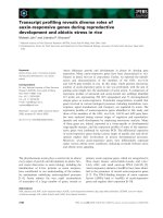
Figure 1 | Inflammation in human breast and prostate cancer. Many types of human carcinomas are characterized by
abundant infiltrations of immune cells that are not revealed by standard histochemical analyses. Representative sections
of normal, pre-malignant and malignant breast and prostate tissues that are stained with haematoxylin and eosin are
shown (upper panels of each pair). When adjacent tissue sections are assessed for CD45
+
leukocytes (lower, brown stained
panels), the extent of immune-cell infiltration into pre-malignant and malignant stroma is revealed.
REVIEWS
NATURE REVIEWS
|
CANCER VOLUME 6
|
JANUARY 2006
|
27
© 2006 Nature Publishing Group
failure of keratinocytes to achieve hyperproliferative
growth characteristics
27
(TABLE 1). This indicates that
activation and/or degranulation of immune cells in
neoplastic tissue upsets a crucial balance and thereby
promotes cancer development. More significantly,
studies such as these indicate that limiting or alter-
ing the presence of harmful innate immune cells in
pre-malignant tissue minimizes oncogene-induced
primary cancer development.
Are all tissue microenvironments susceptible to
immune-cell-potentiated primary cancer develop-
ment? Taking a similar approach, Lin and colleagues,
using polyoma-middle-T-antigen (PyMT) transgenic
mice as a model of mammary carcinogenesis, attenu-
ated macrophage recruitment and found that failure
to recruit macrophages into neoplastic tissue did not
alter the hallmarks of pre-malignancy, but instead
significantly delayed development of invasive carci-
nomas and reduced pulmonary metastasis formation
29
(TABLE 1). Metastatic potential was restored by trans-
genic expression of colony-stimulating factor 1 (
CSF1)
in mammary epithelium of CSF1-deficient/PyMT
mice
29
. These experimental data, combined with the
positive correlation in human cancers between CSF1
levels, macrophage recruitment and poor prognosis
30
,
indicate that macrophages are crucial for facilitating
late-stage metastatic progression of tumours.
Other cells of the myeloid lineage have also been
reported to contribute to tumour development
31
.
However, some types of innate immune cells — in par-
ticular, NK cells — can protect against experimental
tumour growth, in part by producing mediators with
anti-angiogenic properties
32,33
. Together, these studies
have induced a paradigm shift about the role of immune
cells during malignant progression. Whereas the histori-
cal viewpoint was that host immunity is protective with
regards to cancer, it is now clear that certain subsets
of chronically activated innate immune cells promote
growth and/or facilitate survival of neoplastic cells. Such
an unexpected crucial role for innate immune cells as
enhancers of tumour physiology raises questions about
how they convey their tumour-promoting effects and
whether, if understood, they can be harnessed to prevent
or block immune-cell tumour-promoting properties
while simultaneously activating antitumour immune
responses?
Table 1 | Immunomodulation of cancer incidence in mouse models of de novo carcinogenesis
Mouse cancer
model
Target organ Immune modulation Result
*
References
K14-HPV16 Skin Mast-cell deficiency
(Kit
W/WV
)
Decreased keratinocyte proliferation;
decreased angiogenesis
27
K14-HPV16 Skin CD4
+
T-cell deficiency Decreased CD11b
+
infiltration;
decreased cancer incidence
71
K14-HPV16 Skin CD8
+
T-cell deficiency No effect 71
K14-HPV16 Skin T- and B-cell deficiency
(RAG1-deficient mice)
Decreased CD45
+
infiltration; decreased
angiogenesis; decreased keratinocyte
proliferation; decreased cancer
incidence
28
K14-HPV16 and
Mmp9-null
Skin Transplantation with
bone marrow cells that
express MMP9
Increased keratinocyte proliferation;
increased angiogenesis; increased
cancer incidence
37
K14-HPV16/E2 Cervix CD4
+
T-cell deficiency Increased cancer burden; increased
cancer incidence;
72
K14-HPV16/E2 Cervix Mmp9-null Decreased angiogenesis; decreased
cancer incidence
26
K14-HPV16/E2 Cervix Bisphosphonate
treatment
Decreased macrophage MMP9
expression; decreased angiogenesis;
decreased cancer burden; decreased
cancer incidence
26
RIP1-TAG2 Pancreas Mmp9-null Decreased angiogenesis; decreased
cancer burden; decreased cancer
incidence
40
MMTV-PyMT Mammary
gland
CSF1-null mutant
mice (Csf1
op
/Csf1
op
);
macrophage deficiency
Decreased late-stage mammary
carcinoma; decreased pulmonary
metastases
29
Apc
∆716
Colon/small
intestine
COX2 deficiency
(Ptgs2-null mice)
Decreased cancer incidence; decreased
cancer burden
47
Apc
∆716
Colon/small
intestine
COX2 inhibitor Decreased tumour multiplicity;
decreased tumour volume
46
*Results are reported as compared with transgenic littermate controls. Apc
∆716
, adenomatous polyposis coli ∆716; COX2,
cyclooxygenase 2; CSF1, colony-stimulating factor 1; E2, 17β-estradiol; HPV16, human papillomavirus 16; K14, keratin 14; Mmp9,
matrix metalloproteinase 9; MMTV, mouse mammary tumor virus; Ptgs2, prostaglandin endoperoxide synthase 2; PyMT, polyoma
middle T antigen; RAG1, recombinase-activating gene 1; RIP1, rat insulin promoter 1; TAG2, simian-virus-40 large T antigen 2.
REVIEWS
28
|
JANUARY 2006
|
VOLUME 6 www.nature.com/reviews/cancer
© 2006 Nature Publishing Group
Normal Dysplastic
Blood vessels
e
d
e
d
CD45
+
Nuclei
Immunoglobin deposition
in dermal stroma
Nuclei
Basement membrane
Cancer development and innate immune cells
How then do chronically activated innate immune cells
participate in cancer development? Which mechanisms
and which inflammatory-cell-derived mediators are
relevant for specific human malignancies — do these
depend on organ, tumour stage or aetiology? Many of
these questions remain unanswered; however, experi-
mental models are beginning to elucidate molecular
mechanisms by which innate immune cells regulate cancer
processes
(BOX 1). Because of their enormous plasticity and
capacity to produce a myriad of cytokines, chemokines,
metalloproteinases, ROS, histamine and other bioactive
mediators, chronically activated innate immune cells
are key modulators of cell survival (both proliferation
and cell death) as well as regulators of ECM metabolism.
Therefore, several physiological processes that are neces-
sary for tumour development, such as increased cell sur-
vival, tissue remodelling, angiogenesis and suppression of
antitumour adaptive immune responses, are regulated by
leukocytic infiltrates in neoplastic environments. This is
exemplified by a positive correlation between the num-
ber of innate immune cells (macrophages, mast cells and
granulocytes) infiltrating human tumours and the number
of blood vessels
34,35
, and also by experimental findings in
mouse models in which attenuating innate-immune-cell
infiltration of pre-malignant tissue reduces angiogenesis
and limits tumour development
27,28,31
.
Matrix metalloproteinases. Numerous studies have docu-
mented increased expression of matrix metalloproteinases
(MMPs) in human malignant tissue, often correlating
with poor prognosis
36
. MMPs regulate tissue homeostasis
and disease pathogenesis through pleiotropic biological
effects, including remodelling of soluble and insoluble
ECM components and cell–cell and cell–matrix adhesion
molecules, that together alter crucial intracellular signal-
ling pathways
36
. In both human and mouse models of
cancer development, although some MMPs are produced
by epithelial cells, the major source of MMPs is activated
stromal cells — for example, fibroblasts, vascular cells and
in particular, innate immune cells
36
.
During skin and cervical carcinogenesis in HPV16
mice,
MMP9 has been identified as a crucial immune-
cell-derived mediator because of its ability to regulate
epithelial proliferation, angiogenesis and overall cancer
development
26,37
. Although amino-bisphosphonate-
mediated blockade of MMP9 production by macro-
phages and genetic elimination of MMP9 significantly
reduce cancer development in HPV16 mice
(TABLE 1),
infiltration of neoplastic tissue by immune cells is unper-
turbed by MMP9 absence
25,26
. This indicates that one
mechanism by which inflammation potentiates cancer
risk is the local delivery of MMP9.
Other experimental mouse models of cancer devel-
opment have similarly identified MMP9 as a key inflam-
matory-cell-derived mediator of tumour-associated
angiogenesis
38–40
. During pancreatic-islet carcinogenesis,
for example, Bergers and colleagues determined that
MMP9, which is produced predominantly by macro-
phages, regulates angiogenesis by mobilizing ECM-
sequestered vascular endothelial growth factor (VEGF)
and stimulating vascular endothelial cell proliferation
and subsequent angiogenesis
40
(TABLE 1). The processing
of pro-growth factors is not a unique property of MMP9
— in fact, several MMP family members are known to
possess this property, and some of them also regulate
acute inflammation through their ability to process che-
mokines
41
. MMP7 that is produced by osteoclasts has
emerged as a significant regulator of
prostate-cancer
bone metastases by virtue of its ability to process
RANKL
(receptor-activator-of-nuclear-factor-κB ligand) and
induce osteolysis
42
.
As osteoclasts and macrophages are similarly derived
from monocyte precursors, it will be interesting to
determine if bisphosphonate therapy attenuates MMP7
production by osteoclasts in the same way that it inhibits
macrophage MMP9 production during cervical carcin-
ogenesis
26
. Bisphosphonates are known to significantly
reduce the incidence of skeletal-related events during
breast-cancer metastases to bone
43
. Therefore, perhaps
the mechanisms by which this is achieved are monocyte
blockade of MMP production and subsequent inhibi-
tion of the skeletal complications that result from bone
metastases.
Figure 2 | Inflammation and angiogenesis are hallmarks of squamous
carcinogenesis in HPV16 transgenic mice. Fluorescent angiography and
immunohistochemical staining for CD45
+
leukocytes in whole-tissue pieces (upper
panels) shows parallel activation of blood vasculature (angiogenesis, shown in green) and
immune-cell infiltration (red) of pre-malignant (dysplastic) skin tissue from HPV16
transgenic mice, compared with normal skin (cell nuclei are shown in blue; the scale bar
represents 20µm). Interstitial immunoglobulin deposition (green) in the stroma of
pre-malignant dysplastic skin from HPV16 transgenic mice, compared with normal skin,
indicates robust humoral immune response during neoplastic progression (bottom
panels; the scale bar represents 50µm). The dashed lines indicate the position of the
epidermal basement membrane. d, dermis; e, epidermis.
REVIEWS
NATURE REVIEWS
|
CANCER VOLUME 6
|
JANUARY 2006
|
29
© 2006 Nature Publishing Group
COX2. Epidemiological studies have revealed that long-
term usage of non-steroidal anti-inflammatory drugs
(NSAIDs) reduces cancer risk
17,44
. This is probably due
to their inhibition of COX2, which is a multifunctional
enzyme that is involved in prostaglandin biosynthesis,
the expression of which is upregulated in inflamed and
neoplastic tissues
17
. In several human epithelial cancers,
expression of COX2 correlates with poor prognosis, and
pharmacological inhibition of COX2 reduces cancer
incidence
17
. Similar results have been found in rodent
models of cancer development — in the mammary
gland, COX2 overexpression induces carcinogenesis
45
,
whereas pharmacological inhibition and/or genetic
deletion of COX2 reduces cancer development
46,47
(see
Supplementary information S1 (table)).
Stromal cells — in particular, immune cells — as
well as neoplastic cells are known to upregulate COX2
expression during malignant progression. Therefore,
the efficacy of COX2-inhibitor-based therapies might be
achieved by the regulation or the normalization of distinct
biochemical and/or signalling pathways that are unique
to each individual cell type
48,49
. COX2 is believed to exert
its tumour-promoting effects by increasing cell survival
and regulating signalling pathways that are involved in
angiogenesis, inflammation and immune surveillance.
However, the crucial molecules that mediate these effects
remain largely undefined, though they might include the
prostaglandin E
2
receptor EP2 subtype (PTGER2)
50
.
Pro-inflammatory cytokines. Tumour microenviron-
ments are also rich in immune-cell-derived cytokines,
chemokines and pro-angiogenic mediators — for exam-
ple, tumour necrosis factor-α (
TNFα), transforming
growth factor-β (
TGFβ), VEGF, and interleukins 1 (IL-1)
and 6 (
IL-6)
12
. Production of VEGF is one mechanism
by which tumour-infiltrating leukocytes increase angio-
genesis and foster tumour development
34,51
. Similarly,
TNFα, a key cytokine that is mobilized during acute
inflammation, mediates cancer development
52
. Mice
that are deficient for either TNFα or TNFα receptors
have reduced susceptibility to chemically induced skin
cancers and develop fewer experimental metastases (see
Supplementary information S1 (table)). As TNFα recep-
tors are expressed on both epithelial and stromal cells,
TNFα facilitates cancer development directly, by regulat-
ing the proliferation and survival of neoplastic cells, as
well as indirectly, by exerting its effects on endothelial
cells, fibroblasts and immune cells in tumour microen-
vironments
12
. Clinical trials are currently underway to
assess the efficacy of TNFα antagonists in patients with
breast and
ovarian cancer
52,53
.
Recently, a functional link was elucidated between
TNFα and the pro-inflammatory transcription factor
nuclear factor κB (
NFκB), revealing the complexity
of paracrine signalling mechanisms between innate
immune cells and evolving neoplastic cells. Using a
mouse model of cholestatic hepatitis that predisposes
mice to
hepatocellular carcinoma, Pikarsky and col-
leagues reported that hepatocyte survival and malignant
progression are regulated by NFκB, the activation state
and cellular localization of which was under paracrine
TNFα control
54
. Inhibition of TNFα or induction of the
super-repressor mutant of
IκB (inhibitor of NFκB) in
transgenic animals during the later stages of neoplastic
progression resulted in failure to progress to hepato-
cellular carcinoma
54
. This indicates that TNFα, which
is produced by surrounding parenchyma, activates an
NFκB-dependent anti-apoptotic pathway during the
time at which the foci of pre-malignant hepatocytes
develop into tumours.
Karin and colleagues came to a similar conclusion
using a mouse model of colitis-associated cancer
55
.
However, in these studies deletion of the inhibitor of
NFκB kinase (
IKKβ) — a key intermediary of NFκB
— in intestinal epithelial cells did not decrease intestinal
inflammation, as measured by inflammatory cytokine
production, but instead reduced susceptibility to inflam-
mation-induced intestinal tumours
55
. Moreover, specific
deletion of IKKβ in myeloid cells resulted in formation of
smaller inflammation-associated colon cancers and cor-
related with reduced production of tumour-promoting
paracrine factors, including TNFα
55
.
An important feature of these studies was that NFκB
activation was selectively ablated in different cell-
ular compartments of the developing tumour masses
and/or at different stages of tumour development (see
Supplementary information S1 (table)). This revealed
that the NFκB pathway has a dual role in tumour promo-
tion — first, by preventing apoptosis of cells with malig-
nant potential, and second, by stimulating production of
pro-inflammatory cytokines by cells of myeloid origin in
tumours. These pro-inflammatory cytokines then con-
tribute in a paracrine fashion to neoplastic cell prolifera-
tion and increase survival of initiated, and/or otherwise
‘damaged’, epithelial cells. These elegant approaches
offer novel insights into differential regulation of pre-
malignant and malignant states by inflammation, and
the complexities and downstream activities of NFκB in
distinct cellular compartments.
Antitumour adaptive immunity. Chronically activated
innate immune cells can also indirectly contribute to
cancer development through suppression of antitumour
adaptive immune responses, allowing tumour escape from
immune surveillance. A subset of innate immune cells (for
example, myeloid suppressor GR
+
CD11b
+
cells) accu-
mulate in tumours and lymphoid organs
18,21,56
. Myeloid
suppressor cells are known to induce T-lymphocyte
dysfunction by direct cell–cell contact and by production
of immunosuppressive mediators, and therefore actively
inhibit antitumour adaptive immunity
21,56
. In addition,
malignant lesions attract regulatory T cells that are also
known to suppress effector functions of cytotoxic T cells
18
.
Classic regulatory T cells are CD4
+
CD25
+
FOXP3
+
; how-
ever, different subtypes might also exist. Initial investiga-
tions have revealed that in vivo depletion of regulatory
T cells using antibodies against CD25 enhance antitumour
T-cell responses and induce regression of experimental
tumours
57,58
. An elegant study by Curiel and colleagues
revealed that tumour-derived macrophages from patients
with ovarian cancer produce CCL22, a chemokine that
mediates trafficking of regulatory T cells to tumours
20
.
REVIEWS
30
|
JANUARY 2006
|
VOLUME 6 www.nature.com/reviews/cancer
© 2006 Nature Publishing Group
These regulatory T cells in patients with ovarian cancer
suppressed tumour-specific T-cell immunity, and their
presence correlated with reduced survival. Therefore, in
the vicinity of a growing neoplasm, the balance between
innate and adaptive immunity is often disturbed in favour
of cancer progression.
Different tissue, different target. Many types of chroni-
cally activated immune cells therefore exert tumour-
promoting effects directly by influencing proliferation
and survival of neoplastic cells, as well as by indirectly
modulating neoplastic microenvironments to favour
tumour progression
(BOX 1). How can these diverse
mechanisms be translated into the development of
broadly applicable therapeutical approaches? Should
future anticancer strategies focus on regulating COX2
activity, NFκB activation, TNFα bioavailability or cru-
cial extracellular protease actvity? When addressing
these questions, it is important to remember that all
organs are endowed with unique cell-death and damage-
response pathways that typically invoke acute activation
of innate immune cells. In skin, for example, terminal
differentiation is the mode by which keratinocytes die,
and in contrast to colitis-associated and hepatocellular
carcinoma, inhibiting NFκB in skin keratinocytes pro-
motes epidermal hyperplasia and the development of
spontaneous squamous cell carcinomas
59,60
On the other hand, blockade of TNFα attenuates
skin-tumour formation
61
. Therapeutically regulating
multifunctional immunomodulators such as NFκB,
TNFα, COX2 or MMPs requires careful risk assess-
ments as systemic inhibition might have unfavour-
able outcomes, which are the result of cell-type and
environment-dependent activities that differentially
regulate tissue homeostasis. If systemic modulation can
be tolerated without adverse side-effects, combining
immunomodulating cytostatic therapies with radiation
or cytotoxic drugs might be beneficial. Some success
has been demonstrated with this approach both in cell
lines, where overexpression of a ‘super-repressor’ of
NFκB enhanced the activity of cytotoxic drugs
62
, and
in the clinic, where proteasome inhibitors increased the
efficacy of chemotherapy in some patients
63
.
Adaptive immunity and cancer
Whereas it has become generally accepted that chronic
activation of innate immune cells contributes to cancer
development, the role of adaptive immune cells is still
a matter of debate. This is perhaps best exemplified by
epidemiological studies of cancer incidence in patients
with either AIDS or organ transplants who have chronic
suppression of their adaptive immune compartment
(TABLE 2). In these two groups, the relative risk (RR) of can-
cer development varies considerably depending on organ
site and cancer aetiology. It is well known that the RR
for viral-associated cancers, in particular human-herpes-
virus-8-associated Kaposi sarcoma, Epstein–Barr-virus-
associated
non-Hodgkin lymphoma and HPV-associated
squamous carcinoma, are elevated in immune-suppressed
individuals
(TABLE 2), owing largely to the fact that chronic
immune suppression fails to provide protection against
viral infections or viral re-activation
64
. Overall, the RR
for invasive malignancies, other than Kaposi sarcoma,
non-Hodgkin lymphoma and
non-melanoma squamous
cancers
, is approximately 2.5; however, the RR varies
considerably between individual cancers.
Some cancer types occur with increased frequency
in selected groups of immune-compromised patients
for reasons that are unrelated to immune suppression.
For example, chronic exposure to tobacco carcinogens is
associated with an increased RR for cancers of the lung,
lip, mouth and pharynx in AIDS patients
65
. Similarly, the
RR of lung cancer,
head and neck cancer, oesophageal
cancer, gastrointestinal cancer and pancreatic cancer
in patients who have had liver transplants is increased
when there is a previous history of alcohol and tobacco
use
66,67
. On the other hand, the RR for the most common
non-viral-associated solid tumours of epithelial origin is
decreased in immune-suppressed patients; some of these
in fact have an RR less than 1.0
(TABLE 2). In particular,
the RRs for breast, prostate and
bladder cancer are sig-
nificantly reduced in both patients who have had organ
transplants and patients with AIDS.
Although it is not surprising that immune sup-
pression in the adaptive compartment fails to provide
protection from the development of viral-associated or
carcinogen-induced tumours, it is difficult to explain
why immune suppression correlates with a decreased RR
for some non-viral-associated cancers. Mouse models
of cancer have similarly revealed that tumour develop-
ment in immune-suppressed rodents varies depend-
ing on cancer type and cancer aetiology (
TABLE 1; see
Supplementary information S1 (table)). Some studies
have reported an increased susceptibility to chemically
induced cancers in mice with defined immunological
defects, whereas others have reported a higher incidence
of spontaneous tumours in immunocompromised mice
that varies between organs and/or depends on the pres-
ence of persistent bacterial infection (see
Supplementary
information S1
(table)).
These studies, together with the identification
of tumour-associated antigens, have in part fuelled
the development of antitumour immunotherapy
approaches
68
. Although these approaches seem
efficacious in principle, the reality is that, for well-
established bulky tumours, activation of endogenous
antitumour T-cell responses is often insufficient to
induce full tumour regression
68
. Moreover, cancer
vaccines have generally elicited low numbers of
tumour-specific immune cells, and tumour-targeted
T cells often fail to infiltrate solid tumours or they
show a low avidity for tumour antigens
68,69
. Therefore,
they suboptimally recognize target cells that express
specific antigens. Furthermore, cancer vaccines must
overcome the systemic immune suppression that is
exerted by tumours. Some of these problems can be
circumvented by immunotherapy approaches that
make use of adoptive transfer, in which autologous
anticancer T cells from the patient are generated and/
or expanded ex vivo before being transferred back into
the patient
70
. Therefore, despite the many challenges
of cancer immunotherapy, it is clear that sufficient
REVIEWS
NATURE REVIEWS
|
CANCER VOLUME 6
|
JANUARY 2006
|
31
© 2006 Nature Publishing Group
αβ T cells
Lymphocytes that express
T-cell receptors consisting of
heterodimers of α and β
chains. αβ T cells recognize
antigens when they are
presented in association with
major histocompatibility
molecules.
numbers of adequately activated T lymphocytes can
be beneficial for some patients. However, surpassing
the hurdle of adequacy appears to be a difficulty.
Experimental rodent studies have also provided con-
tradictory findings regarding the elimination of adaptive
immune components and the activation of tumour-spe-
cific adaptive immune responses in cancer development
(
TABLE 1; see Supplementary information S1 (table)).
These seemingly paradoxical statements are perhaps best
exemplified by recent studies in which the functional
significance of CD4
+
T lymphocytes was assessed dur-
ing skin and cervical carcinogenesis in HPV16 mice
71,72
.
Genetic elimination of CD4
+
T lymphocytes resulted in
slightly delayed development of late-stage skin dysplasias
and a modest reduction in skin cancer incidence
71
. By
contrast, genetic elimination of CD4
+
T cells in female
HPV16 mice that were undergoing oestrogen treatment
to predispose them to cervical carcinogenesis resulted in
a 10-fold increased tumour burden and a 20% increase in
carcinoma incidence compared with oestrogen-treated
HPV16 controls
71,72
.
The adaptive immune system can also differentially
regulate cancer development within the same epithelial
microenvironment, as a function of varied initiation.
For example, mice that are deficient for
αβ T cells have
an increased incidence of methylcholantrene (MCA)-
induced carcinomas compared with mice that contain
αβ T cells. However, when the same cohorts were
treated with 7,12-dimethylbenz[a]anthracene (DMBA)
and 12-O-tetradecanoylphorbol-13-acetate (TPA), the
αβ-T-cell-deficient mice had a reduced susceptibility to
carcinoma development compared with the controls
73
(see Supplementary information S1 (table)).
Likewise, paradoxical roles for NK T cells have been
reported during cancer development
74
. NK T cells are
CD3
+
T cells that also express some NK-cell markers
and recognize glycolipid ligands that are presented
by the major-histocompatibility-complex class-I-like
molecule, CD1d
75
. It has been reported that NK T cells
are involved in natural host immunity against chemi-
cally induced sarcomas
32
. On the other hand, it has also
been reported that NK T cells can downregulate tumour
Table 2 | Human immune-deficient status and cancer risk
Immune
deficiency
Cohort size Cancer type Relative risk References
AIDS-defining cancers/viral-associated cancers
AIDS 122,993 Kaposi sarcoma 97.5 (male) 202.7 (female) 102
Non-Hodgkin lymphoma 37.4 (male) 54.6 (female)
Skin (excluding Kaposi sarcoma) 20.9 (male) 7.5 (female)
Cervical 9.1
AIDS 8,828 Kaposi sarcoma 545 103
Non-Hodgkin lymphoma 24.6
AIDS 302,834 Kaposi sarcoma 177.7 104
Non-Hodgkin lymphoma 72.8
Cervical 5.2
Liver transplant 187 Cutaneous 16.9 66
Liver transplant 174 Skin (non-melanoma) 70 105
Non-AIDS-defining cancers (with reduced RR)
AIDS 302,834 Breast 0.5 104
Prostate 0.5
AIDS 122,993 Prostate 0.7 102
Bladder 0.5
Breast 0.8 (HIV positive)
0.2 (post-AIDS onset)
AIDS 8,828 Prostate 0.8 103
AIDS 62,157 Ovarian 0.58 106
Breast 0.55
Uterine 0.28
Kidney/heart
transplant
25,914 Breast (year 1) 0.49 107
Breast (year 2–11) 0.84
Liver transplant 1,000 Breast, ovary, uterus and cervical 0.53 67
Genitourinary 0.68
REVIEWS
32
|
JANUARY 2006
|
VOLUME 6 www.nature.com/reviews/cancer
© 2006 Nature Publishing Group
Breast
Prostate
Normal Pre-malignant Malignant
Immunoglobulins (deposited in interstitial
stroma or present in phagocytes)
Nuclei
Chronic idiopathic
thrombocytopaenia
An autoimmune disease that
involves autoantibody-
mediated eradication of
platelets, resulting in a reduced
overall number of platelets.
The primary clinical symptom
is increased and prolonged
bleeding.
Autoimmune haemolytic
anaemia
An autoimmune disease that
involves autoantibody-
mediated premature
destruction of erythrocytes,
resulting in anaemia.
Systemic lupus
erythematosus
A multi-system inflammatory
disease that is characterized by
autoantibody production and
deposition of immune
complexes in many organs,
causing a broad spectrum of
manifestations.
Fc receptors
A family of receptors that are
involved in recognition of the
Fc portion of antibodies. Fc
receptors are expressed on the
surface of various immune
cells. Depending on the type of
Fc receptor that is expressed,
crosslinking can result in
degranulation, activation of
phagocytosis, and cytokine
release.
Complement cascades
The complement system is
made up of more than 25
components that are present
in serum. Foreign antigens and
immune complexes activate
the complement activation
cascade, resulting in formation
of lytic membrane-attack
complexes and liberation of
potent pro-inflammatory
factors.
immunosurveillance against transplanted tumours
76,77
.
This paradoxical influence of NK T cells during cancer
development is probably a consequence of their inherent
capacity to produce both pro-inflammatory T-helper-1
cytokines and anti-inflammatory T-helper-2 cytokines
74
;
therefore, the nature and balance of surrounding stimuli
might determine which type of NK-T-cell-induced
T-helper response dominates and contributes to malig-
nant outcome.
In this way, rodent models parallel human cancer
and indicate that the adaptive immune system differ-
entially modulates de novo cancer development in an
organ-dependent and aetiology-dependent manner.
The paradoxical influence of the adaptive immune sys-
tem during these processes raises many questions, the
understanding of which is crucial if treatment modalities
involve adaptive-immune-based therapies. Depending
on the microenvironment or cancer aetiology, adap-
tive-immune-based therapies could either exacerbate
or arrest neoplastic disease.
Adaptive immunity, inflammation and cancer
Recent advances in understanding underlying mecha-
nisms of chronic autoimmune diseases have revealed
that adaptive immunity has a crucial role in regulating
and activating innate immune cells in affected tissues
78
.
For example, interstitial immunoglobulin (Ig) deposition
has been observed in tissues that are heavily infiltrated
by innate immune cells in patients with
rheumatoid
arthritis
79
. B-lymphocyte depletion in these patients
decreases disease severity, as it also does in
chronic idio-
pathic thrombocytopaenia
, autoimmune haemolytic anaemia
and
systemic lupus erythematosus
80
. This indicates that
immunoglobulin deposition contributes to chronic
inflammation and disease pathogenesis rather than
merely correlating with it.
Antibodies that are deposited into interstitial tissues
can trigger activation of innate immune cells by the cross-
linking of
Fc receptors or the activation of complement
cascades
78
. As CD4
+
and CD8
+
T lymphocytes are impor-
tant modulators of such tissue-damaging B-lymphocyte
responses, and because Ig deposition is found in human
pre-malignant and malignant tissues
(FIG. 3), it is possible
that imbalanced or altered adaptive-immune-cell interac-
tions represent underlying mechanisms that regulate the
onset and/or maintenance of chronic inflammation that
is associated with cancer development.
To address this possibility, we evaluated the role of
adaptive immune cells in HPV16 mice and found that
combined B- and T-lymphocyte deficiency attenuated
innate-immune-cell infiltration of pre-malignant skin
28
.
As a consequence, blood vasculature remained quies-
cent, keratinocytes failed to attain a hyperproliferative
phenotype and the overall incidence of invasive carci-
nomas decreased to ~6%, compared with ~50% in the
controls
28
. Adoptive transfer of B lymphocytes or serum
from HPV16 mice into B- and T-cell-deficient/HPV16
mice restored characteristic chronic inflammation in
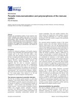
Figure 3 | Humoral immune response in breast and prostate cancer. Robust humoral immune responses are found
during human breast and prostate tumorigenesis compared with normal tissue. The fluorescent images show that
immunoglobulin deposits (green) are prominent in the interstitial stroma of pre-malignant and malignant prostate tissues
(bottom panels). By contrast, during breast tumorigenesis (top panels), immunoglobulins are also found within phagocytes
in pre-malignant and malignant tissues (shown by the colocalization of green fluorescence and the blue flourescence of
the cell nuclei)
101
.
REVIEWS
NATURE REVIEWS
|
CANCER VOLUME 6
|
JANUARY 2006
|
33
© 2006 Nature Publishing Group
Pro-inflammatory
cytokines
DC migration and
antigen presentation
Serum proteins
Tumour-promoting
Pro-growth
Tissue expansion
Malignant conversion
Cell-death inhibition
Genomic instability
Fibroblast activation
Matrix metabolism
Angiogenesis
T- and B-
lymphocyte
activation
Secondary lymphoid organs
Adaptive immune response
Initiation
Neoplastic progression
Innate immune
response
Anti-tumour
T-cell-mediated cytotoxicity
(FAS, perforin and/or cytokine pathways)
Antibody-dependent cell-mediated cytotoxicity
Antibody-induced complement-mediated lysis
pre-malignant skin and reinstated angiogenesis and
keratinocyte hyperproliferation, which are parameters
that are essential for full malignancy
28
.
These data indicate that B lymphocytes, and factors
that are present in serum, are essential for establishing
chronic inflammatory states that are associated with
pre-malignant progression
(FIG. 2), and are therefore
involved in enhancing the neoplastic pathways that are
necessary for skin cancer development. B lymphocytes
do not significantly infiltrate pre-malignant skin of
HPV16 mice, indicating that priming of B lymphocytes
occurs in draining lymph nodes by skin-derived anti-
gen-presenting DCs. It is not yet clear which signals
induce DC trafficking to draining lymph nodes. The
prevailing model for DC migration from inflamed tis-
sue to lymph nodes involves expression of chemokine
(C-C motif) receptor 7 (
CCR7) and specific integrins by
DCs, and the existence of a chemotactic gradient from
the periphery to lymphatic vessels
81
. However, whether
similar pathways are involved during cancer progression
is still unclear. In addition, it remains to be investigated
which skin-derived antigens trigger tumour-promoting
B-lymphocyte responses. Nor is it known whether these
are antigens that are derived from the HPV16 early-region
genes or antigens that are derived from stromal molecules
undergoing remodelling in pre-malignant skin. Are these
results a unique feature of skin carcinogenesis in HPV16
mice, or do other experimental models or clinical data
support a crucial role for B lymphocytes during epithelial
cancer development?
Potential tumour-promoting roles for B lym-
phocytes and/or antibodies were described over 50
years ago, albeit without elucidation of any under-
lying mechanisms. These early studies demonstrated
that passive transfer of tumour-specific antibodies
increased outgrowth of transplanted tumour cells and
chemically induced tumours
82–85
, whereas absence of
B lymphocytes limited tumour formation
86,87
. More
recently, it was reported that low antibody-responder
mice were less susceptible to DMBA/TPA-induced
Figure 4 | A model of innate and adaptive immune-cell function during inflammation-associated cancer
development. Antigens that are present in early neoplastic tissues are transported to lymphoid organs by dendritic cells
(DCs) that activate adaptive immune responses resulting in both tumour-promoting and antitumour effects. The pathways
that regulate DC trafficking during early cancer development and the exact nature of the antigen(s) remains to be
established. Activation of B cells and humoral immune responses results in chronic activation of innate immune cells in
neoplastic tissues. Activated innate immune cells, such as mast cells, granulocytes and macrophages, promote tumour
development by the release of potent pro-survival soluble molecules that modulate gene-expression programmes in
initiated neoplastic cells, culminating in altered cell-cycle progression and increased survival. Inflammatory cells positively
influence tissue remodelling and development of the angiogenic vasculature by production of pro-angiogenic mediators
and extracellular proteases. Tissues in which these pathways are chronically engaged exhibit an increased risk of tumour
development. By contrast, activation of adaptive immunity also elicits antitumour responses through T-cell-mediated
toxicity (by induction of FAS, perforin and/or cytokine pathways) in addition to antibody-dependent cell-mediated
cytotoxicity and antibody-induced complement-mediated lysis.
REVIEWS
34
|
JANUARY 2006
|
VOLUME 6 www.nature.com/reviews/cancer
© 2006 Nature Publishing Group
skin carcinogenesis as compared with high antibody-
responder counterparts
88
(see Supplementary infor-
mation S1
(table)), and that active immunization of
cancer-prone immune-proficient mice resulted in
tumour-specific antibody responses and increased
carcinogenesis after chemical promotion
89
. Consistent
with the link between B lymphocytes and the initia-
tion of chronic inflammation in HPV16 mice, Barbera-
Guillem and colleagues demonstrated that antitumour
humoral immune responses increase outgrowth and
invasion of murine and human tumour-cell xenografts
through recruitment and activation of granulocytes
and macrophages
90
.
In the clinical arena, there is a vast literature that
describes the occurrence of autoantibodies in the serum
of cancer patients, and interstitial antibody deposition in
human tumours
91
(FIG. 3). Moreover, the early presence of
autoantibodies (in particular, antinuclear and smooth-
muscle antibodies) in the serum correlates with an
unfavourable prognosis
92
. Does this correlation indicate
that individuals with tumours that progress harbour a
higher antigen load and therefore trigger greater anti-
body production, or does it indicate that the presence of
antibodies predisposes patients to development of more
advanced or recurrent cancers? Although the answer is
not clear, these data indicate that B lymphocytes are also
involved in human cancer development and therefore
necessitate a more mechanistic evaluation of their role
and specificity to determine if they represent tractable
targets for anticancer therapy.
Conclusion and Perspective
Clinical and experimental studies now indicate that
innate and adaptive immune cells are significant, albeit
sometimes paradoxical, determinants of epithelial
tumorigenesis
(FIG. 4). On the basis of classical theories
of immune surveillance and more recent awareness of
the tumour-promoting properties of innate immune
cells, researchers are now investigating the efficacy of
novel anticancer strategies that are based on immuno-
therapeutics that can either bolster antitumour adaptive
immunity or neutralize cancer-promoting properties of
innate immune cells.
An issue to be resolved with these powerful
approaches is how therapeutically manipulating one
arm of the immune system affects the anticancer or
cancer-promoting properties of the other. For example,
induction of anticancer humoral immune responses
might be beneficial for patients with established can-
cer; however, given the observation that inflammation-
associated epithelial carcinogenesis is B-lymphocyte-
dependent
28
, activation of humoral immune responses
in patients who are predisposed to cancer development
or in patients with latent or pre-malignant disease might
enhance neoplastic programming of tissue rather than
eradicating it. In these latter patients, it might be benefi-
cial to monitor their serum for indications of B-cell acti-
vation and/or humoral immunity, as this might reveal
a therapeutic window for anti-B-lymphocyte-based
therapies or for modalities that are aimed at neutralizing
tumour-promoting properties of innate immune cells.
In addition to NSAIDs and COX2 inhibitors having
been shown to be efficacious
17
, and clinical trials with
GMCSF
16
, TNFα antagonists
52,53
and adoptive transfer
of autologous anticancer T cells
70
being encouraging,
recent successes with B-lymphocyte depletion for relief
of rheumatoid arthritis and systemic lupus erythema-
tosus
93,94
have demonstrated safety and instill optimism
for the approach. Although one or other of these host-
targeted strategies might provide a therapeutic advan-
tage, it seems reasonable to consider combinatorial
immunomodulating strategies that target cancer-pro-
moting properties of both innate and adaptive immune-
cell populations while simultaneously exploiting their
unique anticancer attributes without interfering with
their normal functions.
Clinical use of protease and COX2 inhibitors as
anticancer therapeutics has been minimized
44,95
, largely
because of the failure to take into account the crucial
role that these powerful mediators have in maintaining
and normalizing tissue homeostasis. Nevertheless, once
risk assessments have been carried out, a prediction
for these new host-targeted approaches is that immu-
nomodulating therapies will be used to the patient’s
advantage and might offer the possibility of normalizing
cancer-prone tissues prior to the appearance of bulky
disease. Alternatively, if they are used in combination
with standard antitumour approaches (for example,
cytotoxic drugs), they could provide an overwhelming
assault on malignant cancer cells. Therefore, the goal
should be to determine optimal and tolerable combi-
nations of immunomodulating and cytotoxic therapies
that will result in significant survival and quality-of-life
improvements for patients with cancer.
1. Hamerman, J. A., Ogasawara, K. & Lanier, L. L. NK
cells in innate immunity. Curr. Opin. Immunol. 17,
29–35 (2005).
2. Degli-Esposti, M. A. & Smyth, M. J. Close encounters
of different kinds: dendritic cells and NK cells take
centre stage. Nature Rev. Immunol. 5, 112–124
(2005).
3. Raulet, D. H. Interplay of natural killer cells and their
receptors with the adaptive immune response. Nature
Immunol. 5, 1038–1114 (2004).
References 1–3 are comprehensive reviews on the
role of dendritic and natural killer cells during
tumour development.
4. Finch, C. E. & Crimmins, E. M. Inflammatory exposure
and historical changes in human life-spans. Science
305, 1736–1739 (2004).
A thoughtful analysis of historical changes in
human exposure to inflammatory agents that are
correlated with cancer incidence and lifespan.
5. Balkwill, F. & Mantovani, A. Inflammation and cancer:
back to Virchow? Lancet 357, 539–545 (2001).
6. Coca, S. et al. The prognostic significance of
intratumoral natural killer cells in patients with
colorectal carcinoma. Cancer 79, 2320–2328
(1997).
7. Ishigami, S. et al. Prognostic value of intratumoral
natural killer cells in gastric carcinoma. Cancer 88,
577–583 (2000).
8. Ribatti, D. et al. Tumor vascularity and tryptase-
positive mast cells correlate with a poor prognosis in
melanoma. Eur. J. Clin. Invest. 33, 420–425
(2003).
9. Imada, A., Shijubo, N., Kojima, H. & Abe, S. Mast cells
correlate with angiogenesis and poor outcome in
stage I lung adenocarcinoma. Eur. Respir. J. 15,
1087–1093 (2000).
10. Leek, R. D. et al. Association of macrophage
infiltration with angiogenesis and prognosis in invasive
breast carcinoma. Cancer Res. 56, 4625–4629
(1996).
11. Leek, R. D., Landers, R. J., Harris, A. L. & Lewis, C. E.
Necrosis correlates with high vascular density and
focal macrophage infiltration in invasive carcinoma of
the breast. Br. J. Cancer 79, 991–995 (1999).
12. Balkwill, F., Charles, K. A. & Mantovani, A. Smoldering
and polarized inflammation in the initiation and
promotion of malignant disease. Cancer Cell
7,
211–217 (2005).
REVIEWS
NATURE REVIEWS
|
CANCER VOLUME 6
|
JANUARY 2006
|
35
© 2006 Nature Publishing Group
A timely and detailed review of clinical and
experimental literature that discusses paracrine
communication networks between immune cells
and cells at risk for cancer development.
13. Pagano, J. S. et al. Infectious agents and cancer:
criteria for a causal relation. Semin. Cancer Biol. 14,
453–471 (2004).
14. Velicer, C. M. et al. Antibiotic use in relation to the risk
of breast cancer. JAMA 291, 827–835 (2004).
15. Enzler, T. et al. Deficiencies of GM-CSF and interferon
γ link inflammation and cancer. J. Exp. Med. 197,
1213–1219 (2003).
A detailed analysis of immune-competent mice that
are deficient for GMCSF and IFNγ, which
demonstrates that failure to normalize tissue
homeostasis and clear low-level microbial
infections results in chronic inflammation that
underlies an increased incidence of haematologic
and solid-tissue cancers.
16. Korzenik, J. R., Dieckgraefe, B. K., Valentine, J. F.,
Hausman, D. F. & Gilbert, M. J. Sargramostim for
active Crohn’s disease. N. Engl. J. Med. 352,
2193–2201 (2005).
17. Dannenberg, A. & Subbaramaiah. Targeting
cyclooxygenase-2 in human neoplasia: rationale and
promise. Cancer Cell 4, 431–436 (2003).
18. Zou, W. Immunosuppressive networks in the tumour
environment and their therapeutic relevance. Nature
Rev. Cancer 5, 263–274 (2005).
An excellent review that provides an extensive
overview of immunosuppressive networks in the
microenvironment of developing tumours.
19. Finke, J., Ferrone, S., Frey, A., Mufson, A. & Ochoa, A.
Where have all the T cells gone? Mechanisms of
immune evasion by tumors. Immunol. Today 20,
158–160 (1999).
20. Curiel, T. J. et al. Specific recruitment of regulatory
T cells in ovarian carcinoma fosters immune privilege
and predicts reduced survival. Nature Med. 10,
942–94 (2004).
21. Serafini, P. et al. Derangement of immune responses
by myeloid suppressor cells. Cancer Immunol.
Immunother. 53, 64–72 (2004).
22. Coussens, L. M., Hanahan, D. & Arbeit, J. M. Genetic
predisposition and parameters of malignant
progression in K14-HPV16 transgenic mice. Am. J.
Pathol. 149, 1899–1917 (1996).
23. Arbeit, J., Howley, P. & Hanahan, D. Chronic estrogen-
induced cervical and vaginal squamous carcinogenesis
in HPV16 transgenic mice. Proc. Natl Acad. Sci. USA
93, 2930–2935 (1996).
24. Smith-McCune, K., Zhu, Y. H., Hanahan, D. & Arbeit,
J. Cross-species comparison of angiogenesis during
the premalignant stages of squamous carcinogenesis
in the human cervix and K14-HPV16 transgenic mice.
Cancer Res. 57, 1294–1300 (1997).
25. van Kempen, L. C. L. et al. Epithelial carcinogenesis:
dynamic interplay between neoplastic cells and their
microenvironment. Differentiation 70, 501–623
(2002).
26. Giraudo, E., Inoue, M. & Hanahan, D. An amino-
bisphosphonate targets MMP-9-expressing
macrophages and angiogenesis to impair cervical
carcinogenesis. J. Clin. Invest. 114 , 623–633
(2004).
27. Coussens, L. M. et al. Inflammatory mast cells
up-regulate angiogenesis during squamous epithelial
carcinogenesis. Genes Dev. 13, 1382–1397 (1999).
28. de Visser, K. E., Korets, L. V. & Coussens, L. M.
De novo carcinogenesis promoted by chronic
inflammation is B lymphocyte dependent. Cancer Cell
7, 411–423 (2005).
A study from the authors’ laboratory that reports
that inflammation-associated cancer development
in skin-cancer-prone transgenic mice is dependent
upon serum factors that are derived from activated
peripheral B lymphocytes, so revealing the
complexity of interactions between innate and
adaptive immune cells that regulate epithelial
cancer development.
29. Lin, E. Y., Nguyen, A. V., Russell, R. G. & Pollard, J. W.
Colony-stimulating factor 1 promotes progression of
mammary tumors to malignancy. J. Exp. Med. 193,
727–740 (2001).
Using the well-characterized MMTV-PyMT model of
mammary carcinogenesis, the authors rendered
mice CSF1-deficient to block macrophage
recruitment into neoplastic tissue and therefore
demonstrated that macrophages deliver permissive
factors necessary for development of late-stage
carcinomas and pulmonary metastases.
30. Pollard, J. W. Tumour-educated macrophages promote
tumour progression and metastasis. Nature Rev.
Cancer 4, 71–78 (2004).
A detailed review on the role of macrophages
during mammary carcinogenesis.
31. Sparmann, A. & Bar-Sagi, D. Ras-induced interleukin-
8 expression plays a critical role in tumor growth and
angiogenesis. Cancer Cell 6, 447–458 (2004).
32. Smyth, M. J., Crowe, N. Y. & Godfrey, D. I. NK cells
and NKT cells collaborate in host protection from
methylcholanthrene-induced fibrosarcoma. Int.
Immunol. 13, 459–463 (2001).
33. Hayakawa, Y. et al. IFN-γ-mediated inhibition of tumor
angiogenesis by natural killer T-cell ligand,
α-galactosylceramide. Blood 100, 1728–1733 (2002).
34. Esposito, I. et al. Inflammatory cells contribute to the
generation of an angiogenic phenotype in pancreatic
ductal adenocarcinoma. J. Clin. Pathol. 57, 630–636
(2004).
35. Yano, H. et al. Mast cell infiltration around gastric
cancer cells correlates with tumor angiogenesis and
metastasis. Gastric Cancer 2, 26–32 (1999).
36. Egeblad, M. & Werb, Z. New functions for the matrix
metalloproteinases in cancer progression. Nature Rev.
Cancer 2, 161–174 (2002).
37. Coussens, L. M., Tinkle, C. L., Hanahan, D. & Werb, Z.
MMP-9 supplied by bone marrow-derived cells
contributes to skin carcinogenesis. Cell 103,
481–490 (2000).
38. Huang, S. et al. Contributions of stromal
metalloproteinase-9 to angiogenesis and growth of
human ovarian carcinoma in mice. J. Natl Cancer Inst.
94, 1134–1142 (2002).
39. Hiratsuka, S. et al. MMP9 induction by vascular
endothelial growth factor receptor-1 is involved in
lung-specific metastasis. Cancer Cell 2, 289–300
(2002).
40. Bergers, G. et al. Matrix metalloproteinase-9 triggers
the angiogenic switch during carcinogenesis. Nature
Cell Biol. 2, 737–744 (2000).
41. Overall, C. M., McQuibban, G. A. & Clark-Lewis, I.
Discovery of chemokine substrates for matrix
metalloproteinases by exosite scanning: a new tool for
degradomics. Biol. Chem. 383, 1059–1066 (2002).
42. Lynch, C. C. et al. MMP-7 promotes prostate cancer-
induced osteolysis via the solubilization of RANKL.
Cancer Cell 7, 485–496 (2005).
A provocative study that reveals a previously
unknown role for MMP7/matrilysin as a crucial
regulator of osteolysis during prostate-cancer bone
metastases by virtue of its ability to regulate
bioavailability of RANKL.
43. Coleman, R. E. Bisphosphonates in breast cancer.
Ann. Oncol. 16, 687–695 (2005).
44. Psaty, B. M. & Furberg, C. D. COX-2 inhibitors —
lessons in drug safety. N. Engl. J. Med. 352,
1133–1135 (2005).
45. Liu, C. H. et al. Overexpression of cyclooxygenase-2 is
sufficient to induce tumorigenesis in transgenic mice.
J. Biol. Chem. 276, 18563–18569 (2001).
46. Jacoby, R. F., Seibert, K., Cole, C. E., Kelloff, G. &
Lubet, R. A. The cyclooxygenase-2 inhibitor celecoxib
is a potent preventive and therapeutic agent in the
min mouse model of adenomatous polyposis. Cancer
Res. 60, 5040–5044 (2000).
47.
Oshima, M. et al. Suppression of intestinal polyposis
in Apc
∆716
knockout mice by inhibition of
cyclooxygenase 2 (COX-2). Cell 87, 803–809 (1996).
48. Zha, S., Yegnasubramanian, V., Nelson, W. G., Isaacs,
W. B. & De Marzo, A. M. Cyclooxygenases in cancer:
progress and perspective. Cancer Lett. 215, 1–20
(2004).
49. Basu, G. D. et al. Cyclooxygenase-2 inhibitor induces
apoptosis in breast cancer cells in an in vivo model of
spontaneous metastatic breast cancer. Mol. Cancer
Res. 2,632–642 (2004).
50. Chang, S. H., Ai, Y., Breyer, R. M., Lane, T. F. & Hla, T.
The prostaglandin E2 receptor EP2 is required for
cyclooxygenase 2-mediated mammary hyperplasia.
Cancer Res. 65, 4496–4499 (2005).
51. Barbera-Guillem, E., Nyhus, J. K., Wolford, C. C.,
Friece, C. R. & Sampsel, J. W. Vascular endothelial
growth factor secretion by tumor-infiltrating
macrophages essentially supports tumor
angiogenesis, and IgG immune complexes potentiate
the process. Cancer Res. 62, 7042–7049 (2002).
52. Szlosarek, P. W. & Balkwill, F. R. Tumour necrosis
factor α: a potential target for the therapy of solid
tumours. Lancet Oncol. 4, 565–573 (2003).
53. Madhusudan, S. et al. A phase II study of etanercept
(Enbrel), a tumor necrosis factor α inhibitor in patients
with metastatic breast cancer. Clin. Cancer Res. 10,
6528–6534 (2004).
54. Pikarsky, E. et al. NF-κB functions as a tumour
promoter in inflammation-associated cancer. Nature
431, 461–466 (2004).
55. Greten, F. R. et al. IKKβ links inflammation and
tumorigenesis in a mouse model of colitis-associated
cancer. Cell 118 , 285–296 (2004).
These two elegant studies use state-of-the-art
conditional-knockout mice in combination with
different chemical carcinogenesis strategies to
cause inflammation-induced hepatocellular
carcinoma or colitis-associated cancer, thereby
demonstrating that inactivation of NFκB signalling
in cells at risk for malignant conversion or in
infiltrating inflammatory cells results in attenuated
cancer formation. This highlights the necessity of
paracrine interactions for cancer development.
56. Gabrilovich, D. I., Velders, M. P., Sotomayor, E. M. &
Kast, W. M. Mechanism of immune dysfunction in
cancer mediated by immature Gr
-
1
+
myeloid cells. J.
Immunol. 166, 5398–5406 (2001).
57. Shimizu, J., Yamazaki, S. & Sakaguchi, S. Induction of
tumor immunity by removing CD25
+
CD4
+
T cells: a
common basis between tumor immunity and
autoimmunity. J. Immunol. 163, 5211–5218 (1999).
58. Onizuka, S. et al. Tumor rejection by in vivo
administration of anti-CD25 (interleukin-2 receptor α)
monoclonal antibody. Cancer Res. 59, 3128–3133
(1999).
59. Seitz, C. S., Lin, Q., Deng, H. & Khavari, P. A.
Alterations in NF-κB function in transgenic epithelial
tissue demonstrate a growth inhibitory role for NF-κB.
Proc. Natl Acad. Sci. USA 95, 2307–2312 (1998).
60. Dajee, M. et al. NF-κB blockade and oncogenic Ras
trigger invasive human epidermal neoplasia. Nature
421, 639–643 (2003).
61. Scott, K. A. et al. An anti-tumor necrosis factor-α
antibody inhibits the development of experimental
skin tumors. Mol. Cancer Ther. 2, 445–451 (2003).
62. Wang, C. Y., Cusack, J. C. Jr, Liu, R. & Baldwin, A. S.
Jr. Control of inducible chemoresistance: enhanced
anti-tumor therapy through increased apoptosis by
inhibition of NF-κB. Nature Med. 5, 412–417
(1999).
63. Lenz, H. J. Clinical update: proteasome inhibitors in
solid tumors. Cancer Treat. Rev. 29, 41–48 (2003).
64. Boshoff, C. & Weiss, R. AIDS-related malignancies.
Nature Rev. Cancer 2, 373–382 (2002).
65. Clifford, G. M. et al. Cancer risk in the Swiss HIV
Cohort Study: associations with immunodeficiency,
smoking, and highly active antiretroviral therapy. J.
Natl Cancer Inst. 97, 425–432 (2005).
66. Herrero, J. I. et al. De novo neoplasia after liver
transplantation: an analysis of risk factors and
influence on survival. Liver Transpl. 11, 89–97 (2005).
67. Fung, J. J. et al. De novo malignancies after liver
transplantation: a major cause of late death. Liver
Transpl. 7, S109–S118 (2001).
68. Rosenberg, S. A., Yang, J. C. & Restifo, N. P. Cancer
immunotherapy: moving beyond current vaccines.
Nature Med. 10, 909–915 (2004).
A thoughtful assessment of current anticancer
immunotherapeutical approaches and critical
analysis of successes and failures with current
strategies.
69. de Visser, K. E., Schumacher, T. N. & Kruisbeek, A. M.
CD8
+
T cell tolerance and cancer immunotherapy. J.
Immunother. 26, 1–11 (2003).
70. Overwijk, W. W. Breaking tolerance in cancer
immunotherapy: time to ACT. Curr. Opin. Immunol.
17, 187–194 (2005).
71. Daniel, D. et al. Immune enhancement of skin
carcinogenesis by CD4
+
T cells. J. Exp. Med. 197,
1017–1028 (2003).
72. Daniel, D. et al. CD4
+
T Cell-mediated antigen-specific
immunotherapy in a mouse model of cervical cancer.
Cancer Res. 65, 2018–2025 (2005).
73. Girardi, M. et al. Regulation of cutaneous malignancy
by γδ T cells. Science 294, 605–609 (2001).
This study highlights a dual role for adaptive
immunity during chemically induced skin
carcinogenesis and reveals that the absence of γδ
T cells results in increased susceptibility to MCA-
and DMBA/TPA-induced carcinomas, whereas the
absence of αβ T cells only increases suceptibility to
MCA-induced carcinomas, and reduces
susceptibility to DMBA/TPA-induced carcinomas.
74. Smyth, M. J. & Godfrey, D. I. NKT cells and tumor
immunity — a double-edged sword. Nature Immunol.
1, 459–460 (2000).
REVIEWS
36
|
JANUARY 2006
|
VOLUME 6 www.nature.com/reviews/cancer
© 2006 Nature Publishing Group
75. Papamichail, M., Perez, S. A., Gritzapis, A. D. &
Baxevanis, C. N. Natural killer lymphocytes: biology,
development, and function. Cancer Immunol.
Immunother. 53, 176–186 (2004).
76. Terabe, M. et al. NKT cell-mediated repression of
tumor immunosurveillance by IL-13 and the IL-4R-
STAT6 pathway. Nature Immunol. 1, 515–520 (2000).
77. Moodycliffe, A. M., Nghiem, D., Clydesdale, G. &
Ullrich, S. E. Immune suppression and skin cancer
development: regulation by NKT cells. Nature
Immunol. 1, 521–525 (2000).
78. Hoebe, K., Janssen, E. & Beutler, B. The interface
between innate and adaptive immunity. Nature
Immunol. 5, 971–974 (2004).
An excellent review of the complexities of
interactions that regulate efficient and appropriate
adaptive and innate immune-cell interactions.
79. Firestein, G. S. Evolving concepts of rheumatoid
arthritis. Nature 423, 356–361 (2003).
80. Gorman, C., Leandro, M. & Isenberg, D. B cell
depletion in autoimmune disease. Arthritis Res. Ther.
5, 17–21 (2003).
81. Randolph, G. J., Angeli, V. & Swartz, M. A. Dendritic-
cell trafficking to lymph nodes through lymphatic
vessels. Nature Rev. Immunol. 5, 617–628 (2005).
82. Snell, G. D. Incompatibility reactions to tumor
homotransplants with particular reference to the role
of the tumor: a review. Cancer Res. 17, 2–10 (1957).
83. Kaliss, N. Immunological enhancement of tumor
homografts in mice: a review. Cancer Res. 18, 992–
1003 (1958).
84. Ran, M. & Witz, I. P. Tumor-associated
immunoglobulins. Enhancement of syngeneic tumors
by IgG2-containing tumor eluates. Int. J. Cancer 9,
242–247 (1972).
85. Agassy-Cahalon, L., Yaakubowicz, M., Witz, I. P. &
Smorodinsky, N. I. The immune system during the
precancer period: naturally-occurring tumor reactive
monoclonal antibodies and urethane carcinogenesis.
Immunol. Lett. 18, 181–189 (1988).
86. Brodt, P. & Gordon, J. Natural resistance mechanisms
may play a role in protection against chemical
carcinogenesis. Cancer Immunol. Immunother. 13,
125–127 (1982).
87. Monach, P. A., Schreiber, H. & Rowley, D. A. CD4
+
and
B lymphocytes in transplantation immunity. II.
Augmented rejection of tumor allografts by mice
lacking B cells. Transplantation 55, 1356–1361
(1993).
88. Ibanez, O. M. et al. Low antibody responsiveness is
found to be associated with resistance to chemical
skin tumorigenesis in several lines of Biozzi mice.
Cancer Lett. 136, 153–158 (1999).
89. Siegel, C. T. et al. Enhanced growth of primary tumors
in cancer-prone mice after immunization against the
mutant region of an inherited oncoprotein. J. Exp.
Med. 191, 1945–1956 (2000).
Using classical immunological approaches, this is a
clear study reporting that active immunization can
result in enhancement of chemically induced
tumors, which correlates with induction of humoral
immune responses in experimental mouse models.
This indicates that immunotherapeutical
approaches must consider the duality of
immunomodulation therapies.
90. Barbera-Guillem, E., May, K. F. Jr, Nyhus, J. K. &
Nelson, M. B. Promotion of tumor invasion by
cooperation of granulocytes and macrophages
activated by anti-tumor antibodies. Neoplasia 1,
453–460 (1999).
91. Tomer, Y., Sherer, Y. & Shoenfeld, Y. Autoantibodies,
autoimmunity and cancer. Oncol. Rep. 5, 753–761
(1998).
92. Tan, E. M. & Shi, F. D. Relative paradigms between
autoantibodies in lupus and autoantibodies in cancer.
Clin. Exp. Immunol. 134, 169–177 (2003).
93. Edwards, J. C. et al. Efficacy of B-cell-targeted therapy
with rituximab in patients with rheumatoid arthritis.
N. Engl. J. Med. 350, 2572–2581 (2004).
94. Oligino, T. J. & Dalrymple, S. A. Targeting B cells for
the treatment of rheumatoid arthritis. Arthritis Res.
Ther. 5, 7–11 (2003).
95. Coussens, L. M., Fingleton, B. & Matrisian, L. M.
Matrix metalloproteinase inhibitors and cancer:
trials and tribulations. Science 295, 2387–2392
(2002).
96. Whitcomb, D. C. Inflammation and cancer V. Chronic
pancreatitis and pancreatic cancer. Am. J. Physiol.
Gastrointest. Liver Physiol. 287, G315–G319 (2004).
97. De Luca, A. & Iaquinto, G. Helicobacter pylori and
gastric diseases: a dangerous association. Cancer
Lett. 213, 1–10 (2004).
98. Brechot, C. Pathogenesis of hepatitis B virus-related
hepatocellular carcinoma: old and new paradigms.
Gastroenterology 127, 56–61 (2004).
99. Parsonnet, J. Molecular mechanisms for inflammation-
promoted pathogenesis of cancer — The Sixteenth
International Symposium of the Sapporo Cancer
Seminar. Cancer Res.
57, 3620–3624 (1997).
100. Schmielau, J. & Finn, O. J. Activated granulocytes and
granulocyte-derived hydrogen peroxide are the
underlying mechanism of suppression of T-cell function
in advanced cancer patients. Cancer Res. 61, 4756–
4760 (2001).
101. Stuart, L. M. & Ezekowitz, R. A. Phagocytosis: elegant
complexity. Immunity 22, 539–550 (2005).
102. Gallagher, B., Wang, Z., Schymura, M. J., Kahn, A. &
Fordyce, E. J. Cancer incidence in New York State
acquired immunodeficiency syndrome patients. Am. J.
Epidemiol. 154, 544–556 (2001).
103. Biggar, R. J., Kirby, K. A., Atkinson, J., McNeel, T. S. &
Engels, E. Cancer risk in elderly persons with HIV/
AIDS. J. Acquir. Immune Defic. Syndr. 36, 861–868
(2004).
104. Frisch, M., Biggar, R. J., Engels, E. A. & Goedert, J. J.
Association of cancer with AIDS-related
immunosuppression in adults. JAMA 285, 1736–
1745 (2001).
105. Haagsma, E. B. et al. Increased cancer risk after liver
transplantation: a population-based study. J. Hepatol.
34, 84–91 (2001).
106. Engels, E. A. & Goedert, J. J. Human
immunodeficiency virus/acquired immunodeficiency
syndrome and cancer: past, present, and future. J.
Natl Cancer Inst. 97, 407–409 (2005).
107. Stewart, T., Tsai, S. C., Grayson, H., Henderson, R. &
Opelz, G. Incidence of de-novo breast cancer in
women chronically immunosuppressed after organ
transplantation. Lancet 346, 796–798
(1995).
Acknowledgements
We acknowledge all the scientists who made contributions to
the areas of research that are reviewed here but were not
cited owing to space constraints. We are grateful to T. Tlsty
and the University of California, San Francisco, Breast and
Prostate Specialized Programs of Research Excellence for
providing human tissue sections. The authors were supported
by grants from the Dutch Cancer Society (K.E.d.V.), the
Serono Foundation for Advancement of Medical Science
(A.E.), the National Institutes of Health, the Sandler Program
in Basic Sciences, the National Technology Center for
Networks and Pathways and a Department of Defense Breast
Cancer Center of Excellence grant.
Competing interests statement
The authors declare no competing financial interests.
DATABASES
The following terms in this article are linked online to:
National Cancer Institute:
Bladder cancer | breast carcinoma | colorectal carcinoma |
gastrointestinal cancer | head and neck cancer |
hepatocellular carcinoma | lung adenocarcinoma | melanoma |
non-Hodgkin lymphoma | non-melanoma squamous cancers |
ovarian cancer | pancreatic cancer | prostate cancer
OMIM: />fcgi?db=OMIM
Alzheimer disease | Crohn disease
| rheumatoid arthritis
UniProtKB: />CCR7 | COX2 | CSF1 | GMCSF | IFNγ | IKKβ | IκB | IL-1 | IL-6 |
MMP7 | MMP9 | NFκB | PTGER | RANKL | TGFβ | TNFα
FURTHER INFORMATION
Lisa M. Coussens’s homepage: />index.html
SUPPLEMENTARY INFORMATION
See online article: S1 (table)
Access to this links box is available online.
REVIEWS
NATURE REVIEWS
|
CANCER VOLUME 6
|
JANUARY 2006
|
37
© 2006 Nature Publishing Group
SUPPLEMENTARY INFORMATION In format provided by COUSSENS (JANUARY 2006)
NATURE REVIEWS | CANCER www.n ature.com/revi ews/cancer
Supplementary Table 1. Immune modulation of spontaneous, chemically and
bacterially-induced cancers
Immune modulator
Target
Organ
Initiation Result
a
Ref
K5-COX-2 skin DMBA ↑ carcinoma incidence
1
K5-COX-2 urinary
bladder
spontaneous ↑ inflammation
↑ angiogenesis
↑ epithelial proliferation
↑ carcinoma incidence
2
MMTV-COX-2 mammary
gland
spontaneous ↓ epithelial cell apoptosis
↑ adenocarcinoma incidence
3
K19-COX-2/mPGES-1 gastric
mucosa
bacteria ↑ macrophage infiltration
gastric mucosal hyperplasia
4
K19-COX-2/mPGES-1 gastric
mucosa
antibiotic therapy normal
4
Tnfα
–/–
skin DMBA/TPA ↓ carcinoma incidence
5
Tnf
α
–/–
skin repetitive DMBA ↓ tumour multiplicity
5
Tnf
α
R
–/–
skin DMBA/TPA ↓ carcinoma incidence
6
TNFα antagonist skin DMBA/TPA ↓ carcinoma incidence
7
K14-IκBαM (DN) skin spontaneous ↑epidermal hyperplasia
8
K5-I
κ
B
α
skin spontaneous ↑ inflammation
↑ cancer incidence
9,10
K5-I
κ
B
α
skin TNFR1
-/-
↓ inflammation
↓ cancer incidence
9,10
enterocyte IKKβ-deletion colon azoxymethane/dextran
sulfate
↓ adenocarcinoma incidence
11
myeloid IKKβ-deletion colon azoxymethane/dextran
sulfate
↓ inflammatory cytokines
↓ tumor burden
11
Mdr2
−/−
liver spontaneous Non-suppurative inflammatory
cholangitis
↑ metastatic hepatocellular
carcinoma
12
Mdr2
−/−
+ hepatocyte-specific
NFκB super-repressor
liver
spontaneous
↓ inflammation-associated
hepatocellular carcinoma
13
bacteria ↑ spontaneous cancers
14
Gmcsf
–/–
/Ifnγ
–/–
systemic
antibiotic therapy no cancer
14
Ifnγ-R
–/–
skin MCA ↑ carcinoma incidence
15
Stat1
–/–
skin MCA ↑ carcinoma incidence
15
Trp53
–/–
/IfnγR
–/–
systemic spontaneous ↑ carcinoma incidence
15
Pfp
–/–
systemic spontaneous ↑ lymphoma incidence
↑ adenocarcinoma incidence
16
Ifnγ
–/–
systemic spontaneous ↑ lymphoma incidence
↑ adenocarcinoma incidence
16
Pfp
–/–
/ Ifnγ
–/–
systemic spontaneous ↑↑ adenocarcinoma incidence
16
Pfp
–/–
skin MCA ↑ carcinoma incidence
17
Pfp
–/–
skin DMBA/TPA no change in cancer incidence
17
Pfp
–/–
muscle MSV infection ↓ regression of cancer
17
NK-T cell-deficiency skin MCA ↑ carcinoma incidence
18,19
NK cell-deficiency skin MCA ↑ carcinoma incidence
18,19
Rag2
–/–
skin MCA ↑ carcinoma incidence
20
Rag2
–/–
/Stat1
–/–
systemic spontaneous ↑ carcinoma incidence
20
© 2006 Nature Publishing Group
SUPPLEMENTARY INFORMATION In format provided by COUSSENS (JANUARY 2006)
NATURE REVIEWS | CANCER www.n ature.com/revi ews/cancer
TCRδ-deficiency skin MCA ↑ cancer incidence
21
TCRδ-deficiency skin DMBA/TPA ↑ cancer incidence
21
skin MCA ↑ cancer incidence
21
TCRβ-deficiency
TCRβ-deficiency
skin DMBA/TPA ↓ cancer incidence
21
cKit
W/WV
colon 1,2-dimethylhydrazine mast cell-deficiency
↓ adenocarcinoma incidence
22
L-Biozzi mice (low antibody
response) vs. H-Biozzi mice
(high antibody response)
skin DMBA/TPA or DMBA
↓ tumour incidence in L-mice vs
H-mice
23
a, results reported as compared to wildtype littermate controls
COX, cyclooxygenase; DMBA, 7,12-dimethylbenz[a]anthracene; DN, dominant
negative; GM-CSF, granulocyte-macrophage colony stimulating factor. IκB, inhibitor
kappa B; Ifn, interferon; K5, keratin 5; K14, keratin 14; K19, keratin 19; MCA,
methylcholantrene; MSV, Moloney sarcoma virus; NK, natural killer; NF-κB, nuclear
factor kappa B; Pfp, perforin; mPGES-1, microsomal prostaglandin E synthase; Rag,
recombinase activating gene; TCR, T cell receptor; TNFα, tumour necrosis factor-
alpha; TPA, 12-O-tetradecanoylphorbol-13-acetate
1. Muller-Decker, K. et al. Transgenic cyclooxygenase-2 overexpression
sensitizes mouse skin for carcinogenesis. Proc Natl Acad Sci U S A 99, 12483-
12488 (2002).
2. Klein, R.D. et al. Transitional cell hyperplasia and carcinomas in urinary
bladders of transgenic mice with keratin 5 promoter-driven cyclooxygenase-2
overexpression. Cancer Res 65, 1808-1813 (2005).
3. Liu, C.H. et al. Overexpression of cyclooxygenase-2 is sufficient to induce
tumorigenesis in transgenic mice. J Biol Chem 276, 18563-18569 (2001).
4. Oshima, H., Oshima, M., Inaba, K. & Taketo, M.M. Hyperplastic gastric
tumors induced by activated macrophages in COX-2/mPGES-1 transgenic
mice. Embo J 23, 1669-16678 (2004).
5. Moore, R.J. et al. Mice deficient in tumor necrosis factor-alpha are resistant to
skin carcinogenesis. Nat Med 5, 828-831 (1999).
6. Arnott, C.H. et al. Expression of both TNF-alpha receptor subtypes is
essential for optimal skin tumour development. Oncogene 23, 1902-1910
(2004).
7. Scott, K.A. et al. An anti-tumor necrosis factor-alpha antibody inhibits the
development of experimental skin tumors. Mol Cancer Ther 2, 445-451
(2003).
8. Seitz, C.S., Lin, Q., Deng, H. & Khavari, P.A. Alterations in NF-kappaB
function in transgenic epithelial tissue demonstrate a growth inhibitory role for
NF-kappaB. Proc Natl Acad Sci U S A 95, 2307-2312 (1998).
9. van Hogerlinden, M., Rozell, B.L., Ahrlund-Richter, L. & Toftgard, R.
Squamous cell carcinomas and increased apoptosis in skin with inhibited
Rel/nuclear factor-kappaB signaling. Cancer Res 59, 3299-3303 (1999).
10. Lind, M.H. et al. Tumor necrosis factor receptor 1-mediated signaling is
required for skin cancer development induced by NF-kappaB inhibition. Proc
Natl Acad Sci U S A 101, 4972-4977 (2004).
11. Greten, F.R. et al. IKKbeta links inflammation and tumorigenesis in a mouse
model of colitis-associated cancer. Cell 118, 285-296 (2004).
© 2006 Nature Publishing Group
SUPPLEMENTARY INFORMATION In format provided by COUSSENS (JANUARY 2006)
NATURE REVIEWS | CANCER www.n ature.com/revi ews/cancer
12. Mauad, T.H. et al. Mice with homozygous disruption of the mdr2 P-
glycoprotein gene. A novel animal model for studies of nonsuppurative
inflammatory cholangitis and hepatocarcinogenesis. Am J Pathol 145, 1237-
1245 (1994).
13. Pikarsky, E. et al. NF-kappaB functions as a tumour promoter in
inflammation-associated cancer. Nature 431, 461-466 (2004).
14. Enzler, T. et al. Deficiencies of GM-CSF and interferon gamma link
inflammation and cancer. J Exp Med 197, 1213-1219 (2003).
15. Kaplan, D.H. et al. Demonstration of an interferon gamma-dependent tumor
surveillance system in immunocompetent mice. Proc Natl Acad Sci U S A 95,
7556-7561 (1998).
16. Street, S.E., Trapani, J.A., MacGregor, D. & Smyth, M.J. Suppression of
lymphoma and epithelial malignancies effected by interferon gamma. J Exp
Med 196, 129-134 (2002).
17. van den Broek, M.E. et al. Decreased tumor surveillance in perforin-deficient
mice. J Exp Med 184, 1781-1790 (1996).
18. Smyth, M.J. et al. Differential tumor surveillance by natural killer (NK) and
NKT cells. J Exp Med 191, 661-668 (2000).
19. Smyth, M.J., Crowe, N.Y. & Godfrey, D.I. NK cells and NKT cells
collaborate in host protection from methylcholanthrene-induced fibrosarcoma.
Int Immunol 13, 459-463 (2001).
20. Shankaran, V. et al. IFNgamma and lymphocytes prevent primary tumour
development and shape tumour immunogenicity. Nature 410, 1107-1111
(2001).
21. Girardi, M. et al. Regulation of cutaneous malignancy by gammadelta T cells.
Science 294, 605-609 (2001).
22. Wedemeyer, J. & Galli, S.J. Decreased susceptibility of mast cell-deficient
Kit(W)/Kit(W-v) mice to the development of 1, 2-dimethylhydrazine-induced
intestinal tumors. Lab Invest 85, 388-396 (2005).
23. Ibanez, O.M. et al. Low antibody responsiveness is found to be associated
with resistance to chemical skin tumorigenesis in several lines of Biozzi mice.
Cancer Lett 136, 153-158 (1999).
© 2006 Nature Publishing Group