Hunk/Mak-v is a negative regulator of intestinal cell proliferation
Bạn đang xem bản rút gọn của tài liệu. Xem và tải ngay bản đầy đủ của tài liệu tại đây (1.28 MB, 9 trang )
Reed et al. BMC Cancer (2015) 15:110
DOI 10.1186/s12885-015-1087-2
RESEARCH ARTICLE
Open Access
Hunk/Mak-v is a negative regulator of intestinal
cell proliferation
Karen R Reed1*†, Igor V Korobko2,3†, Natalia Ninkina2,4, Elena V Korobko3, Ben R Hopkins2, James L Platt2,
Vladimir Buchman2 and Alan R Clarke1
Abstract
Background: Conditional deletion of the tumour suppressor gene Apc within the murine intestine results in
acute Wnt signalling activation. The associated over-expression of a myriad of Wnt signalling target genes yields
phenotypic alterations that encompass many of the hallmarks of neoplasia. Previous transcriptomic analysis aimed
at identifying genes that potentially play an important role in this process, inferred the Hormonally upregulated
Neu-associated kinase (HUNK/Mak-v/Bstk1) gene as a possible candidate. Hunk is a SNF1 (sucrose non fermenting
1)-related serine/threonine kinase with a proposed association with many different tumour types, including
colorectal cancer.
Methods: Here we describe the generation of a novel Hunk kinase deficient mouse which has been used to
investigate the involvement of Hunk-kinase activity in intestinal homeostasis and tumourigenesis.
Results: We show that in the morphologically normal intestine, Hunk-kinase negatively regulates epithelial cell
proliferation. However, the increase in cell proliferation observed in the Hunk kinase deficient intestine is counteracted
by increased cell migration, thereby maintaining intestinal homeostasis. Using qRT-PCR, we further demonstrate that
Hunk is significantly over-expressed in Apc deficient / Wnt-signalling activated intestinal tissue. Using the classical
intestinal tumourigenesis ApcMin mouse model we show that loss of Hunk-kinase activity significantly reduced
tumour initiation rates in the small intestine. However, an accompanying increase in the size of the tumours
counteracts the impact this has on overall tumour burden or subsequently survival.
Conclusions: In the intestinal setting we demonstrate that Hunk has a role in normal intestinal proliferation and
homeostasis and, although it does not alter overall survival rates, activity of this kinase does impact on tumour
initiation rates during the early stages in tumourigenesis in the small intestine.
Keywords: Hunk, Mak-V, Intestine, Wnt signalling, Cancer, ApcMin microarray
Background
Activation of the Wnt signalling pathway is a recognised
early event in many intestinal cancers. Mouse models of
intestinal neoplasia have proven to be invaluable in increasing our knowledge and understanding relating to the
contribution of individual genes in this process. We
have previously used Cre-Lox technology to conditionally
delete the Apc gene in the mouse intestine and characterised the phenotypic and transcriptional changes that
occur following the acute activation of Wnt signalling in
* Correspondence:
†
Equal contributors
1
University of Cardiff, European Cancer Stem Cell Research Institute, School
of Biosciences, Cardiff CF10 3AX, UK
Full list of author information is available at the end of the article
this tissue [1]. Our microarray analysis demonstrated transcriptional activation of the Hormonally upregulated Neuassociated kinase (HUNK/Mak-v/Bstk1) gene immediately
following Apc loss, indicating that Hunk is potentially a
Wnt signalling target gene which could play a role during
the initial stages of intestinal neoplasia. Hunk is a SNF1
(sucrose non fermenting 1)-related serine/ threonine
kinase that was originally cloned by Korobko et al. [2,3]
and Gardner et al. [4] but its function still remains largely
unknown. A variety of binding partners for Hunk have
been identified including Nedd4 E3 ubiquitin ligase [5],
Synaptopodin [6], Rabaptin-5 [7] and cofilin-1 [8], although the molecular mechanisms of Hunk action remain
unclear. Hunk has been shown to be expressed in a variety
© 2015 Reed et al.; licensee BioMed Central. This is an Open Access article distributed under the terms of the Creative
Commons Attribution License ( which permits unrestricted use, distribution, and
reproduction in any medium, provided the original work is properly credited. The Creative Commons Public Domain
Dedication waiver ( applies to the data made available in this article,
unless otherwise stated.
Reed et al. BMC Cancer (2015) 15:110
of tissues but is most notably associated with pregnancyinduced alterations in the mammary gland and high levels
of expression within the brain [4,9]. Two independent
studies have shown that Hunk is able to negatively
regulate proliferation in normal epithelial cells. Gainof-function and loss-of-function studies within mouse
distal convoluted tubule (mDCT) cells, demonstrated
that Hunk negatively regulates ANG II-induced c-fos
gene expression and mDCT proliferation [10]. Furthermore, MMTV-driven Hunk over-expression within mammary epithelium, inhibits proliferation of alveolar epithelial
cells during mid-pregnancy [9]. However, within the cancer
setting, both pro- and antitumourigenic properties for
Hunk have been described. Overexpression of Hunk has
been shown in a number of different cancers, and it is
thought to be associated with the more aggressive subset
of carcinomas [11,12], probably due to its ability to support cell viability and survival [3,13,14]. Using transgenic
mouse models, Yeh et al. [13] have shown that Hunk plays
a role in tumour initiation and is required to facilitate
HER2/neu-induced mammary tumourigensis. Contrary to
this, Wertheim et al. [12] demonstrated that Hunk was
dispensable for tumour initiation in a MMTV-cMyc
driven model of mammary tumourigenesis, but was essential for tumour metastasis, and therefore impacted on
overall survival in this mouse tumour model. Both of these
studies suggest Hunk functions in a pro-tumourigenic
manner. Conversely, in a xenograft model of mammary
tumourigenesis using a basal breast cancer cell line in
which Hunk was over expressed, Quintela-Fandino et al.
[8] demonstrate that Hunk overexpression suppresses metastasis, suggesting a tumour suppressor role for Hunk.
However, the differences in the experimental setup of
these studies make it difficult to draw any firm conclusions as to the role of Hunk in tumourigenesis. Although
over-expression of Hunk has been shown to be associated
with advanced and aggressive forms of carcinoma [12],
no one to date has studied the importance of Hunk in
intestinal tumourigenesis. Indeed, analysis of the Oncomine database confirmed the association of Hunk expression and intestinal cancer. For breast cancer, the cancer
conventionally associated with Hunk, 1 out of 27 analyses
(3.7%) within the Oncomine database demonstrate
greater than 1.5 fold over-expression of Hunk (p < 0.01).
However, using the same cut-off criteria, 5 out of 24
analyses (28.8%) associated with colorectal cancer. This
clearly indicates that overexpression of Hunk is potentially
important in intestinal cancer and warrants further
investigation.
Loss of the tumour suppressor protein adenomatosis
polyposis coli (APC) and activation of the Wnt signalling
pathway is recognised as an early key event in the
majority of intestinal neoplasia. Work within Xenopus
embryos has demonstrated that Hunk has the ability to
Page 2 of 9
modulate Wnt signalling, which is presumed to be via
Hunk directed phosphorylation of Disheveled [15]. Here
we describe the generation of a novel Hunk-kinase
deficient mouse, and the subsequent investigation of the
contribution of Hunk-kinase in normal intestinal homeostasis and tumourigenesis using this novel knock-out.
Methods
Targeting construct
A plasmid construct for targeting exon 4 of the mouse
Hunk gene (Figure 1D) was generated using pPNT
vector [16] as a backbone with 0.95 kb “short arm”
cloned into EcoRI site and 4.84 kb “long arm” cloned between XhoI and NotI sites. “Short” and “long arms” were
obtained by PCR amplification of E14Tg2a embryonic
stem (ES) cell genomic DNA with high-fidelity Platinum
Pfx or AccuPrime polymerases (Invitrogen) and specific
primers designed on the basis of C57Bl/6 J genome sequence (GeneBank Acc.No. NT 039625 2).
Targeting Hunk gene in ES cells
E14Tg2a mouse ES cells were used to target Hunk allele
as described in our previous publications [17]. Briefly,
ES cells were transfected with NotI-linearised targeting
plasmid by electroporation and clones were selected
with G418 positive and gancyclovir negative selections.
Clones were screened by PCR on genomic DNA template
with primers MK1S 5′-tgagttgagggcttggtgttctttg-3′ located
upstream of the “short arm” and neoB 5′-aagaacgagatcagc
agcc-3′ located inside neomycin phosphotransferase expression sequence. Clones with homologous recombination of the “short arm” were identified by amplification of
1.1 kb fragment (Figure 1D). Homologous recombination
of the “long arm” was analyzed by Southern blot analysis
of genomic DNA digested with BamHI with P32-labeled
probe “L”. While digestion of the wild type allele results in
10.3 kb fragment detected by hybridization, homologous
recombination of the “long arm” lead to the appearance of
an additional 9.3 kb fragment (Figure 1D).
Generation of mice with targeted Hunk gene
Successfully targeted ES cell clones with normal chromosome complement were used for generating mouse chimeras by blastocyst (C57Bl/6 J) injection. The germ-line
transfer was assessed by breeding male mouse chimeras
with C57Bl/6 female mice. Presence of Hunk + and
Hunk- alleles was confirmed by PCR with primers MKK
(5′-tagtctggttggcatcaccg-3′), MK1A (5′-cagaatccagctag
acctaacagtg-3′) and neoB on templates of genomic
DNA isolated from mouse tail biopsies. Amplification
with primers MK1A and MKK on Hunk + allele resulted
in 383 bp amplification product while PCR with neoB
and MKK primers on Hunk allele resulted in amplification of a 476 bp fragment (Figure 1D). PCR reaction
Reed et al. BMC Cancer (2015) 15:110
Page 3 of 9
I
562
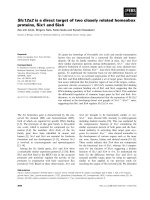
Figure 1 Strategy for targeting the Hunk allele. A) Nucleotide sequence of mouse Hunk cDNA corresponding to targeted exon 4 (highlighted). Arrows
mark exon/intron junctions. Subdomains VII (single-underline) and VIII (double-underline) of the catalytic domain are indicated. B, C Nucleotide sequences
of Hunk cDNA with spliced out exon 4 (B) or exons 4 and 5 (C). Note, a frame shift and truncation protein truncation occurs following splicing between
exons 3 and 5. However, splicing between exons 3 and 6 (which doesn’t induce a frame shift), produces a protein lacking subdomains important for
kinase activity. D Schematic of the targeting strategy. Hatched boxes represent exons. EcoRI (R), XhoI (X), NotI (N) sites (for cloning “short” and “long arms”
into pPNT vector) and BamHI (B) sites (for “long arm” recombination analysis) are shown. Arrows represent primers (used for ES clone analysis and
genotyping of mice). Nucleotide positions are shown according to mouse chromosome 16 sequence GeneBank Acc. No. NT 039625 2. Neomycin
phopshotransferase (dark grey arrow) and HSV thymidine kinase (black arrow) expression cassettes, are also shown. E PCR analysis of ES clones (primers
MK1S and neoB) demonstrating homologous recombination of the “short arm” in clones 194, 292 and 328 but not in negative clone N or parental
E14Tg2a ES cells (ES). F Southern blot analysis using P32-labeled probe “L” (white box in panel D) and BamHI-digested genomic DNA from clones 194 and
328, negative clone N and parental E14Tg2a ES cells (ES). G PCR genotyping of Hunk−/−(lane1), Hunk+/−(lane2) and Hunk+/+(lane3) mice (primers MK1A,
MKK and neoB). H Scheme of exons 2 through 7 of Hunk cDNA, demonstrating amplification products from wild type allele (wt), targeted allele missing
exon 4 (KODex4) and with additionally spliced out exon 5 (KODex4&5). Arrows represent RT-PCR Primers. I RT-PCR amplification from cerebellum RNA
of Hunk+/+(lane1), Hunk+/−(lane2) and Hunk−/−(lane3) mice. MW – DNA molecular weight markers.
Reed et al. BMC Cancer (2015) 15:110
contained 1 mM MgCl2 in Taq DNA polymerase reaction buffer, 0.2 mM dNTP, 2 U of Taq DNA polymerase
(Fermentas), primers MKK, MK1A, neoB and genomic
DNA template in the final volume of 25 μl. 35 cycles of
15 sec at 94°C, 30 sec at 63°C, and 60 sec at 72°C were
carried out on DNA EngineDyad amplifier (BioRad).
Mice and sample preparation
This study received ethical approval from Cardiff
University’s Animal Welfare and Ethical Review Body
(previously known as the ERP), and all animal procedures
were conducted in accordance with UK Home Office regulations. AhCre + Apc+/+ and AhCre + Apcfl/fl mice were
generated and maintained on an outbred background as
previously described [1]. Cre-recombinase activity was
induced from the Ah-Cre transgene by three intraperitoneal (IP) injections of 80 mg/kg β-naphthoflavone
within 24 h. Mice were sacrificed at day 4. Cohorts containing the ApcMin allele were sacrificed when animals
displayed symptoms of intestinal disease, including
weight loss, rectal bleeding and criteria of anaemia (as
assessed by pale feet). Tissues were harvested, fixed and
processed according to standard protocols as previously
described [1].
Page 4 of 9
group, in order to provide the relative fold change.
Thus, figures representing relative fold change do not
possess error bars, although statistical significance between
the ΔCT values was tested using the Mann–Whitney U test
and deemed significant when p < 0.05. Primers used were:
Hunk 5′atcacacagctccagagtacca3′ and 5′ggttggtgtggctcta
gtttct3′, β-actin 5′caccacaccttctacaatgagc3′ and 5′gtacga
ccagaggcatacagg3′, Axin2 5′gcagctcagcaaaaagggaaat3′ and
5′tacatggggagcactgtctcgt3′, Wif1 5′aacaagtgccagtgtcgaga
gg3′ and 5′gcctttttaagtgaaggcgtgtg3′.
Affymetrix microarray analysis
Normal colonic and paired polyp tissue was collected
from symptomatic ApcMin mice. Biotinylated target cRNA
was generated from these tissues as previously described
[1,20]. Affymetrix MOE430_2 gene arrays were run at the
CRUK facility at the Paterson Institute for Cancer Research, and the data has been deposited in NCBI’s Gene
Expression Omnibus and are accessible through GEO
Series accession number GSE65461 (.
nih.gov/geo/query/acc.cgi?acc=GSE65461). Arrays from
AhCre + Apc+/+ and AhCre + Apcfl/fl mice have previously been published for intestinal tissue [1] and liver
tissues [20].
BrdU labelling
Results
To achieve BrdU labelling for proliferation and migration studies, mice were administered with 250 μl BrdU
(Amersham) via an IP injection either 2 hrs or 24 hrs
prior to culling (n = 3 in all cases). Immunohistochemical
(IHC) staining for BrdU was performed using an antiBrdU antibody (BD biosciences 1:500). BrdU-positive cell
position and number were scored. Kolmogorov–Smirnov
test proved a significant difference between the distributions of BrdU-positive epithelial cells in crypts, 24 hr post
BrdU administration.
Wnt signalling activation results in up-regulated Hunk
expression
RT-PCR analysis
Total RNA extraction and first-strand cDNA synthesis
were carried out as described previously [18]. For analysis
of Hunk expression in mouse tissues one μl of cDNA was
used as a template for PCR amplification with primers 5′agatccagcagatgatccgac-3′ and 5′-tagcgctcaagtttcttgttcaa-3′
and Platinum AccuPrime DNA polymerase (Invitrogen).
35 cycles of 15 sec at 95°C and 90 sec at 68°C were carried
out on DNA Engine Dyad amplifier (BioRad). qPCR was
performed using Applied Biosystems TaqMan Universal
PCR mix and Steponeplus machine. The 2 − ΔΔCT
method [19] was used to calculate relative fold change
in expression levels, with β-Actin expression being used
as the housekeeping gene, which we can confirm amplified with an equivalent efficiency to the test primers.
The mean ΔCT values for the experimental groups were
compared to the mean ΔCT values for the control
Apc is a known key regulator of Wnt signalling, and
critically important in regulating normal intestinal
homeostasis. Conditional deletion of Apc within the
mouse intestine using an Ah-Cre recombinase to drive
recombination of LoxP flanked Apc alleles, has previously been shown to result in acute activation of Wnt
signalling and many hallmarks of neoplasia, including
increased proliferation and apoptosis and loss of differentiation and migration [1]. Affymetrix microarray analysis indicates an acute transcriptional activation of Hunk
following the loss of Apc in the intestine and liver and in
colonic adenomas from the ApcMin mouse (Figure 2).
qRT-PCR analysis confirms the transcriptional activation
of Hunk in these settings (Figure 1), indicating that Hunk
transcription is coincident with Wnt signalling activation
and tumour formation. Indeed, a Tcf/LEF consensus binding site can be found within the promoter region of Hunk,
and significant up-regulation of HUNK has been shown to
occur in human colorectal cancer cell lines [12].
Hunk-kinase deficiency results in increased intestinal cell
proliferation
To assist in our quest to investigate the importance of
Hunk-kinase in intestinal tumourigenesis, we generated a
novel mouse line carrying a Hunk-kinase deficient allele.
To do this exon 4 of the mouse Hunk gene was targeted.
Reed et al. BMC Cancer (2015) 15:110
Page 5 of 9
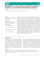
Figure 2 A Relative fold change in the levels of Hunk expression compared to the appropriate normal tissue assessed using qRT-PCR
analysis and Affymetrix micro-array (Intestinal array [1], liver array [20]). In all cases Mann–Whitney U test reveals a significant difference
between ΔCT values (p < 0.05). Small intestine (S. Int) and liver samples were collected Day 4 post Ah-Cre induction. Normal colonic tissue and
adjacent adenoma tissue (polyp) were taken from ApcMin animals.
A protein fragment encoded by this exon (amino acids
204–249) contains a part of subdomain VII (starting from
conserved Asp204 which is important for γ-phosphate of
MgATP orientation), the entire subdomain VIII, which is
critical for substrate recognition, and a portion of subdomain IX of the Hunk protein kinase catalytic domain.
Therefore, deletion of exon 4 results in the production of
a catalytically inactive Hunk protein. Moreover, deletion of
the exon 4 results in a shift of the open reading frame in
the transcript after joining exons 3 and 5. As a result, the
translated protein would consist of only 203 amino acids
of the Hunk polypeptide with translation terminating 2
codons downstream of the codon encoding Ile203
(Figure 1А and B). Following successful targeting in ES
cells (Figure 1D, F) and the production of chimeric
animals after ES cell injection into blastocysts, successful
germ line transmission of the targeted allele was confirmed by PCR (Figure 1G). Transgenic mice were further
back-crossed for 6 generation to obtain a mouse line on a
pure C57Bl/6 J genetic background.
Due to the lack of suitable antibodies for the detection
of endogenous Hunk protein in mouse tissue, RT-PCR
was used to analyze Hunk transcripts in wild type, Hunkheterozygous and homozygous mice (Figure 1 H,I). The
Hunk+/+ yielded the expected 826 bp PCR fragment
corresponding to the wild type Hunk allele, while it was
completely absent in samples of Hunk−/− animals
(Figure 1I). However, along with a 690 bp PCR fragment
corresponding to mRNA lacking exon 4, an additional
prominent amplification product, a 562 bp fragment, was
detected in Hunk−/− animals (Figure 1I). Cloning and sequencing revealed that this fragment represents a Hunk
transcript lacking not only targeted exon 4 but also exon 5
sequences. Importantly, deletion of both exons 4 and 5
(Figure 1H), while resulting in transcript encoding
catalytically inactive protein due to deletion of catalytic
domain portion, does not lead to a frame-shift and the
translated protein should be identical to full-length Hunk
except for the deletion of amino acids 204–291 (Figure 1C).
In heterozygous Hunk+/− mice, the wild type allele transcript was substantially more abundant than both variants
of the mutant allele transcript (Figure 1I).
Given our interests in the role of Hunk in intestinal
tumourigenesis, a detailed examination of the phenotype
in Hunk−/− intestine was performed. No differences
were found in the representation of the different cell
types of the intestine (assessed using alcian blue staining
for goblet cells, lysozyme IHC for paneth cells and grimelius staining for enteroendocrine cells), suggesting
Hunk-kinase activity is not involved in lineage specification in the intestine (Figure 3A). However, although the
gross morphology remained unaltered, with the number
of cells within the crypt remaining the same, Hunk−/−
intestine displayed a significant increase in crypt cell
proliferation within the small intestines (scored using
BrdU incorporation and histological examination of
intestinal crypts, Figure 3B). This was not accompanied
by any alteration in the rates of apoptosis (Figure 3B),
although migration rates along the crypt-villus axis were
significantly perturbed; Hunk−/− intestinal cells display
an increased rate of migration (Figure 3C). Consequently,
these data demonstrate that within a normal intestinal
setting, loss of kinase active Hunk was sufficient to induce
alterations in the normal intestinal kinetics, but this did
not alter normal intestinal physiology.
Hunk-kinase deficiency alters tumour initiation rates but
not survival in ApcMin mice
In order to address the importance of Hunk-kinase in
Wnt driven intestinal tumourigenesis, the Hunk-kinase
Reed et al. BMC Cancer (2015) 15:110
Page 6 of 9
A
Hunk +/+
Hunk -/-
Hunk +/+
Alcian Blue staining to
identify Goblet Cells
Hunk +/+
Hunk -/-
Lysozyme IHC to
identify Paneth Cells
Hunk -/-
Hunk +/+
Grimelius silver stain to
identify Enteroendocrine Cells
Hunk -/-
BRDU IHC to identify Cells
in S Phase
B
% within the crypt
*
3
2
1
Hunk +/+ Hunk -/-
Hunk +/+ Hunk -/-
60
BrdU
*
50
40
30
20
10
Hunk +/+
Hunk -/-
1.2
1
0.8
WT 2hr
0.6
WT 24 hr
Hunk-/- 2hr
0.4
Hunk -/-24hr
0.2
0
1
9
17
25
33
41
49
57
65
73
81
89
97
105
113
121
129
137
145
153
161
169
177
185
Accumulative frequency
C
Apoptosis
% of within the crypt
Mitosis
4
Cell position along the crypt-villus axis
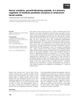
Figure 3 Characterisation of the intestine following Hunk-kinase loss. A Representative images showing no difference in Alcain Blue staining
(Goblet cells) and Lysozyme IHC (Paneth cells) in the different genotypes, and an increase in BRDU incorporation 2 hours post administration.
B Haematoxylin and eosin stained intestinal sections were used to score the percentage of Mitotic and Apoptotic bodies within intestinal crypts, while
BrdU IHC stained sections were used to score BrdU incorporation 2 hours post administration. Bar charts show means ± SD determined by scoring at
least 50 half crypts within 4 individuals from each cohort. * denotes p < 0.05 Mann–Whitney U test. C Accumulative frequency of BrdU positive cell
position along the crypt-villus axis, 2 hours and 24 hours post administration. Significant differences between the genotypes were detected at both
time points using Kolmogorov–Smirnov test, a test designed to examine probability distribution patterns.
deficient mice were inter-crossed with the established
ApcMin mouse model of intestinal cancer. Cohorts of
ApcMinHunk+/+, ApcMinHunk+/− and ApcMinHunk−/− littermates were generated and aged and monitored until
the animals displayed overt symptoms of intestinal disease, at which stage they were culled using the appropriate schedule 1 method, and tissues were harvested.
Kaplan-Meier survival analysis demonstrated that loss of
kinase active Hunk does not alter the survival of ApcMin
mice (Figure 4A). However, macroscopic scoring of
tumours at dissection showed significantly fewer (p = 0.025
Mann–Whitney), yet larger tumours in the ApcMinHunk−/−
cohort (mean number 21.4per animal +/− 2.7SEM, mean
size 7.1 mm2 +/− 0.3SEM) compared to the ApcMin
Hunk+/+ cohort (mean number 36.2 per animal +/−
4.8SEM, mean size 5.7 mm2 +/− 0.3SEM). Furthermore,
this difference was restricted to the small intestine
(Figure 4B). Detailed microscopic analysis of these tumours
did not reveal any differences in the stage, types or characteristics of the tumours occurring in the different cohorts.
Reed et al. BMC Cancer (2015) 15:110
A
Page 7 of 9
Kaplan-Meier Survival
% survival
ApcMinHunk+/+
Apc Min Hunk+/Apc MinHunk -/-
Time in Days
Number of Tumours
per animal
B
100
**
80
60
40
20
0
S.Int
Apc Min
Hunk +/+
S.Int
Apc Min
Hunk -/-
Colon
Apc Min
Hunk +/+
Colon
Apc Min
Hunk -/-
S.Int
S.Int
Apc Min
Apc Min
+/+
Hunk
Hunk -/-
Colon
Apc Min
Hunk +/+
Number of Tumours
per animal
C
15
Colon
Apc Min
Hunk -/-
Relative Fold Change
D
10
5
0
-5
Colon Colon Polyp Polyp
Apc Min Apc Min Apc Min Apc Min
Hunk +/+ Hunk -/- Hunk +/+ Hunk -/-
Colon Colon Polyp Polyp
Apc Min Apc Min Apc Min ApcMin
Hunk +/+ Hunk -/- Hunk +/+Hunk -/-
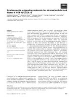
Figure 4 Survival and tumour burden analysis. A Kaplan-Meier
survival curve of aged cohorts of ApcMinHunk+/+ (n = 23), ApcMinHunk+/−
(n = 27) and ApcMinHunk−/− (n = 23) mice, demonstrating that no
significant differences in survival between the cohorts. B Box plot
displaying the total number of tumours found at death in the
aged cohorts of ApcMinHunk+/+ and ApcMinHunk−/− mice. The box
encompasses the first quartile (at bottom) to the third quartile
(at top) of the data set and the horizontal boxed line represents the
median. ** p < 0.01 Mann–Whitney U test. C Box plot displaying the
size of tumours found at death in the aged cohorts of ApcMinHunk+/+
and ApcMinHunk−/− mice. D qRT-PCR analysis showing relative
expression levels of Axin2 and Wif1 in normal colonic tissue (colon) and
adjacent adenoma tissue (polyp) taken from 4 aged matched animals
from the different genotypes. * p < 0.05 Mann–Whitney U test between
ΔCT values.
Neither the proliferation rates nor apoptosis rates within
tumours differed with Hunk-kinase status (Additional file
1: Figure S1). Furthermore, qRT-PCR analysis for Wnt target genes (including cMyc, Ascl2, Axin2, CD44, CD1,
Sox17, Wif1 and Tiam1) did not identify any significant
differences between the colonic tumours isolated from
both cohorts of mice. However, it is pertinent to note that
both Wif1 and Axin2 were significantly down-regulated
within the normal colonic ApcMinHunk−/− tissue compared to normal colon samples from ApcMinHunk+/+ mice
(Figure 4D). Thus, kinase active Hunk positively contributes toward the normal expression of two negative regulators of canonical Wnt signalling within normal intestinal
tissue, but loss of Hunk-kinase and the subsequent down
regulation of Axin2 and Wif1 does not confer a generic
mis-regulation of Wnt signalling. A precise molecular
characterisation following the loss of Hunk-kinase is required to ascertain the exact mechanism by which this
kinase influences gene transcription and cell proliferation.
Overall, in relation to intestinal tumourigenesis our results
show that Hunk-kinase activity does impact on intestinal
tumour initiation within the small intestine, but this is not
sufficient to alter the overall tumour burden or survival in
the ApcMin mouse model of intestinal cancer.
Discussion
The exact role of the SNF1-related serine/threonine kinase
Hunk (Mak-V) remains unclear. Here we have generated a
novel kinase-deficient Hunk allele, and produced homozygous Hunk-kinase deficient mice, Hunk−/−. Two previous
studies have shown Hunk to be a negative regulator of
proliferation within normal epithelial cells [9,10] and our
findings support this notion. We have shown that loss of
Hunk-kinase activity within the normal intestinal setting
results in an increase in proliferation within epithelial
cells. This is accompanied by an increase in cell migration
rates, thereby maintaining normal physiology despite altered kinetics.
An increasing body of evidence links the function of
Hunk with cancer initiation, progression and metastasis
[3,8,11-13], although there remains uncertainty regarding the precise involvement of Hunk in tumourigenesis.
To date, most studies have analysed the role of Hunk in
mammary tumourigenesis, largely due to the known role
of this protein in mammary gland development [4].
However, our microarray findings implicated Hunk in intestinal tumourigenesis, a role we wished to elucidate
further. We have shown that Hunk expression becomes
significantly up-regulated from the earliest stages of
tumour initiation following Apc loss, indicating this gene
is probably a Wnt signalling target gene. Indeed a Tcf/LEF
binding motif can be found within the promoter region of
Hunk. We appreciate this evidence is circumstantial, and a
Reed et al. BMC Cancer (2015) 15:110
more detailed interrogation is required to confirm Hunk
as a Wnt target gene.
Studies using Xenopus embryos have shown that
Hunk has the ability to modulate Wnt signalling. We
used qRT-PCR analysis to examine this in the intestine
and demonstrated a significant reduction in expression
levels of two negative regulators of Wnt signalling, Wif1
and Axin2, accompanying the loss of kinase active Hunk.
However, this did not translate to a generic de-regulation
of Wnt signalling. Interestingly, contrary to a recent publication by Yeh et al. [14] who described Hunk as a negative
regulator of cMyc expression, we did not observe any
significant alteration in the levels of cMyc transcription
accompanying Hunk-kinase loss. Discrepancies in the
examined tissues and experimental setup might account for these differences. In an attempt to explain
the mis-regulation of both Wif1 and Axin2 which
can be regulated by components of the BMP/
TGFβpathway [21,22], qRT-PCR analysis of components and targets of this pathway was performed.
However once more, a generic mis-regulation of this
pathway was not confirmed by qRT-PCR analysis. A
more detailed genomic wide study would be required
to confidently identify the mechanism through which
Hunk-kinase is able to negatively regulate proliferation in the intestine.
Intercrossing Hunk−/− mice with ApcMin mice allowed
us to determine the role of Hunk-kinase in Wnt signalling driven intestinal tumourigenesis. We have shown a
significant reduction in the tumour initiation rate within
the small intestine in ApcMinHunk−/− mice, but this does
not impact on overall survival due to an accompanying
increase in the size of those tumours that do form. It is
possible that the reduced tumour initiation rate is associated with the increased cell turnover rate along the
crypt-villus axis (increased proliferation and migration)
seen following the loss of Hunk-kinase, although the
exact mechanisms for this have not been elucidated. Further studies would be required to determine the significance of these subtle changes associated with the lack of
kinase active Hunk.
Overall, our data confirm Hunk-kinase as a negative
regulator of normal epithelia proliferation, and demonstrate that in the classical ApcMin mouse model of intestinal tumourigenes, Hunk-kinase activity significantly
impacts on tumour initiation rates during the early
stages in tumourigenesis.
Conclusions
Here we describe the production of a new Hunk-kinase
deficient mouse model and use it to examine the importance of this kinase during the early stages of intestinal
tumourigenesis. We show that despite not affecting
overall survival of the ApcMin mice, Hunk-kinase is a
Page 8 of 9
negative regulator of normal intestinal proliferation, and
impacts significantly on small intestinal tumour initiation rates.
Availability of supporting data
The Affymetrix array data has been deposited in NCBI’s
Gene Expression Omnibus and are accessible through
GEO Series accession number GSE65461 (http://www.
ncbi.nlm.nih.gov/geo/query/acc.cgi?acc=GSE65461).
Additional file
Additional file 1: Figure S1. Mitosis and apoptosis levels scored from
H + E stained sections on intestinal adenomas. Bar charts show means SD
of values obtained from at least 6 tumours from three individuals within
each cohort.
Abbreviations
APC: Adenomatosis polyposis coli; ES: Embryonic stem cell; HUNK: Hormonally
upregulated Neu-associated kinase; IHC: Immunohistochemistry; IP: Intraperitoneal; MDCT: Mouse distal convoluted tubule; S. Int: Small intestine.
Competing interests
The authors declare that they have no competing interests.
Authors’ contributions
KRR, IVK, VB and ARC designed the research, IVK, NN and EVK generated the
novel Hunk- allele, KRR managed the mouse intercrosses, BH and JLP performed
IHC and qRT-PCR, KRR analysed data. The manuscript was drafted by KRR and
all authors critically reviewed. All authors read and approved the final
manuscript.
Acknowledgements
This work was supported by a Cancer Research UK program grant awarded
to ARC, a Wellcome Trust VIP award awarded to KRR, a Wellcome Trust Short
Term Travel Grant 071269 and a INTAS Young scientist Fellowship awarded
to IV, and Nuffield Bursary summer studentships awarded to JLP and BH. We
thank Mark Bishop and Derek Scarborough for technical services and support
with genotyping and histology.
Author details
1
University of Cardiff, European Cancer Stem Cell Research Institute, School
of Biosciences, Cardiff CF10 3AX, UK. 2School of Biosciences, University of
Cardiff, Cardiff CF10 3AX, UK. 3Russian Academy of Sciences, Institute of Gene
Biology, 34/5 Vavilov street, Moscow 119334, Russia Federation. 4Institute of
General Pathology and Pathophysiology of Russian Academy of Medical
Science, 8 Baltijskaya str, Moscow 125315, Russian Federation.
Received: 14 August 2014 Accepted: 19 February 2015
References
1. Sansom OJ, Reed KR, Hayes AJ, Ireland H, Brinkmann H, Newton IP, et al.
Loss of Apc in vivo immediately perturbs Wnt signaling, differentiation, and
migration. Genes Dev. 2004;18:1385–90.
2. Korobko IV, Kabishev AA, Kiselev SL. Identification of the new protein kinase
specifically transcribed in mouse tumors with high metastatic potential.
Dokl Akad Nauk. 1997;354(4):554–6.
3. Korobko IV, Kalinichenko SV, Korobko EV, Ninkina NN, Kiselev SL, Buchman
VL. Pro-survival activity of the MAK-V protein kinase in PC12 cells. Cell Cycle.
2010;9(20):4248–9.
4. Gardner HP, Wertheim GBW, Ha SI, Copeland NG, Gilbert DJ, Jenkins NA,
et al. Cloning and characterization of Hunk, a novel mammalian SNF1-related
protein kinase. Genomics. 2000;63:46–59.
5. Kalinichenko SV, Itoh K, Korobko EV, Sokol SY, Buchman VL. Korobko IV
identification of Nedd4 E3 ubiquitin ligase as a binding partner and
regulator of MAK-V protein kinase. PLoS One. 2012;7(6):e39505.
Reed et al. BMC Cancer (2015) 15:110
6.
7.
8.
9.
10.
11.
12.
13.
14.
15.
16.
17.
18.
19.
20.
21.
22.
Page 9 of 9
Kalinichenko SV, Vikhreva PN, Korobko IV. Interaction between MAK-V protein
kinase and synaptopodin. Biochemistry (Mosc). 2011;76(2):196–201.
Korobko IV, Korobko EV, Kiselev SL. The MAK-V protein kinase regulates
endocytosis in mouse. Mol Gen Genet. 2000;264(4):411–8.
Quintela-Fandino M, Arpaia E, Brenner D, Goh T, Yeung FA, Blaser H. HUNK
suppresses metastasis of basal type breast cancers by disrupting the
interaction between PP2A and cofilin-1. Proc Natl Acad Sci U S A.
2010;107(6):2622–7.
Gardner HP, Belka GK, Wertheim GB, Hartman JL, Ha SI, Gimotty PA, et al.
Developmental role of the SNF1-related kinase Hunk in pregnancy-induced
changes in the mammary gland. Development. 2000;127(20):4493–509.
Saki M, Tamura K, Tsurumi Y, Tanaka Y, Koide Y, Matsuda M, et al. Renal
Expression of MAK-V/Hunk in renal distal tubules and its possible involvement
in proliferative suppression. Am J Physiol. 2007;292(5):F1526–36.
Korobko IV, Zavalishina LE, Kiselev SL, Raĭkhlin NT, Frank GA. Proteinkinase
MAK-V/Hunk as a possible diagnostic and prognostic marker of human
breast carcinoma. Arkh Patol. 2004;66(5):6–9.
Wertheim GBW, Yang TW, Pan T, Ramne A, Liu Z, Gardner HP, et al. The
Snf1-related kinase, Hunk, is essential for mammary tumor metastasis. Proc
Natl Acad Sci U S A. 2009;106:15855–60.
Yeh ES, Yang TW, Jung JJ, Gardner HP, Cardiff RD, Chodosh LA. Hunk is
required for HER2/neu-induced mammary tumorigenesis. J Clin Invest.
2011;121(3):866–79.
Yeh ES, Belka GK, Vernon AE, Chen CC, Jung JJ. Chodosh LA Hunk
negatively regulates c-myc to promote Akt-mediated cell survival and
mammary tumorigenesis induced by loss of Pten. Proc Natl Acad Sci U S A.
2013;110(15):6103–8.
Kibardin A, Ossipova O, Sokol SY. Metastasis associated kinase modulates
Wnt signaling to regulate brain patterning and morphogenesis.
Development. 2006;133(15):2845–54.
Tybulewicz VL, Crawford CE, Jackson PK, Bronson RT, Mulligan RC. Neonatal
lethality and lymphopenia in mice with a homozygous disruption of the
c-abl proto-oncogene. Cell. 1991;65:1153–63.
Ninkina N, Papachroni K, Robertson DC, Schmidt O, Delaney L, O’Neil F,
et al. Neurons expressing the highest levels of γ-synuclein are unaffected by
targeted inactivation of the gene. Mol Cel Biology. 2003;23:8233–45.
Buchman VL, Luke C, Borthwick EB, Ninkina N. Organisation of the mouse
Ruk locus and expression of isoforms in mouse tissues. Gene. 2002;295:13–7.
Livak KJ, Schmittgen TD. Analysis of relative gene expression data using
real-time quantitative PCR and the 2(−delta delta C(T)) method. Methods.
2001;25:402–8.
Reed KR, Athineos D, Meniel VS, Wilkins JA, Ridgway RA, Burke ZD, et al.
B-catenin deficiency, but not Myc deletion, suppresses the immediate
phenotypes of Apc loss in the liver. Proc Natl Acad Sci U S A.
2008;105(48):18919–23s.
Xu B, Chen C, Chen H, Zheng SG, Bringas Jr P, et al. Smad1 and its target
gene Wif1 coordinate BMP and Wnt signaling activities to regulate fetal
lung development. Development. 2011;138(5):925–35.
Dao DY, Yang X, Chen D, Zuscik M, O’Keefe RJ. Axin1 andAxin2 are
regulated by TGF- and mediate cross-talk between TGF- and Wnt signaling
pathways. Ann N Y Acad Sci. 2007;1116:82–99.
Submit your next manuscript to BioMed Central
and take full advantage of:
• Convenient online submission
• Thorough peer review
• No space constraints or color figure charges
• Immediate publication on acceptance
• Inclusion in PubMed, CAS, Scopus and Google Scholar
• Research which is freely available for redistribution
Submit your manuscript at
www.biomedcentral.com/submit