Multimodal therapy in treatment of rectal cancer is associated with improved survival and reduced local recurrence - a retrospective analysis over two decades
Bạn đang xem bản rút gọn của tài liệu. Xem và tải ngay bản đầy đủ của tài liệu tại đây (542.44 KB, 10 trang )
Wiegering et al. BMC Cancer 2014, 14:816
/>
RESEARCH ARTICLE
Open Access
Multimodal therapy in treatment of rectal cancer
is associated with improved survival and reduced
local recurrence - a retrospective analysis over
two decades
Armin Wiegering1,2*, Christoph Isbert1, Ulrich A Dietz1, Volker Kunzmann3, Sabine Ackermann1,
Alexander Kerscher1,4, Uwe Maeder4, Michael Flentje5, Nicolas Schlegel1, Joachim Reibetanz1,
Christoph-Thomas Germer1,4 and Ingo Klein1
Abstract
Background: The management of rectal cancer (RC) has substantially changed over the last decades with the
implementation of neoadjuvant chemoradiotherapy, adjuvant therapy and improved surgery such as total
mesorectal excision (TME). It remains unclear in which way these approaches overall influenced the rate of local
recurrence and overall survival.
Methods: Clinical, histological and survival data of 658 out of 662 consecutive patients with RC were analyzed for
treatment and prognostic factors from a prospectively expanded single-institutional database. Findings were then
stratified according to time of diagnosis in patient groups treated between 1993 and 2001 and 2002 and 2010.
Results: The study population included 658 consecutive patients with rectal cancer between 1993 and 2010.
Follow up data was available for 99.6% of all 662 treated patients. During the time period between 2002 and 2010
significantly more patients underwent neoadjuvant chemoradiotherapy (17.6% vs. 60%) and adjuvant chemotherapy
(37.9% vs. 58.4%). Also, the rate of reported TME during surgery increased. The rate of local or distant metastasis
decreased over time, and tumor related 5-year survival increased significantly with from 60% to 79%.
Conclusion: In our study population, the implementation of treatment changes over the last decade improved the
patient’s outcome significantly. Improvements were most evident for UICC stage III rectal cancer.
Keywords: Rectal cancer, Improved survival, TME
Background
Colorectal cancer (CRC) is the second leading cancer in
the western world, accounting for about 500,000 deaths
annually worldwide [1]. About half of the CRC are located in the rectum [2,3]. Rectal carcinoma (RC) has
been considered and treated as an independent disease
due to its primarily extra peritoneal location, the potential, impairment of anorectal continence and the
* Correspondence:
1
Department of General, Visceral, Vascular and Pediatric Surgery, University
Hospital, University of Wuerzburg, Oberduerrbacherstr. 2, 97080 Wuerzburg,
Germany
2
Department of Biochemistry and Molecular Biology, University of
Wuerzburg, Am Hubland, 97074 Wuerzburg, Germany
Full list of author information is available at the end of the article
differences in metastatic behavior. Over the last decades
numerous studies extensively investigated different treatment options in chemo-, radio-, chemoradiotherapy and
surgery to improve the outcome, leading to significant
changes in the management of RC [4,5].
Today the treatment can be divided in four phases:
First, the preoperative diagnostic phase with the staging
based on rectoscopy, endosonography, MRI and CT
scan, followed by a second phase of neoadjuvant therapy
for locally advanced and nodal-positive cancer in the
middle and lower rectum [6,7]. The third phase consists
of surgical removal of the cancer, which is performed by
central ligation of the lower mesenteric vessels, systemic
lymph-node dissection and rectal resection including the
© 2014 Wiegering et al.; licensee BioMed Central Ltd. This is an Open Access article distributed under the terms of the
Creative Commons Attribution License ( which permits unrestricted use,
distribution, and reproduction in any medium, provided the original work is properly credited. The Creative Commons Public
Domain Dedication waiver ( applies to the data made available in this
article, unless otherwise stated.
Wiegering et al. BMC Cancer 2014, 14:816
/>
total mesorectal excision (TME) [8-11]. The fourth phase
consists of adjuvant therapy depending on the definitive
histopathological stage with 5-fluorouracil, leucovorin and
oxaliplatin [12,13]. In the fifth phase, multimodal chemotherapy and/or resection of metastases are performed if
recurrent disease is detected during a structured followup [14-16].
While each individual modification of the disease management has been described in detail with respect to its
specific effect and clinical outcome, little is known about
the synergistic effects of all modifications together. The
presumed additive effect has led to multimodal treatment suggestions in the current guidelines (NCIE CG131
( NCCN rectal
cancer (); ESMO (o.
org); AWMF (www.AWMF.de)). Recently also the European
consensus guidelines for treatment of patients with colorectal cancer has been published to achieve an equivalent
treatment for patients across Europe and to address open
questions [17].
We performed a single center retrospective analysis of
patients with rectal cancer from 1993 to 2010. The aim
was to compare how the combination of multi factorial
changes has improved the cancer-related outcome in
terms of local recurrence, distant metastasis and survival.
Methods
Patient population
All patients with rectal cancer treated at the University of
Wuerzburg Medical Centre (UKW) between January 1993
and December 2010 were chosen from the Wuerzburg
Institutional Database (WID). Patients were grouped into
categories according to the time of diagnosis (January
1993 to December 2001 and January 2002 and December
2010).
Data source
The WID is a central data repository that has been expanded on a daily basis since 1984 with clinical, operative
and research data of patients who were evaluated and
treated at the UKW. Data available within the WID include patient demographics, histological diagnoses based
on International Classification of Diseases coding standards, physician data, inpatient admission and outpatient
registration data, operative procedures, laboratory results
and computerized pharmacy records. Continuous cross
platform integration with the Wuerzburg Comprehensive
Cancer Registry ensures updated follow-up information
for identification of deceased patients. Inpatient and outpatient records of all identified patients were reviewed
retrospectively to extract information regarding type and
duration of chemotherapy, sites of metastatic disease at
presentation and disease status at last follow-up. Missing
data were retrieved from patient case notes when possible.
Page 2 of 10
Demographic details, along with clinical data at the
time of primary diagnosis and during the surgery (tumor
site and the presence of metastases) as well as histologic
results (tumor (T) stage, nodal (N) stage, tumor differentiation (G) and evidence of microscopic venous (V) and
lymphatic vessel invasion (L)) were correlated with survival data obtained from prospective follow-up registry.
Follow-up
Postoperative follow-up consisted of quarterly outpatient
assessments or gathering complete information from the
patient’s primary care physician in 3-month intervals, for
10 years. After 10 years, information was obtained on an
annual basis retrospectively. Depending on the postoperative staging, follow-up included abdominal ultrasound at 3, 6, 12 and 18 months and after that on a
yearly basis. Computed tomography and surveillance
colonoscopy were routinely performed 3 to 6 months
after the resection and repeated every year. After 5 years,
no structured follow-up was performed and diagnostic
tests where based on symptoms or incidental findings.
Ethics
The University of Wurzburg ethics committee has approved this study for full ethics waiver due to its retrospective and anonymised nature. The head of the board
for internal data requests, Dr. U. Maeder granted permission to access data from the registry.
Statistical analysis
The data were analyzed with statistical software set up
in Linux by an-house biostatistician (M.U.). Clinical and
histological parameters were compared with the Mann–
Whitney U or Kruskal–Wallis test for continuous data
and with the χ2 test for categorical variables. P < 0.05
was considered statistically significant. Cox proportional
hazard modeling or ‘Cox regression’ was used for multivariate testing [18,19]. Survival curves were drawn according to Kaplan–Meier methods.
Results
Patient cohort, demographics and tumor stage
From January 1993 until December 2010 a total of 662 patients were diagnosed with rectal cancer; only 4 patients
(0.6%) had to be excluded from further analysis secondary
to missing follow-up data. The remaining cohort consisted
of 426 men and 232 women, with an average age of
66 years (+/− 11.7). 301 of these patients were diagnosed
before 2002, 357 between January 2002 and December
2010. Tumors located in the distal 4 cm from the anal
verge increased from 19.6% to 33.9% (p < 0.001). In contrast, tumors located 8-12 cm from the anal verge decreased from 34.6 to 22.7% (p < 0.001). Whereas the
pathological UICC stage (post surgical therapy) did not
Wiegering et al. BMC Cancer 2014, 14:816
/>
change between both periods, the clinical (pre-treatment)
cUICC stage differed significantly and shifted towards
more advanced disease. Patients with cUICC stage III increased from 23.3% to 37.8% (p < 0.001). Also, patients
with cT3&4 increased from 59.5% to 69.5% (p = 0.007) and
cN + from 30.2% to 51.0% (p < 0.001). The post-resection
pathological examination, in the more recent period between 2002 and 2010 revealed an overall reduced tumor
size and significantly less tumor-infiltrated lymph nodes
(p = 0.005). The comparison of limited (pUICC 0;I;II) to
advanced tumor stage (pUICC III; IV) showed that significantly more patients were in pathological limited stage
during the second time period (p = 0.048).
Demographics, tumor stage and size, tumor localization
and lymph node status are summarized in Table 1.
Therapeutic management
Overall the proportion of patients undergoing any additional therapy to surgery (neoadjuvant and adjuvant) increased over time. For neoadjuvant treatment the rate
increased from 17.6% to 60%. Neoadjuvant radiotherapy
(RT) independent of the protocol (short term 5×5Gy
or long term 25×1.8Gy), doubled from 12% to 23.3%
(p = 0.011). However, changes were most prominent for
neoadjuvant chemoradiotherapy (RCT), which increased
from 5.3% to 35.3% (p < 0.001). When analyzing the
changes in neoadjuvant treatment they were most prominent for patients in clinical stage cUICC II/III. The percentage of patients without any preoperative treatment in
this group dropped from 71.8% in the first time frame to
15.7%. While the proportion of patients undergoing radiotherapy alone more then doubled from 20.4% to
50.0% (p < 0.001), patients undergoing chemoradiotherapy
increased even more by five times from 7,0% to 34.3%
(p < 0.001) (Tables 2 and 3). When comparing patients in
the clinical cUICC stage I there was no difference in the
proportion of patients receiving neoadjuvant treatment
(2.5% vs. 5.1%; p = n.s.). Neoadjuvant radiochemotherapy
resulted in 11.9% of patients with a complete pathological
response, 73.3% of these patients had been in clinical UICC
stage III previous to neoadjuvant treatment. Still more than
20% of all patients did not receive preoperative treatment
in the later time period, which was either secondary to patient refusal or to tumors located above 12 cm from the
anal verge in 7% of all rectal cancers who were not enrolled
in neoadjuvant treatment.
Also significantly more patients underwent any adjuvant treatment in the second time period (38% vs. 58%,
p < 0.001). Whereas adjuvant radiation therapy alone
(6.3% vs. 2.2% p = 0.009) or in combination with chemotherapy (11.0% vs. 5.9% p = 0.02) was more common between 1993 and 2001, the rate of adjuvant chemotherapy
increased three-fold in the second period from 16% to
45.3% (p < 0.001) (Table 3).
Page 3 of 10
For adjuvant treatment in pUICC stage III the percentage of patients receiving any therapy did not change significantly, whereas the distribution shifted from radiotherapy
with (29% vs. 11.4% p = 0.008) or without chemotherapy
(8.1% vs. 1.3% p = 0.047) (total 37.1% vs. 12.7% p < 0.001)
towards chemotherapy only (22.6% vs. 53.2% p < 0.001).
Differences were more pronounced in stage pUICC II: in
the first time period 22% of all patients received chemo or
chemoradiotherapy, whereas it was 67% in the second
period (p < 0.001).
Overall, more than 90% of the patients underwent any
form of surgical intervention (resection or extirpation)
(92% vs. 91.6%). The proportion undergoing low anterior
rectum resection increased from 59.5% to 64.1% (p < 0.001)
whereas patients undergoing rectum extirpation decreased
(22.3% to 18.2%; n.s.). The rate of patients undergoing
transanal resection increased slightly from 4% to 7.6%.
Also, the rate of patients receiving enterostomy increased
from 64.8% to 75.1% (p = 0.004). TME was reported for
only two patients before 2002, whereas in the second time
period TME was documented in 124 patients (34.7%,
p < 0.001; Table 4).
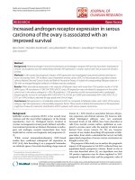
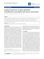
Recurrence rate
A significantly lower rate of tumor recurrence (local and
metastatic) was found in the second period (Figure 1A).
Five-year recurrence rate was 32% in the first period,
whereas it was 19% between 2002 and 2010 (p = 0.0035).
The five-year local recurrence rate decreased from 14.3%
to 5.3% after 2002 (Figure 1B). In addition, a decreased
five-year distant metastasis was observed (25,5% to
15,2%; p < 0.015). (Figure 1C). When preforming a stageby-stage analysis for the occurrence of distant metastasis, especially patients in UICC stage III had a significant
lower 5 year rate in the second time period (40.8% vs
17.5% p = 0.0075). Comparing the neoadjuvant and adjuvant treatment for this subgroup, in the second timeframe patients were more commonly treated with
neoadjuvant radio- (17.7% vs 37.7% p = 0.01) or radiochemotherapy (5.2% vs. 39% p < 0.001) whereas adjuvant treatment was not significantly different (data not
shown). To determine the effect of radiotherapy or radiochemotherapy an analysis independent of the timeframe was performed. The five-year distant metastasis
rate differed significantly from 39.1% for patients without any treatment, to 22.1% for patients with radiotherapy only and 7.3% for patients with radiochemotherapy
(p = 0.028).
Treatment of metastatic disease
During the first period, 38 out of 67 patients with stage
UICC IV had synchronous liver metastasis only. Three
patients (7.9%) underwent liver resection. Two remained
without recurrent disease. In the later period, 39 out of
Wiegering et al. BMC Cancer 2014, 14:816
/>
Page 4 of 10
Table 1 Characteristics of 658 patients treated between 1993–2010 for rectal cancer at the University hospital of
Wuerzburg
Characteristic
1993-2001 (n=301)
No.
2002..2010 (n=357)
%
No.
p-value
%
Sex
0.035
Male
182
244
Female
119
113
Age, years
n.s.
Median
66.16 (+/−11.88)
66.83 (+/−11.5)
Range
22.06-93.6
27.7-93.6
pUICC
0
0
0
15
4.2
<0.0001
I
95
28.6
127
35.6
n.s.
II
58
19.3
67
18.8
n.s.
III
62
20.6
79
22.1
n.s.
IV
67
22.3
64
17.9
n.s.
X
19
6.3
5
1.4
<0.001
I
81
26.9
79
22.1
n.s.
cUICC
II
75
24.9
69
19.3
n.s.
III
67
22.3
135
37.8
<0.001
IV
66
21.9
69
19.3
n.s.
X
12
4
5
1.4
0.037
0
0
18
5
<0.001
Patho. T-stage
pT0
pT 1,2
118
39.2
163
45.7
n.s.
pT3
124
41.2
129
36.1
n.s.
pT4
29
9.6
13
3.6
0.002
pTx
30
10
30
8.4
n.s.
pTis
0
0
3
0.8
n.s.
pN0
149
49.5
216
60.5
0.005
pN1
52
17.3
65
18.2
n.s.
pN2
61
20.2
38
10.6
<0.001
pNx
39
13
37
10.4
n.s.
59
19.6
121
33.9
<0.001
Patho. N-stage
Distance to anal verge
<4cm
4-8cm
96
31.9
129
36.1
n.s.
8-12cm
104
34.6
81
22.7
<0.001
>12cm
36
12
24
6.7
0.02
x
6
2
1
0.5
0.033
cT1,2
95
31.6
81
22.7
0.01
cT3,4
179
59.5
248
69.5
0.007
cTx
27
9
28
7.8
n.s.
Clinical T-stage
Wiegering et al. BMC Cancer 2014, 14:816
/>
Page 5 of 10
Table 1 Characteristics of 658 patients treated between 1993–2010 for rectal cancer at the University hospital of
Wuerzburg (Continued)
Clinical N-stage
N0
140
46.5
128
35.9
0.006
cN+
91
30.3
182
51
<0.001
cNx
70
23.2
47
13.2
<0.001
64 patients had synchronous liver metastasis only. 12 patients (30.8%) underwent liver resection and 6 developed
recurrent diseases. (Rate of liver resection p = 0.011)
During follow up of patients diagnosed before 2002
(n = 234), 31 developed metachronous liver metastases
and 9 underwent liver resection. In contrast, out of the
293 patients diagnosed from 2002–2010, 20 patients developed liver metastasis. In this cohort, 12 (60%) underwent liver resection (p = 0.028) (Table 5).
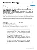
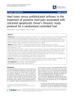
Survival
The overall survival rate improved significantly in patients who were diagnosed between 2002 and 2010
(5 year 60.5% vs. 79.8% p < 0.0001) (Figure 2). When
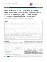
comparing patients according to the stage at diagnosis,
those in UICC I did not show any differences between
both time periods. Interestingly, all other patients (UICC
stage II, III and IV) demonstrated a significantly improved survival (Figure 3A-D).
Multivariate testing
In a multivariate analysis of epidemiological and clinical
features, presence of distant metastases (HR = 3,627, CI:
1,338-9,833, P = 0.011), presence of locoregional lymph
node metastases (HR: 2.38; CI:1.49-3.82, P < 0.001) and
decade of tumor incidence (HR = 2.280, CI: 1,649 3,153, P < 0.001) were independent predictors of tumorrelated death.
Discussion
By analyzing the patient treatment and outcome from a
prospective institutional based database (WID) we found
a significantly improved survival of patients treated for
rectal cancer in the last two decades. This was eminent
and therefore attributable to patients who were treated
with newly implemented strategies for rectal cancer.
Major changes as neoadjuvant radiochemotherapy and
TME have been introduced at our institution between
1999–2003. Consequently, improvements in outcome
comparing the time periods between 1993–2001 with
2002–2010 were to be expected. Unfortunately we cannot attribute the improved survival directly to special
change in treatment. It seams very likely to be an additional and potentially synergistic effect of improved
surgery, neoadjuvant and adjuvant treatment rather than
coexistence of the several effects.
Historically, surgical resection for rectal cancer has
been burdened by a high local recurrence rate and concomitant or consecutive distant metastatic disease resulting in a moderate 5-year survival rate. With progress
in surgical technique, supportive management and new
insights in the understanding of oncological principles
improved outcome was observed [20]. Especially in the
last two decades, the therapeutic management has changed dramatically in terms of pre- and postoperative
treatment, as well as surgical strategy. Each individual
change has demonstrated advantages in terms of outcome (survival, recurrence etc.) or quality of life (sphincter preservation, fecal continence, etc.).
To our knowledge, this is the first study comparing
survival and recurrence rates including all implemented
changes over the past two decades, rather than focusing
on a single aspect in the change of management in a
large case series with over 600 patients. We have deliberately included all patients, irrespective of cancer
stage, age or treatment intention to reflect the clinical
daily live reality in this cancer. Since this is a longitudinal study of a single institution within the same region,
a selection bias by massive socioeconomic changes in
the study population appears to be unlikely.
We observed a significant shift towards more patients
with clinical stage UICC III and less clinical stage UICC
II, probably due to a more detailed diagnostic work-up
via MRI and endoluminal ultrasound in the second time
Table 2 Percentage of neoadjuvant therapy performed in each time period in clinical stage UICC III patients
Neoadjuvant-Therapy in clinical stage
1993-2001 (n=142)
2002. 2010 (n=204)
p-value
cUICC II/III
No.
%
No.
%
No
102
71.8
32
15.7
<0.001
Chemo
1
0.7
0
0
n.s.
Radio
29
20.4
102
50.0
<0.001
Radiochemo
10
7.0
70
34.3
<0.001
Wiegering et al. BMC Cancer 2014, 14:816
/>
Page 6 of 10
Table 3 Percentage of neoadjuvant and adjuvant therapy
performed in each time period over all patients
Therapy
all patients
1993-2001 (n=301)
2002. 2010 (n=357)
p-value
No.
%
No.
%
No
248
82.4
143
40
<0.001
Chemo
1
0.3
3
0.8
n.s.
Neoadjuvant
Radio
36
12
83
23.3
0.011
Radiochemo
16
5.3
126
35.3
<0.001
Unknown
0
0
2
0.6
n.s.
No
187
62.1
149
41.6
<0.001
Chemo
48
16
162
45.3
<0.001
Adjuvant
Radio
19
6.3
8
2.2
0.009
Radiochemo
33
11
20
5.9
0.02
Unknown
14
4.7
18
5.0
n.s.
period [21,22]. This also might account for a possible
underrepresentation of clinical UICC stage III patients
in the first treatment period and thereby leading to a
stage migration in the later time period [23]. However,
stage migration alone can hardly explain the observed
major improvement. This is emphasized by the fact that
that the survival of patients in stage UICC III in the
second timeframe is superior to UICC II in the first
timeframe.
Table 4 Type of surgical procedure performed in each
time period over all patients
Characteristics
1993-2001
(n=301)
No.
%
2002. 2010
(n=357)
No.
p-value
%
Operation
n.s.
Yes
277
92
327
91.6
No
24
8
30
8.4
No
24
8
30
8.4
n.s.
Anterior resection
179
59.5
167
64.1
<0.001
Extirpation
67
22.3
65
18.2
n.s.
Trans anal excision
12
4
27
7.6
n.s.
Other
19
6.3
6
1.7
TME/PME reported
0.002
<0.001
Yes
2
0.7
124
34.7
No
299
99.3
233
65.3
Yes
195
64.8
268
75.1
No
88
29.2
66
18.5
Not reported
18
6
23
6.4
Stoma
0.004
When analysing post-operative T and N stage separately,
patients with T1/T2 and the proportion of nodal negative
cancer had increased significantly. Also, comparing the ratio of histologically advanced cancer (pUICC III and IV)
to limited cancer (pUICC 0, I and II) showed a significant
shift towards limited cancer. Since there is no biological
explanation why patients in the second time period should
have different tumor stages, the shift toward lower pathological tumor stages could be attributed to the effects of
neoadjuvant treatment, in the second time period or earlier diagnostic detection.
The effect of neoadjuvant radiochemotherapy is also
supported by the fact that in the second time period a
complete histopathological response was observed in
11.9% of neoadjuvant radiochemotherapy treated patients. This is in line with published complete response
rate between 10 to 30% [24].
The better survival and reduced recurrence rate is not
observed for patients with UICC stage I, with only a
slight improvement in overall survival, which was not
significant. This reflects the fact that introduced changes
were not applied for UICC stage I patients. UICC stage I
did not undergo perioperative radio-chemotherapy. Also
introduction of TME was reported not to change local
recurrence rate, distant recurrence rate or overall survival in UICC stage I patients [25]. Hereby, the group of
UICC stage I patients provides a reference for the patients with more advanced cancer which showed significant changes in treatment and outcome. Also when
comparing a small subgroup of patients in stage UICC
III in both time periods, who did not receive pre- and or
postoperative radio-chemotherapy and TME, no difference in cancer-related survival was observed. This
supports the notion that the improved survival in other
patient populations can be attributed to the implemented therapeutic changes.
The most prominent survival increase was noted in patients stage UICC III. This group received preoperative
treatment in a significant higher percentage since 2002
(24 vs. 77%). In addition to the rate also the modality of
neoadjuvant treatment changed: In the early period more
patients received radiation therapy alone (20% radiotherapy vs. 5% chemoradiotherapy) whereas in the second
period around 78% received radio- or chemoradiotherapy
(36% radiotherapy vs. 43% chemoradiotherapy).
The effect of radiotherapy alone probably had a limited
impact on the overall survival and distant metastasis rate
[26,27]. Also in our analysis radiotherapy alone reduced
the occurrence of distant metastasis but did not reach
statistical significance, whereas patients treated with radiochemotherapy demonstrated significantly lower distant
metastasis rates. Therefore, the observed survival improvement can be attributed to improved surgery, adjuvant therapy and neoadjuvant chemoradiotherapy, which
Wiegering et al. BMC Cancer 2014, 14:816
/>
Page 7 of 10
Figure 1 Kaplan-Meir plot showing influence of diagnosis time point on recurrence risk. (A) Total recurrence risk including local recurrence
and distant metastasis, (B) local recurrence rate, (C) distant metastasis rate (1993–2001 blue; 2002–2010 green).
is supported by recent literature [13]. Taking into account
that adjvant chemotherapy is standard since the early 1990
and the use of 5-FU did not change over time, the
enhanced survival in part could also be referred due to
introduction of new chemotherapeutic agents such as
Oxaliplatin and biological agents [28-30]. The change in
the surgical procedures may also account for the improved
survival. Köckerling et al. showed that the use of TME not
only reduce local recurrence but also improving 5-year
survival rate from 50% to 71% [10]. Similar results were
demonstrated comparing trials using different operative
strategies for rectal cancer resection (CRAB and TME trial)
[25]. Also the introduction of the so-called Holm procedure for abdomino-rectal extirpation with extended resection margins improved the oncological outcome [31,32].
Several studies have shown that resection of liver metastasis increased the 5-year survival from around 4% up
to 40% [33-37]. In line with this, the rate of patients with
liver metastasis undergoing liver resection increased
significant. In addition to the resection of liver metastases, other factors like resection of pulmonary metastases,
multimodal chemotherapy with targeted therapeutics
and HIPEC therapy account for the five-year survival of
nearly 30% in UICC stage IV patients since 2002.
Compared to distant metastases, local recurrence rate
is probably much more influenced by radiotherapy and
surgical procedure [38]. Local recurrence rate decreased
by ~60% from 14% to 5%, which is in accordance with
published data after the introduction of TME [11] and
neoadjuvant radio chemotherapy [7] in the second time
period. The observed local recurrence rate in the first
time period was 14% which is lower than the about 30%
reported elsewhere for the same time period [10]. This
could be explained by surgical procedures in a TME-like
fashion, which have not been termed as such during the
first time period and the relative high number of patients undergoing neoadjuvant radiotherapy in the first
treatment period. With TME being the “gold standard”
Table 5 Number of liver resection due to metachronos liver metastasis according to each time period
Liver operation in case of
metachron liver metastasis
during 5 year follow up
1993-2001 (n=31 of 234)
2002. 2010 (n=20 of 293)
No.
No.
%
%
No
22
71
8
40
Yes
9
29
12
60
p-value
0.028
Wiegering et al. BMC Cancer 2014, 14:816
/>
Page 8 of 10
When comparing our results with the data from the
EUROCARE study which analyzed the progress in survival of patients with CRC in 16 European countries
from the 1980s to the early 21st century, we observed a
slightly better 5-year survival then the 50-60% reported
in Europe diagnosed between 2000–2002 which could
be attributed to the academic setting of our hospital and
the higher volume [44].
In the presented study the time point of diagnosis appeared as an independent factor for cancer related survival, despite a significantly higher number of patients
with advanced tumor stages and lymph node metastases
during this time period. This fact makes it over all very
unlikely that the observed change in survival benefit in
the second time period is coincidental.
Figure 2 Kaplan-Meir plot showing relative survival of patients
treated between 1993–2001 (n= 301) and 2002–2010 (n=357)
(1993–2001 blue; 2002–2010 green).
for rectal cancer surgery the reported TME in only one
third of all patients appears very low. However, the item
“TME” in the database was only set to “yes” if TME is
specifically named in the procedure note, most likely
resulting in a documentation bias [8,39-43].
Conclusion
Survival of patients with stage UICC II-IV rectal cancer
has dramatically improved over the last decade, in terms
of tumor recurrence and patient survival. Our data demonstrates clearly that the current combination-treatment
of perioperative therapy and surgical resection, which is
recommended in the national and international guidelines results in significantly enhanced patient outcome
with synergistic effects compared to each individual
change.
Figure 3 Kaplan-Meir plot showing relative survival of patients treated between 1993–2001 and 2002–2010 according to UICC stage at
diagnosis. (A) UICC I (95 vs. 127) (B) UICC II (58 vs.67) (C) UICC III (62 vs.79) (D) UICC IV (67 vs. 64) (1993–2001 blue; 2002–2010 green).
Wiegering et al. BMC Cancer 2014, 14:816
/>
Competing interests
The authors declare that they have no competing interests.
Authors’ contributions
AW: collected data, performed analyses, interpreted results of analyses,
prepared, reviewed and input into each stage of the manuscript.
CI: interpreted results of analyses, prepared, reviewed and input into each
stage of the manuscript. UAD: interpreted results of analyses, prepared,
reviewed and input into each stage of the manuscript. VK: performed
analyses, interpreted results of analyses, prepared, reviewed and input into
each stage of the manuscript. SA: collected data, performed analyses,
prepared, reviewed and input into each stage of the manuscript.
AK: collected data, performed analyses, prepared, reviewed and input into
each stage of the manuscript. UM: collected data, performed analyses,
prepared, reviewed and input into each stage of the manuscript.
MF: interpreted results of analyses, prepared, reviewed and input into each
stage of the manuscript. NS: interpreted results of analyses, prepared,
reviewed and input into each stage of the manuscript. JR: interpreted results
of analyses, prepared, reviewed and input into each stage of the manuscript.
CTG: interpreted results of analyses, prepared, reviewed and input into each
stage of the manuscript. IK: collected data, performed analyses, interpreted
results of analyses, prepared, reviewed and input into each stage of the
manuscript. All authors read and approved the final manuscript.
Acknowledgment
We thank Mrs Nielsson for excellent collection of data since 1984. This
publication was funded by the German Research Foundation (DFG) and the
University of Wuerzburg in the funding programme Open Access Publishing.
Author details
1
Department of General, Visceral, Vascular and Pediatric Surgery, University
Hospital, University of Wuerzburg, Oberduerrbacherstr. 2, 97080 Wuerzburg,
Germany. 2Department of Biochemistry and Molecular Biology, University of
Wuerzburg, Am Hubland, 97074 Wuerzburg, Germany. 3Department of
Internal Medicine II, University Hospital, University of Wuerzburg,
Oberduerrbacherstr. 2, 97080 Wuerzburg, Germany. 4Comprehensive Cancer
Centre Mainfranken, University Hospital, University of Wuerzburg,
Josef-Schneiderstr. 6, 97080 Wuerzburg, Germany. 5Department of Radiation
Oncology, University Hospital, University of Wuerzburg, Josef-Schneiderstr.
11, 97080 Wuerzburg, Germany.
Received: 24 June 2014 Accepted: 27 October 2014
Published: 6 November 2014
References
1. Jemal A, Bray F, Center MM, Ferlay J, Ward E, Forman D: Global cancer
statistics. CA Cancer J Clin 2011, 61:69–90.
2. American Cancer society. www.cancer.org.
3. Gesellschaft der epidemiologischen Krebsregister in Deutschland/Robert Koch Institut: Krebs in Deutschland 2007–2008, Häufigkeiten und Trends.
Darm 2012, 8:36–39.
4. Heald RJ, Ryall RD: Recurrence and survival after total mesorectal excision
for rectal cancer. Lancet 1986, 1:1479–1482.
5. Weitz J, Koch M, Debus J, Höhler T, Galle PR, Büchler MW: Colorectal
cancer. Lancet 2005, 365:153–165.
6. Kapiteijn E, Marijnen CA, Nagtegaal ID, Putter H, Steup WH, Wiggers T,
Rutten HJ, Pahlman L, Glimelius B, van Krieken JH, Leer JW, van de Velde CJ,
Dutch Colorectal Cancer Group: Preoperative radiotherapy combined with
total mesorectal excision for resectable rectal cancer. N Engl J Med 2001,
345:638–646.
7. Sauer R, Becker H, Hohenberger W, Rödel C, Wittekind C, Fietkau R, Martus
P, Tschmelitsch J, Hager E, Hess CF, Karstens JH, Liersch T, Schmidberger H,
Raab R, German Rectal Cancer Study Group: Preoperative versus
postoperative chemoradiotherapy for rectal cancer. N Engl J Med 2004,
351:1731–1740.
8. Heald RJ, Husband EM, Ryall RD: The mesorectum in rectal cancer
surgery–the clue to pelvic recurrence? Br J Surg 1982, 69:613–616.
9. MacFarlane JK, Ryall RD, Heald RJ: Mesorectal excision for rectal cancer.
Lancet 1993, 341:457–460.
Page 9 of 10
10. Kockerling F, Reymond MA, Altendorf-Hofmann A, Dworak O, Hohenberger
W: Influence of surgery on metachronous distant metastases and
survival in rectal cancer. J Clin Oncol 1998, 16:324–329.
11. Havenga K, Enker WE, Norstein J, Moriya Y, Heald RJ, van Houwelingen HC,
van de Velde CJ: Improved survival and local control after total
mesorectal excision or D3 lymphadenectomy in the treatment of
primary rectal cancer: an international analysis of 1411 patients. Eur J
Surg Oncol 1999, 25:368–374.
12. O’Connell MJ: Current status of adjuvant therapy for colorectal cancer.
Oncology (Williston Park) 2004, 18:751–755. discussion 755–8.
13. Petersen SH, Harling H, Kirkeby LT, Wille-Jørgensen P, Mocellin S: Postoperative
adjuvant chemotherapy in rectal cancer operated for cure. Cochrane
Database Syst Rev 2012, 3:CD004078.
14. Nordlinger B, Van Cutsem E, Gruenberger T, Glimelius B, Poston G, Rougier
P, Sobrero A, Ychou M, European Colorectal Metastases Treatment Group,
Sixth International Colorectal Liver Metastases Workshop: Combination of
surgery and chemotherapy and the role of targeted agents in the
treatment of patients with colorectal liver metastases: recommendations
from an expert panel. Ann Oncol 2009, 20:985–992.
15. Glimelius B, Pahlman L, Cervantes A, ESMO Guidelines Working Group:
Rectal cancer: ESMO clinical practice guidelines for diagnosis, treatment
and follow-up. Ann Oncol 2010, 21(Suppl 5):v82–v86.
16. Schmoll HJ, Van Cutsem E, Stein A, Valentini V, Glimelius B, Haustermans K,
Nordlinger B, van de Velde CJ, Balmana J, Regula J, Nagtegaal ID, Beets-Tan
RG, Arnold D, Ciardiello F, Hoff P, Kerr D, Köhne CH, Labianca R, Price T,
Scheithauer W, Sobrero A, Tabernero J, Aderka D, Barroso S, Bodoky G,
Douillard JY, El Ghazaly H, Gallardo J, Garin A, Glynne-Jones R, et al: ESMO
Consensus Guidelines for management of patients with colon and rectal
cancer. a personalized approach to clinical decision making. Ann Oncol
2012, 23:2479–2516.
17. van de Velde CJ, Boelens PG, Borras JM, Coebergh JW, Cervantes A,
Blomqvist L, Beets-Tan RG, van den Broek CB, Brown G, Van Cutsem E,
Espin E, Haustermans K, Glimelius B, Iversen LH, van Krieken JH, Marijnen
CA, Henning G, Gore-Booth J, Meldolesi E, Mroczkowski P, Nagtegaal I,
Naredi P, Ortiz H, Påhlman L, Quirke P, Rödel C, Roth A, Rutten H, Schmoll
HJ, Smith JJ, et al: EURECCA colorectal: multidisciplinary management:
European consensus conference colon & rectum. Eur J Cancer 2014,
50:1. e1-1 e34.
18. Cox DR: Regression models and life tables (with discussion). J R Stat Soc
Series B 1972, 34:931–940.
19. Hosmer D: Applied Survival Analysis Regression Modelling of Time to Event
Data. New York: Wiley; 1990.
20. Dietz UA, Debus ES: Intestinal anastomoses prior to 1882; a legacy of
ingenuity, persistence, and research form a foundation for modern
gastrointestinal surgery. World J Surg 2005, 29:396–401.
21. Bipat S, Glas AS, Slors FJ, Zwinderman AH, Bossuyt PM, Stoker J: Rectal
cancer: local staging and assessment of lymph node involvement with
endoluminal US, CT, and MR imaging–a meta-analysis. Radiology 2004,
232:773–783.
22. Matsuoka H, Nakamura A, Sugiyama M, Hachiya J, Atomi Y, Masaki T: MRI
diagnosis of mesorectal lymph node metastasis in patients with rectal
carcinoma. what is the optimal criterion? Anticancer Res 2004,
24:4097–4101.
23. Feinstein AR, Sosin DM, Wells CK: The Will Rogers phenomenon, Stage
migration and new diagnostic techniques as a source of misleading
statistics for survival in cancer. N Engl J Med 1985, 312:1604–1608.
24. De Caluwe L, Van Nieuwenhove Y, Ceelen WP: Preoperative
chemoradiation versus radiation alone for stage II and III resectable
rectal cancer. Cochrane Database Syst Rev 2013, 2:CD006041.
25. Kapiteijn E, Putter H, van de Velde CJ, Cooperative investigators of the
Dutch ColoRectal Cancer Group: Impact of the introduction and training
of total mesorectal excision on recurrence and survival in rectal cancer
in The Netherlands. Br J Surg 2002, 89:1142–1149.
26. Wong RK, Tandan V, De Silva S, Figueredo A: Pre-operative radiotherapy
and curative surgery for the management of localized rectal carcinoma.
Cochrane Database Syst Rev 2007. Issue 2. Art. No.: CD002102.
27. Peeters KC, Marijnen CA, Nagtegaal ID, Kranenbarg EK, Putter H, Wiggers T,
Rutten H, Pahlman L, Glimelius B, Leer JW, van de Velde CJ, Dutch
Colorectal Cancer Group: The TME trial after a median follow-up of 6
years: increased local control but no survival benefit in irradiated
patients with resectable rectal carcinoma. Ann Surg 2007, 246:693–701.
Wiegering et al. BMC Cancer 2014, 14:816
/>
28. Bosset JF, Calais G, Daban A, Berger C, Radosevic-Jelic L, Maingon P,
Bardet E, Pierart M, Briffaux A, EORTC Radiotherapy Group: Preoperative
chemoradiotherapy versus preoperative radiotherapy in rectal cancer
patients: assessment of acute toxicity and treatment compliance. Report
of the 22921 randomised trial conducted by the EORTC Radiotherapy
Group. Eur J Cancer 2004, 40:219–224.
29. Gastrointestinal Tumor Study Group: Prolongation of the disease-free
interval in surgically treated rectal carcinoma. N Engl J Med 1985,
312:1465–1472.
30. Krook JE, Moertel CG, Gunderson LL, Wieand HS, Collins RT, Beart RW,
Kubista TP, Poon MA, Meyers WC, Mailliard JA, Twito DI, Morton RF, Veeder
MH, Witzig TE, Cha S, Vidyarthi SC, M: Effective surgical adjuvant therapy
for high-risk rectal carcinoma. N Engl J Med 1991, 324:709–715.
31. West NP, Anderin C, Smith KJ, Holm T, Quirke P, European Extralevator
Abdominoperineal Excision Study Group: Multicentre experience with
extralevator abdominoperineal excision for low rectal cancer. Br J Surg
2010, 97:588–599.
32. Holm T, Ljung A, Haggmark T, Jurell G, Lagergren J: Extended
abdominoperineal resection with gluteus maximus flap reconstruction of
the pelvic floor for rectal cancer. Br J Surg 2007, 94:232–238.
33. Fong Y, Cohen AM, Fortner JG, Enker WE, Turnbull AD, Coit DG, Marrero
AM, Prasad M, Blumgart LH, Brennan MF: Liver resection for colorectal
metastases. J Clin Oncol 1997, 15:938–946.
34. Nordlinger B, Guiguet M, Vaillant JC, Balladur P, Boudjema K, Bachellier P, Jaeck
D: Surgical resection of colorectal carcinoma metastases to the liver. A
prognostic scoring system to improve case selection, based on 1568
patients. Association Francaise de Chirurgie. Cancer 1996, 77:1254–1262.
35. Scheele J, Altendorf-Hofmann A, Grube T, Hohenberger W, Stangl R,
Schmidt K: Resection of colorectal liver metastases. What prognostic
factors determine patient selection? Chirurg 2001, 72:547–560.
36. Kato T, Yasui K, Hirai T, Kanemitsu Y, Mori T, Sugihara K, Mochizuki H,
Yamamoto J: Therapeutic results for hepatic metastasis of colorectal
cancer with special reference to effectiveness of hepatectomy: analysis
of prognostic factors for 763 cases recorded at 18 institutions. Dis Colon
Rectum 2003, 46:S22–S31.
37. Kopetz S, Chang GJ, Overman MJ, Eng C, Sargent DJ, Larson DW, Grothey A,
Vauthey JN, Nagorney DM, McWilliams RR: Improved survival in metastatic
colorectal cancer is associated with adoption of hepatic resection and
improved chemotherapy. J Clin Oncol 2009, 27:3677–3683.
38. Valentini V, van Stiphout RG, Lammering G, Gambacorta MA, Barba MC,
Bebenek M, Bonnetain F, Bosset JF, Bujko K, Cionini L, Gerard JP, Rödel C,
Sainato A, Sauer R, Minsky BD, Collette L, Lambin P: Nomograms for
predicting local recurrence, distant metastases, and overall survival for
patients with locally advanced rectal cancer on the basis of European
randomized clinical trials. J Clin Oncol 2011, 29:3163–3172.
39. Heald RJ, Karanjia ND: Results of radical surgery for rectal cancer. World J
Surg 1992, 16:848–857.
40. Enker WE: Total mesorectal excision–the new golden standard of surgery
for rectal cancer. Ann Med 1997, 29:127–133.
41. Zaheer S, Pemberton JH, Farouk R, Dozois RR, Wolff BG, Ilstrup D: Surgical
treatment of adenocarcinoma of the rectum. Ann Surg 1998, 227:800–811.
42. Leong AF: Selective total mesorectal excision for rectal cancer. Dis Colon
Rectum 2000, 43:1237–1240.
43. Bokey EL, Ojerskog B, Chapuis PH, Dent OF, Newland RC, Sinclair G: Local
recurrence after curative excision of the rectum for cancer without
adjuvant therapy: role of total anatomical dissection. Br J Surg 1999,
86:1164–1170.
44. Brenner H, Bouvier AM, Foschi R, Hackl M, Larsen IK, Lemmens V, Mangone
L, Francisci S, EUROCARE Working Group: Progress in colorectal cancer
survival in Europe from the late 1980s to the early 21st century: the
EUROCARE study. Int J Cancer 2012, 131:1649–1658.
Page 10 of 10
Submit your next manuscript to BioMed Central
and take full advantage of:
• Convenient online submission
doi:10.1186/1471-2407-14-816
Cite this article as: Wiegering et al.: Multimodal therapy in treatment of
rectal cancer is associated with improved survival and reduced local
recurrence - a retrospective analysis over two decades. BMC Cancer
2014 14:816.
• Thorough peer review
• No space constraints or color figure charges
• Immediate publication on acceptance
• Inclusion in PubMed, CAS, Scopus and Google Scholar
• Research which is freely available for redistribution
Submit your manuscript at
www.biomedcentral.com/submit