báo cáo hóa học: " Peripheral reductive capacity is associated with cognitive performance and survival in Alzheimer''''s disease" potx
Bạn đang xem bản rút gọn của tài liệu. Xem và tải ngay bản đầy đủ của tài liệu tại đây (255.61 KB, 6 trang )
BioMed Central
Page 1 of 6
(page number not for citation purposes)
Journal of Neuroinflammation
Open Access
Research
Peripheral reductive capacity is associated with cognitive
performance and survival in Alzheimer's disease
Luisa Minghetti*
1
, Anita Greco
1
, Maria Puopolo
1
, Marc Combrinck
2,3
,
Donald Warden
3,4
and A David Smith
3,4
Address:
1
Department of Cell Biology and Neurosciences, Section of Degenerative and Inflammatory Neurological Diseases, Istituto Superiore di
Sanità, Viale Regina Elena, 299, 00161 Rome, Italy,
2
Neurology Unit, Department of Medicine, University of Cape Town, South Africa,
3
The Oxford
Project to Investigate Memory and Ageing (OPTIMA), University Department of Pharmacology & Radcliffe Infirmary, Oxford, UK and
4
Oxford
Centre for Gene Function, University Department of Physiology, Anatomy & Genetics, Parks Rd, Oxford OX1 3PT, UK
Email: Luisa Minghetti* - ; Anita Greco - ; Maria Puopolo - ;
Marc Combrinck - ; Donald Warden - ; A
David Smith -
* Corresponding author
Abstract
Background: Oxidative stress is believed to be an early event and a key factor in Alzheimer's
disease (AD) pathogenesis and progression. In spite of an intensive search for surrogate markers
to monitor changes related to oxidative stress in the brain, there is as yet no consensus about
which markers to use in clinical studies. The measurement of peripheral anti-oxidants is an
alternative way of evaluating the involvement of oxidative stress in the course of the disease. Given
the complexity of peripheral anti-oxidant defence, variations in the levels of individual anti-oxidant
species may not fully reflect the overall capacity to fight oxidant conditions. We therefore chose
to evaluate the total reductive capacity (herein defined as anti-oxidant capacity, AOC) in serum
from control subjects and AD patients in order to study the association between peripheral anti-
oxidant defence, cognitive impairment and patient survival.
Methods: We measured the levels of AOC in serum samples from 26 cognitively normal controls
and 25 AD patients (12 post-mortem confirmed) who completed the Cambridge Cognitive
Assessment. Cognitive decline was assessed in a subgroup of 19 patients who underwent a second
cognitive assessment 2 years after the initial visit.
Results: Serum AOC levels were lower in AD patients than in controls and were correlated with
their cognitive test scores, although AOC levels were unrelated to cognitive decline assessed two
years later. On the other hand, AOC levels were predictive of the length of patients' survival, with
higher levels giving longer survival.
Conclusion: This study indicates that peripheral anti-oxidant defences are depleted in AD
patients. The results suggest that serum AOC is a good index of the general health status and
prognosis of patients but does not necessarily reflect the extent to which vulnerable neuronal
populations are protected from oxidant processes. Further studies are required to establish
whether peripheral AOC measurements may be useful in identifying asymptomatic individuals or
those with early symptoms at high risk of developing significant cognitive impairment or dementia.
Published: 03 March 2006
Journal of Neuroinflammation2006, 3:4 doi:10.1186/1742-2094-3-4
Received: 10 February 2006
Accepted: 03 March 2006
This article is available from: />© 2006Minghetti et al; licensee BioMed Central Ltd.
This is an Open Access article distributed under the terms of the Creative Commons Attribution License ( />),
which permits unrestricted use, distribution, and reproduction in any medium, provided the original work is properly cited.
Journal of Neuroinflammation 2006, 3:4 />Page 2 of 6
(page number not for citation purposes)
Background
Oxidative stress, a condition in which oxidants over-
whelm anti-oxidant defences, is associated with ageing
and several brain pathologies. In Alzheimer's disease
(AD), oxidative stress is one of the earliest events occur-
ring prior to the onset of symptoms and it has been recog-
nised as an essential contributor to the pathogenesis and
progression of the disease [1,2].
In the last decade, there has been an intensive search for
surrogate markers of brain oxidant injury that could be
used to monitor changes related to oxidative stress as well
as the efficacy of anti-oxidant therapy. Among the pro-
posed biomarkers are products of lipid peroxidation (e.g.
4-hydroxynonenal and F
2
-isoprostanes), protein oxida-
tion (nitrotyrosine), and DNA oxidation (8-hydroxy-2'-
deoxyguanosine, 8-OHdG). However, there is no consen-
sus about which markers can be reliably used in clinical
and epidemiological studies, as many of them lack specif-
icity or sensitivity, are subject to the generation of artefacts
or are not easily measurable in accessible human samples,
such as serum, plasma or urine [3,4].
The measurement of peripheral anti-oxidants is an alter-
native way of analysing the involvement of oxidative
stress in the course of AD and other diseases. Anti-oxidant
defences comprise enzymes [superoxide dismutase (SOD)
1 and 2, catalase, glutathione peroxidase (GPx)], as well as
many small anti-oxidant molecules, including vitamins A,
C and E, carotenoids, glutathione and uric acid. The indi-
vidual components of peripheral anti-oxidant defence
may play similar roles in different cell compartments or
they may act in sequence, as in the case of SOD that trans-
forms superoxide into hydrogen peroxide, which is, in
turn, broken down into harmless products by catalase or
GPx. Some of these components may also be induced by
stress, thus increasing the flexibility of the anti-oxidant
defence system to react to unfavourable conditions.
Decreases in the levels or activities of anti-oxidant
enzymes, such as SOD and GPx, and of both water soluble
and lipophilic antioxidant micronutrients such as vita-
mins A, C and E, and α-carotene, have been reported in
AD patients and elderly subjects with mild cognitive
impairment [5,6]. In AD plasma levels of anti-oxidants
such as lycopene, lutein and α- and β-carotene were
inversely correlated with the content of 8-OHdG in lym-
phocyte DNA [5]. However, the relation between anti-oxi-
dant depletion and clinical outcomes, such as cognitive
deficit and patient survival, were not investigated. Epide-
miological studies considering dietary or vitamin supple-
ment intake and the risk of dementia have produced
conflicting results [7,8]. The evaluation of variations in
the levels of individual anti-oxidant species may not fully
reflect the overall capacity of the subject to fight oxidant
conditions, due to the complexity of peripheral anti-oxi-
dant defences, the induction of compensatory mecha-
nisms and the paradoxical pro-oxidant activity of some
anti-oxidants under certain specific conditions.
In an attempt to circumvent the intrinsic problems of
measuring single anti-oxidant activities, in the present
study we evaluated the total reductive capacity (herein
defined as anti-oxidant capacity, AOC) present in serum
samples obtained from control subjects and a group of AD
patients. The aims of the study were to evaluate the degree
to which AD patients are protected from oxidative stress
by measuring their peripheral AOC and to examine the
association between AOC levels, the severity of cognitive
impairment, and patient survival. We found that serum
AOC is reduced in AD patients and is associated with their
cognitive scores. In addition AOC is predictive of patients'
survival but not of cognitive decline assessed two years
after AOC determination.
Methods
Participants
Participants were volunteers in the Oxford Project to
Investigate Memory and Ageing (OPTIMA), a longitudinal
observational study established in 1988. OPTIMA proto-
cols have been described previously [9], and were
approved by the Central Oxford Research Ethics Commit-
tee. The present study concerned 26 cognitively normal
controls and 25 patients who satisfied NINCDS-ADRDA
criteria for the clinical diagnosis of probable Alzheimer's
disease [10]. All participants underwent a physical exami-
nation, blood tests and neuroimaging assessments. They
also underwent cognitive assessments using the Cam-
bridge Examination for Mental Disorders in the Elderly
(CAMDEX) [11], which includes the Cambridge Cogni-
tive Examination (CAMCOG) and the mini-mental state
examination (MMSE). The CAMCOG learning subscale
(LSS, out of 17) reflects the severity of episodic memory
impairment, the cognitive domain that declines first in
most patients. The MMSE (out of 30) is a test of global
cognitive function that includes questions on orientation
in time and space, attention, language, memory and vis-
ual construction. The present study excluded volunteers
with clinically overt infections, systemic inflammatory
conditions, erythrocyte sedimentation rates (ESRs) over
40 mm/hour and those receiving anti-oxidant and or anti-
inflammatory treatments. Four participants in each group
were sporadic users of low dose aspirin. A subgroup of 19
patients underwent a second cognitive assessment 2.0
(0.2) [median (IQR/2)] years after the initial visit. Of the
13 patients who came to autopsy, 12 were Consortium to
Establish a Registry for Alzheimer's disease (CERAD) "def-
inite" and one was CERAD "probable" AD; 2 had Braak
limbic stage and 9 had neocortical disease. Of the 3 con-
Journal of Neuroinflammation 2006, 3:4 />Page 3 of 6
(page number not for citation purposes)
trols who came to autopsy, 2 were confirmed as CERAD
negative; 1 had cortical neuritic plaques.
Serum collection and AOC assay
Blood was collected in a plain tube without anti-coagu-
lants and left at room temperature for one hour to allow
the clot to retract. The sample was then centrifuged at
1000 xg for 5 minutes, at 4°C. The serum supernatant was
removed and re-spun for 10 minutes at 1000 g and 4°C.
The final supernatant was stored at -70°C prior to assay.
The quantification of total reductive capacity in serum
samples was determined using an assay kit (P.A.O.,
MED.DIA, San Germano Vercellese, Italy), which evalu-
ates the reduction of Cu++ to Cu+ by the activity of all
anti-oxidants present in the sample [12]. The reduced cop-
per (Cu+) forms a stable complex with bathocuproine
that shows an absorption maximum at 490 nm. Values
obtained for serum samples were compared with a stand-
ard curve of uric acid, used as typical reducing agent. Serial
dilutions of each serum sample were analysed in dupli-
cate. Data were expressed as µmoles/L of reducing power.
The value of reductive capacity is obtained by multiplying
the equivalent in concentration of uric acid by a coeffi-
cient that takes into account the oxidation potential of the
couple Cu
++
/Cu
+
. The assay was found to be linear from 1
to 1000 µM of uric acid (r = 0.99, P < 0.001). Sensitivity
was 22 µM of reductive capacity. Both intra- and inter
assay variability showed a coefficient of variance CV that
was lower than 4%. Storage time of serum samples did
not relate to serum AOC levels (r
s
= 0.20, not significant).
In the present study, we used serum samples to avoid any
possible interference of anticoagulants with the Cu++ to
Cu+ reduction. However, in a pilot study involving 10
subjects, AOC levels in serum and EDTA-plasma in the
same subject were significantly correlated (r
s
= 0.772, p =
0.008).
Apolipoprotein E genotyping
DNA was isolated by standard procedures and the method
of Wenham et al. [13] was used for Apolipoprotein E
(APOE) genotyping.
Statistical analysis
Non-parametric analysis of variance (Mann-Whitney U-
test) was applied to evaluate the differences in AOC con-
centrations between groups. Data are expressed as median
and half the interquartile range (IQR/2). Correlation
between experimental variables was measured by the
Spearman rank correlation (r
s
). Differences between
groups in categorical variables were tested by the chi
square test or Fisher's exact probability test. AD patients (n
= 25) were split into two subgroups, above and below the
observed AOC median value. The Kaplan-Meier method
was used to evaluate patient survival for different AOC
levels. Statistical inferences were made using the general-
ised Wilcoxon statistic. A Cox proportional hazards
regression model (CPHR) was then used to determine the
relationship between survival time and AOC levels,
accounting simultaneously for demographic characteris-
tics (sex and age) and MMSE scores. A stepwise procedure
was carried out and the relative hazard of dying (RH) and
95% confidence interval (C.I.) were calculated.
Results
Table 1 shows the demographic and clinical characteris-
tics, cognitive scores and AOC levels of the participants. As
expected, we found significant differences between con-
trols and patients with respect to MMSE, LSS and the pres-
ence of the APOE ε4 allele. Serum albumin
concentrations and ESRs were comparable in controls and
patients. The levels of total anti-oxidant defence measured
by AOC assay in serum samples were significantly lower
in AD patients than in controls (Mann-Whitney U-test, p
= 0.0098). Serum AOC levels were not related to the age
of the subjects.
To investigate the possible relationship between serum
AOC and cognitive deficit, we used the MMSE scores as
proxy of status (control vs AD) and of disease progression.
Subjects were divided into three mutually exclusive
groups ranked by increasing MMSE scores [below 20 (n =
12)], between 21 and 27 (n = 14), and above 28 (n = 25)].
The three groups were homogeneous with respect to age
Table 1: Demographic and clinical characteristics of controls and patients
Controls Patients p
Total number of subjects 26 25
Sex 15F/11M 17F/8M ns
Age (years) 71.1 (6.4) 71.1 (6.0) ns
MMSE 28.0 (0.5) 21.0 (5.0) < 0.0001*
LSS 14.0 (1.0) 4.0 (2.5) < 0.0001*
APOE ε4 (%) 23 68 0.0019**
Serum albumin (g/L) 43 (1.5) 44 (2.5) ns
ESR (mm/hour) 9.5 (5.5) 9.0 (5.0) ns
AOC (µmoles/L) 1197.5 (91.9) 1103.0 (99.5) 0.0098*
Data are expressed as median (IQR/2); ns = not significant; * Mann-Whitney U test, ** Fisher's exact probability test
Journal of Neuroinflammation 2006, 3:4 />Page 4 of 6
(page number not for citation purposes)
[70.8 (5.4), 70.7 (6.8) and 72.0 (7.3) for low, intermedi-
ate and high MMSE scores, respectively], but differed
regarding the presence of at least one APOE ε4 allele (83,
57 and 20% for low, intermediate and high MMSE scores,
respectively). As shown in Figure 1, there was a tendency
towards AOC levels being positively associated with cog-
nitive performance: the median AOC value of the group
with the lowest MMSE score range was significantly lower
than that of the group with the highest MMSE scores
[1012.5 (78.4) and 1209.0 (131.7) µmoles/L, respec-
tively; p = 0.0018]. The middle range group (MMSE scores
between 21 and 27) showed an intermediate AOC level,
which did not differ from the levels of the two extreme
groups. Similar results were obtained when the analysis
was carried out using LSS scores as proxy of status (p <
0.025, lowest vs highest LSS scores).
As shown in Figure 2, high serum AOC levels predicted a
longer survival of AD patients (CPHR, co-varying MMSE
scores, age and sex). To illustrate this, AD patients were
divided into two groups according to their serum AOC
levels, below or above the median value of 1103.0
µmoles/L. The median residual survival of patients (years
of survival since AOC measurement) was 5.9 years for the
subgroup with low AOC levels and 9.1 years for the sub-
group with high AOC level (generalized Wilcoxon test p =
0.032). In the Cox proportional hazards model, the step-
wise procedure accounting simultaneously for demo-
graphic characteristics (sex and age) and MMSE scores,
selected only the AOC level as a factor significantly associ-
ated with survival of patients. The hazard of dying was
lower in patients with elevated AOC levels than in
patients with low AOC levels (RH = 0.34; 95% C.I. =
0.12–0.97). The frequency of subjects with at least one
APOE ε4 allele was not different in the two groups with
low or high AOC levels (69.2 and 66.6%, respectively).
Since a subgroup of patients (n = 19; 13F/6M) underwent
a second cognitive assessment 2.0 (0.2) years after the first
visit, we analysed whether serum AOC level could also
predict the degree of cognitive deficit two years later. The
characteristics of the subgroup at first visit [median age
71.1 (6.4); APOE ε4 68.4%; MMSE 20.0 (5.3), LSS 4.0
(3.5) and AOC 1103.0 (98.0) µmole/L] were not different
from those of the whole AD group. As shown in Figure 3,
serum AOC levels correlated with the MMSE scores at the
time of blood sampling (Fig. 3A, r
s
= 0.567, p = 0.011, n =
19), but not with the cognitive decline over the 2 years of
observation, calculated as difference between MMSE
scores obtained at the two visits (Fig. 3B, r
s
= 0.275, p =
0.253, n = 19). A similar picture was observed for LSS
scores: AOC levels were correlated with LSS scores (r
s
=
0.540, p = 0.016, n = 19) at initial assessment but not with
the cognitive decline (r
s
= 0.003, p = 0.991).
Discussion
This study shows that the peripheral anti-oxidant defence
system in AD patients is significantly impaired. The level
of anti-oxidant defence is predictive of patients' survival
but not the cognitive decline over the subsequent two
years.
The lower levels of AOC in AD patients is consistent with
the lower levels and/or activities of individual compo-
nents of the peripheral anti-oxidant defence system previ-
ously reported in subjects with AD or mild cognitive
impairment [5,6]. However, other studies failed to con-
firm the findings of depletion of specific plasma anti-oxi-
dants such as vitamins A and E [8]. Variations in
individual anti-oxidant activities per se may not, in fact, be
very relevant. In a population based study, Berr et al., [14]
showed that the cognitive decline in subjects whose
MMSE scores dropped by at least three points after a four
year follow-up was accompanied by decreased levels of
selenium-dependent GPx activity but increased levels of
Cu/Zn-SOD activity. The association of the enzymatic
activities with cognitive decline was no longer significant
when adjusting for MMSE, age, sex, education and other
factors related to life style. However, the ratio Cu/Zn-Sod
/ GPx, which was about 10% higher in subjects with cog-
nitive decline, remained significantly associated with cog-
nitive decline after adjustment.
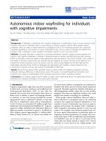
Serum AOC levels in participants grouped according to MMSE scoresFigure 1
Serum AOC levels in participants grouped according to
MMSE scores. Square and circle symbols indicate controls
and Alzheimer's disease patients, respectively. *p = 0.0018
versus >28 MMSE score group.
600
800
1000
1200
1400
1600
*
> 28 27 - 21 <20
AOC (µmoles/L)
MMSE scores
Journal of Neuroinflammation 2006, 3:4 />Page 5 of 6
(page number not for citation purposes)
The evaluation of the total reducing capacity of biological
fluids such as serum or plasma may provide a better esti-
mation of the peripheral resistance to oxidant injury than
the measurement of a set of individual anti-oxidant spe-
cies. The anti-oxidant activity measured by the AOC assay
is the net result of the contribution of all the individual
anti-oxidants, of their interactions and complexities. Total
plasma anti-oxidant activity has been measured previ-
ously, although different methods have been used [15-
17]. One of these methods, measuring the hydrosoluble
anti-oxidant status of biological fluids such as plasma, led
to conflicting results [15,16]. In a recent study using a
spectrophotometric method, total anti-oxidant activity
was decreased in AD patients and showed a tendency
towards a slight negative correlation with clinical duration
as defined by the time between first symptoms and clini-
cal diagnosis [17].
To our knowledge, our study is the first one attempting to
correlate peripheral anti-oxidant defence with the progres-
sion of cognitive deficit in a well defined group of AD
patients. Although AOC levels were positively associated
with MMSE and LSS scores at the time of blood sampling
for AOC determination, they did not predict the cognitive
deficit two years later. Although our study may have been
underpowered to detect such an association, our findings
suggests that the level of peripheral anti-oxidant defence
may be indicative of peripheral oxidant status rather than
of central processes related to neurodegeneration and cog-
nitive decline. However, we cannot rule out the possibility
that the rate of cognitive decline might be associated with
AOC levels at later time points. Longitudinal studies over
extended periods of time (more than 2 years) should help
to clarify this issue.
Another important and novel observation of our study is
the positive relationship between peripheral reductive
capacity and survival of AD subjects. According to the Cox
proportional hazards model, the hazard of dying was
lower in patients with elevated serum AOC levels (above
the median level of 1103.0 µmoles/L) than in patients
with low AOC levels. The median residual survival of
patients with high serum AOC was about three years
longer than that of subjects in the subgroup with low AOC
levels. Since participants taking anti-inflammatory drugs
and/or anti-oxidant supplements were excluded from the
study, these findings suggest that AOC might be a good
indicator of the general health status of the body and of its
capacity to cope with inflammatory and oxidative insults.
The lower levels of peripheral AOC observed in AD
patients is also consistent with the finding of lower oxida-
tive resistance of plasma and CSF from AD patients com-
pared with controls [18]. These findings further indicate
that oxidative stress is a pervasive condition, which not
only affects selectively vulnerable neuronal populations
but also occurs in the periphery, as indicated by the higher
8-OHdG content in the lymphocyte DNA of AD patients
compared with controls [5].
Conclusion
This study indicates that the levels of peripheral anti-oxi-
dant defence, measured by an assay that evaluates the
total reductive capacity present in serum, are decreased in
AD and are positively associated with the survival of
patients. The AOC levels were associated with cognitive
scores, but they were not predictive of the progression of
cognitive deficit. Serum AOC may therefore be a good
index of the general anti-oxidant status of the body but
does not necessarily reflect the body's capacity to protect
vulnerable neuronal populations from oxidant processes.
Our study does not, however, rule out the possibility that
serum AOC measurements may be useful in identifying
asymptomatic subjects or those with early cognitive symp-
toms who are at risk of progressing to more severe cogni-
tive impairment or dementia.
Competing interests
The author(s) declare that they have no competing inter-
ests.
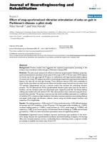
Cumulative survival of patients with Alzheimer's disease with low (solid line) and high (dashed line) initial AOC levelsFigure 2
Cumulative survival of patients with Alzheimer's disease with
low (solid line) and high (dashed line) initial AOC levels. Low
and high AOC levels were defined as below or above the
median AOC levels of the patient group (1103.0 µmoles/L).
Cox proportional hazards regression.
02468101214
0
20
40
60
80
100
Cumulative survival (%)
Time since serum AOC measurment (years)
Journal of Neuroinflammation 2006, 3:4 />Page 6 of 6
(page number not for citation purposes)
Authors' contributions
LM participated in the design of the study, performed
AOC assay and prepared the manuscript; AG performed
AOC assay and help to draft the manuscript; MP per-
formed statistical analysis and help to draft the manu-
script; DW participated in the design of the study, was
involved in collection of blood samples and performed
ApoE genotyping; MC participated in the design of the
study, was involved in clinical examinations and help to
draft the manuscript; ADS participated in the design of the
study and helped to draft the manuscript.
Acknowledgements
We wish to thank all the patients, caregivers and volunteers of OPTIMA.
We acknowledge especially the help of Elizabeth King and the research
nursing team, the projects doctors over several years and, for histopathol-
ogy, Professor M. Esiri, Dr Z. Nagy, Dr C. Joachim and staff of the Neuropa-
thology Department at the Radcliffe Infirmary, Oxford, England. This work
was supported by Italian Ministry of Health, project "Malattie neurodegen-
erative – Progetto finalizzato ex art. 56/03", by Bristol-Myers Squibb, the
Norman Collisson Foundation, the Takayama Foundation and Merck & Co
Inc.
References
1. Nunomura A, Perry G, Aliev G, Hirai K, Takeda A, Balraj EK, Jones
PK, Ghanbari H, Wataya T, Shimohama S, Chiba S, Atwood CS,
Petersen RB, Smith MA: Oxidative damage is the earliest event
in Alzheimer disease. J Neuropathol Exp Neurol 2001, 60:759-767.
2. Zhu X, Raina AK, Perry G, Smith MA: Alzheimer's disease: the
two-hit hypothesis. Lancet Neurol 2004, 3:219-226.
3. Greco A, Minghetti L: Isoprostanes as biomarkers and media-
tors of oxidative injury in infant and adult central nervous
system diseases. Curr Neurovasc Res 2004, 1:341-354.
4. Migliore L, Fontana I, Colognato R, Coppede F, Siciliano G, Murri L:
Searching for the role and the most suitable biomarkers of
oxidative stress in Alzheimer's disease and in other neurode-
generative diseases. Neurobiol Aging 2005, 26:587-595.
5. Mecocci P, Polidori MC, Cherubini A, Ingegni T, Mattioli P, Catani M,
Rinaldi P, Cecchetti R, Stahl W, Senin U, Beal MF: Lymphocyte oxi-
dative DNA damage and plasma antioxidants in Alzheimer
disease. Arch Neurol 2002, 59:794-798.
6. Rinaldi P, Polidori MC, Metastasio A, Mariani E, Mattioli P, Cherubini
A, Catani M, Cecchetti R, Senin U, Mecocci P: Plasma antioxidants
are similarly depleted in mild cognitive impairment and in
Alzheimer's disease. Neurobiol Aging 2003, 24:915-919.
7. Engelhart MJ, Geerlings MI, Ruitenberg A, van Swieten JC, Hofman A,
Witteman JC, Breteler MM: Dietary intake of antioxidants and
risk of Alzheimer disease. JAMA 2002, 287:3223-3229.
8. Engelhart MJ, Ruitenberg A, Meijer J, Kiliaan A, van Swieten JC, Hof-
man A, Witteman JC, Breteler MM: Plasma levels of antioxidants
are not associated with Alzheimer's disease or cognitive
decline. Dement Geriatr Cogn Disord 2005, 19:134-139.
9. Clarke R, Smith AD, Jobst KA, Refsum H, Sutton L, Ueland PM:
Folate, vitamin B12, and serum total homocysteine levels in
confirmed Alzheimer's disease. Arch Neurol 1998, 55:1449-1455.
10. McKhann G, Drachman D, Folstein M, Katzman R, Price D, Stadlan
EM: Clinical diagnosis of Alzheimer's disease: report of the
NINCDS-ADRDA Work Group under the auspices of
Department of Health and Human Services Task Force on
Alzheimer's Disease. Neurology 1984, 34:939-944.
11. Roth M, Huppert FA, Tym E, Mountjoy CQ: CAMDEX: The Cam-
bridge examination for mental disorders of the elderly. Cam-
bridge, Cambridge University Press; 1988.
12. Vassalle C, Petrozzi L, Botto N, Andreassi MG, Zucchelli GC: Oxida-
tive stress and its association with coronary artery disease
and different atherogenic risk factors. J Intern Med 2004,
256:308-315.
13. Wenham PR, Price WH, Blandell G: Apolipoprotein E genotyping
by one-stage PCR. Lancet 1991, 337:1158-1159.
14. Berr C, Richard MJ, Gourlet V, Garrel C, Favier A: Enzymatic anti-
oxidant balance and cognitive decline in aging – the EVA
study. Eur J Epidemiol 2004, 19:133-138.
15. Repetto MG, Reides CG, Evelson P, Kohan S, de Lustig ES, Llesuy SF:
Peripheral markers of oxidative stress in probable Alzhe-
imer patients. Eur J Clin Invest 1999, 29:643-649.
16. Foy CJ, Passmore AP, Vahidassr MD, Young IS, Lawson JT: Plasma
chain-breaking antioxidants in Alzheimer's disease, vascular
dementia and Parkinson's disease. QJM 1999, 92:39-45.
17. Guidi I, Galimberti D, Lonati S, Novembrino C, Bamonti F, Tiriticco
M, Fenoglio C, Venturelli E, Baron P, Bresolin N, Scarpini E: Oxida-
tive imbalance in patients with mild cognitive impairment
and Alzheimer's disease. Neurobiol Aging 2006, 27:262-269.
18. Schippling S, Kontush A, Arlt S, Buhmann C, Sturenburg HJ, Mann U,
Muller-Thomsen T, Beisiegel U: Increased lipoprotein oxidation
in Alzheimer's disease. Free Radic Biol Med 2000, 28:351-60.
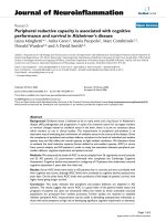
Dependence of serum AOC levels on MMSE scores (A) or on cognitive decline (B), defined as the difference between MMSE scores over a period of 2.0 (0.2) [median(IQR/2)] yearsFigure 3
Dependence of serum AOC levels on MMSE scores (A) or
on cognitive decline (B), defined as the difference between
MMSE scores over a period of 2.0 (0.2) [median(IQR/2)]
years.
600 800 1000 1200 1400 1600
-4
0
4
8
12
16
20
600 800 1000 1200 1400 1600
0
5
10
15
20
25
30
B
A
cognitive decline
AOC (
µ
moles/L)
MMSE scores
AOC (
µ
moles/L)