Clinical characteristics and prognostic factors of bone lymphomas: Focus on the clinical significance of multifocal bone involvement by primary bone large B-cell lymphomas
Bạn đang xem bản rút gọn của tài liệu. Xem và tải ngay bản đầy đủ của tài liệu tại đây (325.11 KB, 9 trang )
Wu et al. BMC Cancer 2014, 14:900
/>
RESEARCH ARTICLE
Open Access
Clinical characteristics and prognostic factors of
bone lymphomas: focus on the clinical significance
of multifocal bone involvement by primary bone
large B-cell lymphomas
Huanwen Wu1, Marilyn M Bui2, Douglas G Leston3, Haipeng Shao4, Lubomir Sokol5, Eduardo M Sotomayor5
and Ling Zhang4*
Abstract
Background: Malignant bone lymphoma can be classified as primary (PBL) or secondary (SBL) bone lymphoma.
However, the clinico-pathological characteristics and prognostic factors of PBL versus SBL have not yet been well
defined. Whether lymphoma with multifocal bone involvement should be considered as stage IV PBL or SBL still
remain controversial throughout the literature.
Methods: In this study, we retrospectively reviewed 127 patients with bone lymphoma diagnosed from1998 to
2013 at the Moffitt Cancer Center. Patients were classified as PBL (81 cases) and SBL (46 cases) using the 2013 WHO
Classification of Bone/Soft Tissue Tumors and PBL patients were further subdivided into: 1) PBL with unifocal bone
disease (uPBL, 46 cases), 2) PBL with multifocal bone involvement (mPBL, 35 cases). Patient characteristics, survival,
and prognostic factors were analyzed.
Results: Diffuse large B-cell lymphoma (DLBCL) was the most common histological subtype in all three groups
(37/46 of uPBL, 23/35 of mPBL, 23/46 of SBL). B symptoms, lymph node involvement, and bone marrow involvement
were found to be more common in mPB-DLBCL and SB-DLBCL groups than in the uPB-DLBCL group. Femur was found
to be the most common affected site in uPB-DLBCL patients, while spine was most commonly involved in the other
two groups. Survival analysis indicated that uPBL-DLBCL patients had a significantly better progression-free survival
(PFS) and overall survival (OS) than those in the other two groups (P < 0.05). We also found by univariate analysis
that multifocality, and stage IV were significantly poor prognostic factors for both PFS and OS in PBL patients.
Using multivariate analysis, multifocality remained an independent prognostic factor for both PFS and OS
(P = 0.0117, RR: 3.789, 95% CI: 1.275-11.256).
Conclusion: Overall, our results suggest that mPBL is more similar to SBL in characteristics and survival rather
than uPBL, and thus should be better classified and treated as SBL.
Keywords: Primary bone lymphoma (PBL), Secondary bone lymphoma (SBL), Diffuse large B-cell lymphoma
(DLBCL), Clinico-pathological characteristics, Prognostic factors, Multifocal bone involvement/multifocality
* Correspondence:
4
Department of Hematopathology and Laboratory Medicine, H. Lee Moffitt
Cancer Center and Research Institute, Tampa, FL, USA
Full list of author information is available at the end of the article
© 2014 Wu et al.; licensee BioMed Central Ltd. This is an Open Access article distributed under the terms of the Creative
Commons Attribution License ( which permits unrestricted use, distribution, and
reproduction in any medium, provided the original work is properly credited. The Creative Commons Public Domain
Dedication waiver ( applies to the data made available in this article,
unless otherwise stated.
Wu et al. BMC Cancer 2014, 14:900
/>
Background
Malignant bone lymphomas are uncommonly encountered clinically. According to the initial extent of disease,
malignant bone lymphomas can be divided into two
groups: primary bone lymphoma (PBL) and secondary
bone lymphoma (SBL) [1,2]. PBL is an extremely rare
entity, accounting for approximately 7% of malignant
bone tumors, 5% of extra-nodal lymphomas, and <1% of
all non-Hodgkin lymphomas [1,3]. It has been considered to have the best prognosis of all primary malignant
bone lesions. However, SBL is more common, seen in
approximately 16% to 20% of patients with lymphoma,
and has a relatively poor prognosis [1]. Given their different clinical outcomes and treatment strategies, subclassification of bone lymphomas into either primary or
secondary bone lymphomas is critical. A review of the
literature shows that there is no consensus regarding
how to accurately distinguish PBL from SBL. The most
common debate falls in how to subclassify and treat
bone lymphoma when it primarily presents with multifocal bone disease with/without regional lymph node
and/or adjacent soft tissue involvement. The reported 5year overall survival (OS) rates vary between different
study groups of PBL patients due to different diagnostic
criteria, ranging from less than 36% to more than 88.3%
[4-8]. In addition, the clinico-pathological characteristics
and prognostic factors of PBL versus SBL have not yet
been well studied.
Recently, the 2013 World Health Organization (WHO)
classification of bone/soft tissue tumors [1] defined PBL
as a neoplasm composed of malignant lymphoid cells,
producing one or more masses within bone, without any
supra-regional lymph-node involvement or other extranodal lesions. According to the criteria, bone lymphoma
with/without regional lymph node and without other
extra-nodal lesions was also classified as PBL clinically, regardless of whether the bone lesion occurred unifocally or
multifocally. We recently identified several potential prognostic factors using the new 2013 WHO classification in a
large study-cohort including 70 PBL cases and showed
that soft tissue extension and IPI score were the most important unfavorable prognostic indicators for both PFS
and OS in PBL [9]. However, limited information was
available per literatures on the prognostic role of multifocality in PBL, and whether PBL with multifocal bone
involvement should be considered as SBL. Here, we conducted a single-center retrospective study in which we
classified bone lymphoma as PBL and SBL using the new
2013 WHO classification of soft tissue neoplasms, further
subcategorized PBL patients into two groups, those with
unifocal bone disease (uPBL) and those with multifocal
bone involvement (≥2 foci) (mPBL), and compared patient
characteristics, treatments and outcome among uPBL,
mPBL and SBL groups, aiming to further explore the
Page 2 of 9
clinical and prognostic significance of multifocal bone
involvement in PBL and to clarify the current definition
of PBL.
Methods
Patients
Chart records of 145 patients with biopsy-proven malignant bone lymphoma were retrieved from the surgical
pathology files of Moffitt Cancer Center diagnosed over
a 15-year-period (1998–2013), following the guidelines
of the Moffitt Cancer Center Scientific Research Committee and with the approval of the Institutional Review
Board at the University of South Florida. After an initial
review of the clinical and pathological data, 18 patient
records were excluded because of the inadequacy of staging and/or follow-up information. Patient characteristics, survival, and prognostic factors were analyzed.
Medical records were reviewed for age, sex, race, involved sites, lactate dehydrogenase (LDH), pathological
diagnosis, treatments, date of diagnosis, lymph node involvement, bone marrow involvement, stage and date of
disease progression, relapse, death, or last follow-up.
Lymph node involvement was demonstrated by a clinical
and imaging enlargement of node (>1.5 cm measured
per the Positron Emission Tomography, PET scan) with
or without an excision biopsy. Our study thus included
127 patients with bone lymphomas. We classified patients as PBL and SBL using the updated 2013 WHO
criteria for bone/soft tissue tumors [1] and then further subcategorized PBL into two subgroups:1) uPBL
(n = 46),2) mPBL(n = 35). Bone marrow involvement
was assessed by an aspiration and bone marrow biopsy
from iliac crest. If the primary lesion is near iliac or
pelvic region, contra-lateral ilic crest is used for the biopsy site. Bone lymphoma with distant bone marrow
involvement as the only other site of extranodal disease
was also classified as PBL (stage IV) in our study, because
a number of previous studies have demonstrated that it
has a similar prognosis to PBL with localized disease
[2,7,10]. Given the relatively rarity of the other histological
subtype, only patients with diffuse large B-cell lymphoma
(DLBCL), were further reviewed for patient characteristics
and analyzed for prognostic factors in the current study.
Histological diagnosis and immunohistochemistry
findings
All patients were diagnosed lymphoma by bone biopsy.
Morphologic assessments in conjunction with flow cytometry (only if fresh tissue had been harvested) or immunohistochemical (IHC) study were conducted. The
IHC markers included CD20, PAX-5, CD10, Bcl-2, Bcl-6,
or MUM-1 for DLBCL or large B-cell lymphoma, unclassifiable, with features between DLBCL and Burkitt
lymphoma (BLUI) and CD30, CD3, CD4, CD8, CD43,
Wu et al. BMC Cancer 2014, 14:900
/>
granzyme B and anaplastic lymphoma kinase (ALK) for
anaplastic large T-cell lymphoma (ALCL).
Staging
Patients were staged retrospectively according to the Ann
Arbor staging system as described before [9]. In all cases,
staging evaluation included 1) a chest X-ray or a computed tomography (CT) scan of the chest, 2) a CT scan or
ultrasonogram of the abdomen and pelvis, 3) whole body
bone scan or positron emission tomography–computed
tomography (PET-CT) scan or magnetic resonance imaging (MRI), and 4) bone marrow biopsy of iliac bone.
Survival analysis
Progression-free survival (PFS) was defined as the interval from the date of diagnosis to the date of disease progression, relapse, or death from any cause. Patients who
showed no progression were censored at the date of
most recent available radiographic imaging. OS was calculated from the date of diagnosis to the date of death
from any cause using the Social Security Death Index
(SSDI). For unknown deaths, patients were censored at
last follow-up. Survival curves were calculated according
to the Kaplan-Meier method and compared using the
log-rank test. Differences were considered significant if
P values were ≤0.05 (two-tailed). Multivariate analysis
was performed using a Cox model using a forward variable selection procedure. Only the variables with significant values (P ≤ 0.05) in univariate analysis were included
in the multivariate analysis. All data analyses were performed by SPSS software for windows, version 20 (SPSS
Inc., Chicago, IL).
Results
Histological diagnosis and patient characteristics
The histological classification of our series is shown in
Table 1. DLBCL was the most common histological subtype in all three groups. However, the proportion of
DLBCL patients in the SBL group was significantly lower
than that in the uPBL group (23/46, 50% versus 60/71,
85.7%) (P < 0.05). Classical Hodgkin lymphoma and follicular lymphoma were more commonly shown in SBL
group, while only 1 classical Hodgkin lymphoma case
was identified in the PBL groups. T-cell lymphoma is
relatively rare, with four of the total six T-cell lymphoma
cases in the mPBL group. All classical Hodgkin lymphoma cases had nodular sclerosis histology. Among the
127 bone lymphoma patients, only two PBL cases were
HIV positive, including one DLBCL and one large B-cell
lymphoma, unclassifiable, with features intermediate between DLBCL and Burkitt lymphoma (BLUI).
Given the histological heterogeneity and the relative
rarity of the other histological types, only DLBCL patients were further explored for demographical and
Page 3 of 9
Table 1 Histopathological subtypes of patients with bone
lymphoma
Classification
uPBL
mPBL
SBL
DLBCL, n (%)
37(80.4)
23(65.7)
23(50.0)
4(8.7)
3(8.6)
9(19.6)
Follicular lymphoma, n (%)
Small lymphocytic lymphoma, n (%)
1(2.2)
1(2.9)
0(0)
Marginal zone lymphoma, n (%)
1(2.2)
1(2.9)
2(4.3)
Not further subclassifieda, n (%)
1(2.2)
2(5.7)
1(2.2)
0
1(2.9)
0(0)
BLUI , n (%)
Classical Hodgkin lymphoma, n (%)
1(2.2)
0(0)
10(21.7)
T cell, n (%)
1(2.2)b
4(11.4)c
1(2.2)d
Total, n (%)
46
35
46
Abbreviations uPBL: primary bone lymphoma with unifocal bone disease;
mPBL: primary bone lymphoma with multifocal bone disease; SBL: secondary
bone lymphoma; DLBCL: diffuse large B-cell lymphoma; Large B-cell lymphoma,
unclassifiable, with features intermediate between DLBCL and Burkitt lymhoma:
BLUI; ALCL: anaplastic large T-cell lymphoma; PTCL, NOS: peripheral T-cell
lymphoma, not otherwise specified.
a
Low-grade, small B-cell lymphoma no further information for subclassification.
b
ALCL(n = 1).
c
ALCL (n = 2), T-lymphoblastic lymphoma (n = 1), and PTCL, NOS (n = 1).
d
ALCL (n = 1).
clinical characteristics as well as survival. The characteristics of the 83 DLBCL patients are summarized in
Tables 2 and 3. Compared with primary bone DLBCL
with unifocal bone disease (uPB-DLBCL), B symptoms,
lymph node involvement, and bone marrow involvement
were more commonly shown in the other two groups:
primary bone DLBCL with multifocal bone disease
(mPB-DLBCL) and secondary bone DLBCL (SB-DLBCL).
No significant differences regarding age distribution were
shown among three groups.
Femur was most commonly involved in the uPBDLBCL group. However, spine was the most common affected site in the other two groups. Pelvis, humerus, and
tibia were also commonly involved in our series.
Most patients in the uPB-DLBCL group were classified
as stage IE (unifocal localized bone lesions without lymph
node involvement). In the mPB-DLBCL group, all patients
were staged as IVE on the basis of multifocal bone involvement. The majority of SB-DLBCL patients were classified as stage II-IVE. Only 1 patient with SB-DLBCL, who
had presented with unifocal bone disease and without
lymph node or other extra-nodal sites involvement when
disease relapsed, was classified as stage I.
IHC findings
IHC study was performed in only a subset of patients with
PB-DLBCL. The available data are summarized as follows:
approximately half (26/43) were CD10-positive. Bcl-2,
Bcl-6, and MUM-1 expression were detected in 19 of 23
(82.6%), 24 of 27 (88.9%), and 2 of 16 (12.5%) patients, respectively. In situ hybridization using Epstein-Barr virus-
Wu et al. BMC Cancer 2014, 14:900
/>
Page 4 of 9
Table 2 Patient demographics and clinical characteristics
of bone DLBCL
Parameter
Total
Age range, years
Table 3 Common involved sites of bone DLBCL
Sites
uPB-DLBCL mPB-DLBCL SB-DLBCL
uPB-DLBCL
(N = 37)
mPB-DLBCL
(N = 23)
SB-DLBCL
(N = 23)
Extremities, n (%)
37
23
23
15-89
17-82
29-82
Femur
12(32.4)
12(52.2)
7(30.4)
6(16.2)
5(21.7)
3(13.0)
Median age, years
53.0
60.0
54.0
Humerus
Mean age, years
52.9
53.4
55.0
Tibia
5(13.5)
6(26.1)
2 (8.7)
Pelvis, n (%)
5(13.5)
13(56.5)
9 (39.1)
Spine, n (%)
7(18.9)
13(56.5)
11 (47.8)
<60, n (%)
22(59.5)
11(47.8)
15(65.2)
≥60, n (%)
15(40.5)
12(52.2)
8(34.8)
Sex
Male, n (%)
22(59.5)
14(60.9)
9(39.1)
Female, n (%)
15(40.5)
9(39.1)
14(60.9)
1.5:1
1.6:1
0.6:1
White, n (%)
33(89.2)
21(91.3)
17(73.9)
Black, n (%)
2(5.4)
1(4.3)
3(13.0)
Unknown, n (%)
2(5.4)
1(4.3)
3(13.0)
B symptoms, n (%)
7(18.9)
8(34.8)
8(34.8)
Male :female ratio
Race
Treatments
LDH
Normal
23(79.3)
5(29.4)
8(57.1)
Elevated
6(20.7)
12(70.6)
6(42.9)
Lymph node involvement,
n (%)
4(10.8)*
9(39.1)*
14(60.9)**
Bone marrow involvement,
n (%)
3(8.1)
5(21.7)
7(30.4)
Number of bone sites, n (%)
Unifocal
Multifocal
Abbreviations uPB-DLBCL: primary bone diffuse large B-cell lymphoma with
unifocal bone disease; mPB-DLBCL: primary bone diffuse large B-cell lymphoma
with multifocal bone disease; SB-DLBCL: secondary bone diffuse large
B-cell lymphoma.
37(100.0)
0(0)
7(30.4)
0(0)
23(100.0)
16(69.6)
Sites, n (%)
10(43.5)
7(30.4)
Treatments of DLBCL patients were summarized (Table 4).
Most patients with uPB-DLBCL with received combined modality therapy (chemotherapy and radiotherapy), whereas more than half of SB-DLBCL patients
with bone involvement at presentation and mPB-DLBCL
patients were treated with chemotherapy alone. Most bone
DLBCL patients received CHOP or CHOP-like chemotherapy with rituximab, and only eight DLBCL patients
received CHOP or CHOP-like chemotherapy alone
without rituximab. R-ESHAP (rituximab plus etoposide,
methylprednisolone, cytarabine, cisplatin) was the main
salvage therapy for SB-DLBCL with recurrent bone
involvement.
Survival analysis of patients with bone lymphoma
Patient follow-up time was calculated using reverse
Kaplan-Meier analysis. For 83 bone DLBCL patients, the
median follow-up times for PFS and OS were 28 months
(range, 1–138 months) and 38 months (range, 1–139
months), respectively. PFS and OS data for uPB-DLBCL,
Appendicular
29(78.4)
Axial
8(21.6)
2(8.7)
9(39.1)
Both
0(0)
11(47.8)
7(30.4)
IE
30(81.1)
0(0)
1(4.3)
Table 4 Treatments of bone DLBCL
IIE
4(10.8)
0(0)
2(8.7)
Parameter
IIIE
0(0)
0(0)
0(0)
IVE
3(8.1)
23(100.0)
20(87.0)
Stage, n (%)
Abbreviations uPB-DLBCL: primary bone diffuse large B-cell lymphoma with
unifocal bone disease; mPB-DLBCL: primary bone diffuse large B-cell lymphoma
with multifocal bone disease; SB-DLBCL: secondary bone diffuse large
B-cell lymphoma.
*Patients with regional lymph node enlargement.
**Patients with both regional and supraregional or systemic lymph
node involvements.
encoded RNA probe was performed in five PB-DLBCL
cases, with all five being negative.
Besides CD30, CD3, CD4, CD8, CD43, and granzyme
B, ALK IHC staining was performed in all three primary
bone ALCL cases, with two of the three being positive.
Number (%)
uPB-DLBCL mPB-DLBCL SB-DLBCL*
Treatment
n = 37
n = 23
n = 12
CMT
11(29.7)
16(69.6)
7(58.3)
1(2.7)
0
0
24(54.1)
6(26.1)
5(41.7)
1(2.7)
1(4.3)
0
Chemotherapy
n = 35
n = 22
n = 12
CHOP or CHOP-like CHOP-like
7(20.0)
0
1(8.3%)
CHOP + rituximab
28(80.0)
22(100.0)
11(91.7%)
RT
CMT and RT
No CMT or RT
Abbreviations uPB-DLBCL: primary bone diffuse large B-cell lymphoma with
unifocal bone disease; mPB-DLBCL: primary bone diffuse large B-cell lymphoma
with multifocal bone disease; SB-DLBCL: secondary bone diffuse large B-cell
lymphoma; CMT: chemotherapy; RT: radiation therapy.
*SB-DLBCL with recurrent bone involvement was not included in the table.
Wu et al. BMC Cancer 2014, 14:900
/>
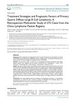
mPB-DLBCL and SB-DLBCL groups are illustrated in
Figure 1. The 5-year PFS rates were 75.7% for uPBDLBCL, 13.4% for mPB-DLBCL, and 22.0% for SBDLBCL (Figure 1A). The 5-year OS rates were 83.4% for
uPB-DLBCL, 36.7% for mPB-DLBCL and 41.9% for SBDLBCL (Figure 1B). uPBL patients had a significantly
better PFS and OS than those in the other two groups
(PFS: P = 0.001 for uPB-DLBCL vs. mPB-DLBCL, P < 0.001
for uPB-DLBCL vs. SB-DLBCL; OS: P < 0.001 for uPBDLBCL vs. mPB-DLBCL, P < 0.001 for uPB-DLBCL vs. SBDLBCL). There were no significant differences in either
PFS or OS between the other two groups (PFS: P = 0.732;
OS: P = 0.572).
Similar results were obtained for our total series of
127 bone lymphoma (Additional file 1: Figure S1).
Prognostic factor analyses
We analyzed the influence of the following individual
factors on survival in PB-DLBCL patients: age, sex, B
symptoms, LDH, lymph node involvement, bone marrow
involvement, involved sites, the number of bone sites,
stage, and IHC markers (CD10, Bcl-2, Bcl-6, and MUM1). In univariate analysis, LDH, involvement of both appendicular and axial sites multifocality, and stage IV
were significant poor prognostic factors for both PFS
and OS (Table 5). Age ≥ 60 years was also a significant
poor prognostic factor for OS (Table 5). None of the
IHC markers were significant predictors for PFS or OS.
Using Cox regression for multivariate analysis, multifocality were independent unfavorable prognostic factors for both PFS and OS (Table 6). Age ≥ 60 years was
again an independent unfavorable prognostic factor
for OS.
Moreover, as for SBL, we found that all three patients
with recurrent lymphoma presenting with unifocal bone
disease as the only involved site (re-stage I) (one DLBCL,
one low-grade B cell lymphoma, and one classical Hodgkin
Page 5 of 9
lymphoma) survived without disease progression until final
follow-up (3, 104, and 136 months, respectively).
Discussion
PBL was first described by Oberling in 1928 [11] and is
thought to be a separate disease entity from conventional nodal or extranodal base lymphoma with an excellent prognosis. Up to now, its definition still remains
controversial, especially regarding whether multifocal
bone involvement by lymphoma at initial presentation
without any supra-regional lymph node involvement and
other extra-nodal disease should be defined as PBL [10].
Of importance, with obvious improvements in imaging
technology in recent decades, the proportion of patients
diagnosed with multifocal bone lymphoma has increased
[7,12]. Thus, these discrepancies in PBL definition and
the improvements in diagnostic procedures have led to
difficulties in the comparison of clinic-pathological characteristics and clinical outcomes between studies. In
addition, it also raises the question regarding whether
multifocality of bone lymphoma should be considered as
an independent prognostic predictor. Although there
have been several studies on malignant bone lymphoma,
these have thus far been limited by small sample sizes
and/or have included only early-stage PBL cases [4,13,14].
Here, we describe a relatively large cohort of PBL patients
(n = 81) and a compared group of SBL patients (n = 46) diagnosed and treated during 1998–2013 at our institution
with modern and contemporary diagnostic and therapeutic modalities. A relatively high proportion of our PBL
patients (43.2%, 35 of 81 cases) presented with multifocal
bone disease. This may be due to the routine use of PET,
CT, MRI, and bone scanning for staging. Because only
bone biopsy-proven cases were selected, the number of
SBL patients was relatively small in our series.
Our initial analysis of patient characteristics (age and sex
distribution) was consistent with previous studies [10,15].
Figure 1 Overall survival (A) and progression-free survival (B) in three groups of bone DLBCL.
Wu et al. BMC Cancer 2014, 14:900
/>
Page 6 of 9
Table 5 Univariate analysis of prognostic factors for survival in patients with PB-DLBCL
Parameter
PFS
3-year
5-year
60.8
45.9
<60 years, %
64.5
46.9
≥60 years, %
50.3
40.2
Overall, %
Age
OS
P
3-year
5-year
78.8
68.0
87.9
80.6
57.9
44.3
0.278
Sex
0.004
0.634
0.410
Male, n (%)
63.9
52.1
80.2
71.0
Female, n (%)
53.4
44.8
71.9
61.4
No, %
62.5
52.4
80.5
74.3
Yes, %
50.0
31.5
66.1
45.8
B symptoms
0.673
LDH
0.289
0.011
0.018
Normal, %
73.6
58.2
89.2
68.2
Elevated, %
34.8
21.9
55.2
38.2
No, %
66.8
60.0
81.4
75.1
Yes, %
15.6
2.2
64.0
44.3
Lymph node involvement
0.143
Bone marrow involvement
0.231
0.719
0.319
No, %
59.1
42.0
80.0
72.3
Yes, %
72.9
72.9
54.9
42.0
Appendicular, %
66.7
51.1
78.4
70.6
Axial, %
59.1
59.1
100.0
100.0
Both, %
5.6
0
61.4
61.4
Unifocal, %
75.7
75.7
89.2
83.5
Multifocal, %
36.8
13.4
53.1
36.7
Sites
0.022
Number of bone sites
0.022
0.001
Stage
<0.001
0.005
0.001
IE/IIE, %
73.8
73.8
97.1
97.1
IVE, %
43.0
18.0
58.5
49.8
Negative, %
52.9
29.8
76.8
65.2
Positive, %
64.4
55.2
95.7
95.7
CD10
P
0.267
0.077
Bold values indicate statistical significance (P<0.05).
Table 6 Multivariate analysis of prognostic factors for patients with PB-DLBCL
Parameter
PFS
RR
95%CI
Number of bone sites
P
RR
95%CI
0.015
Unifocal
Multifocal
OS
Reference group
3.728
1.292-10.754
Reference group
20.061
1.851-217.377
Age
0.035
<60 years
≥60 years
Bold values indicate statistical significance (P<0.05).
P
0.014
Reference group
15.791
1.215-205.200
Wu et al. BMC Cancer 2014, 14:900
/>
However, mPB-DLBCL and SB-DLBCL patients had
higher frequency of B symptoms, lymph node involvement,
and iliac bone marrow involvement than uPB-DLBCL patients. In previous studies, femur has been reported to be
the most commonly involved site in PBL [3,16,17]. In our
series, femur was also found to be the most common affected site in uPB-DLBCL patients. However, spine was
most commonly involved in mPB-DLBCL and SB-DLBCL.
From this point of view, our results suggest that mPBL is
more similar to clinical characteristics of patients with SBL
rather than with uPBL.
Consistent with previous studies, DLBCL was the most
common histological subtype in our bone lymphoma
series. However, the uPBL group had a significantly higher
proportion of DLBCL (80.4%, 37 of 46 cases) than the SBL
group (50.0%, 23 of 46 cases). Our results indicate that the
histological distributions are different between the uPBL
group and the SBL group.
Regarding the subclassification of PBL, several studies
with small sample sizes have described the IHC characteristics of PB-DLBCL [18-21]. In these previous reports,
approximately half of the PB-DLBCL cases demonstrated
a germinal center B-cell (GCB) phenotype by IHC with
high Bcl-2 and/or Bcl-6 expression and relatively low
MUM-1 expression. We also observed high percentages
of Bcl-2 and Bcl-6 expression in our series. However, incomplete IHC data of MUM-1 in our study precluded an
accurate subclassification of our PB-DLBCL cases into
GCB or non-GCB subgroups. Despite so, 26 of 43 patients
were able to be classified with PB-DLBCL in our series according to CD10-positivity, which meant that at least
60.5% of these patients were of GCB phenotype. Prior
studies have yielded conflicting results about the predictive value of these IHC markers, particularly of CD10 and
GCB stubtype [5,18-21]. Although insufficient for subclassification of GCB or non-GCB subtype of PB-DLBCL, our
limited data showed no association between various
markers (CD10, Bcl-6, Bcl-2, MUM-1) and survival in PBDLBCL.
In the study, we temporally subgrouped the patients
with mPBL as stage IV since whether lymphoma with
multifocal bone involvement should be considered as
stage IV PBL or SBL still remain controversial in the literature. Because of the unequivocal definition of PBL,
some previous studies restricted diagnoses to those with
early-stage PBL (stage IE and IIE) [14,22]. Only a few
studies have focused on the significance of multifocal bone
diseases in PBL [10,15]. In our study, patients classified
with uPB-DBLCL had an excellent prognosis, whereas
those with mPB-DLBCL carried a poor prognosis, with
survival being similar to SB-DLBCL. The finding suggests
that those with mPBL would benefit from being classified
as SBL rather than conventional PBL. Further prognostic
factor analyses also revealed that multifocality was an
Page 7 of 9
independent prognostic factor of PB-DLBCL, which also
supports that mPBL may be a different clinical entity from
uPBL. Although unifocal bone lymphoma, in general, can
be eradicated with local radiation in 50% of patients, the
treatment of patients with multifocal osseous disease, especially those presenting with associated soft tissue invasion or generalized adenopathy, is much less satisfactory
[23]. The treatment modality was also somewhat different
among PB-DLBCL and SB-DLBCL groups in our study.
Most patients with uPB-DLBCL were treated with combined modality therapy (chemotherapy and radiotherapy)
for localized lesions, whereas mPB-DLBCL and SB-DLBCL
typically received chemotherapy alone. Given that mPBL
and SBL patients had similar clinical characteristics, prognosis, and treatment modality, our data suggest that it
would be better to classify so-called “mPBL” as SBL in particular under the setting of DLBCL. As known, DLBCL
constitutes the majority of PBL. Thus, we consider that the
current definition for PBL might need further clarification.
In clinically and radiologically advanced-stage PBL patients
having multiple bone site involvement, especially in those
with regional lymph node and/or adjacent soft tissue involvement, it may be impossible to distinguish mPBL from
SBL. According to our results, it might not be necessary to
distinguish mPBL from SBL clinically.
In a study by Jawad et al. [10], it was suggested that
the use of the name “PBL” should be limited to those
with truly local disease with a single osseous lesion. This
is also the reason we limited mPBL to those with stage
IV in the study. Although stage IV itself was also a significant poor prognostic factor for survival in PBL patients by
univariate analysis, it failed to show independent prognostic significance in multivariate analysis, probably due to
the strong correlation between stage IV and multifocal
bone involvement. Ostrowski et al. [15] also reported that
those with malignant lymphoma with multifocal bone disease had a significantly poorer survival than those with
unifocal bone involvement. However, their study demonstrated that prognosis of patients with malignant lymphoma with multifocal bone disease was considerably better
than those having SBL. Two main reasons may explain
the difference from our study. First, their SBL group included a high proportion of patients with malignant
lymphoma with recurrent bone involvement when compared with our data. Second, their patients with regional
lymph node involvement and/or soft tissue extension were
excluded from the group of multifocal bone involvement.
Furthermore, due to the rarity of PBL patients who
present with regional lymph node and/or bone marrow
involvement, there is no consensus regarding the effects
of regional lymph node or bone marrow involvement on
survival in patients with PBL. No significant association
was observed between regional lymph node or bone marrow involvement and survival in our PB-DLBCL patients,
Wu et al. BMC Cancer 2014, 14:900
/>
suggesting that it was reasonable to categorize these cases
into PBL rather than SBL with a relatively worse prognosis. However, our results should be interpreted with caution given the small sample size.
As for SBL, although recurrent lymphoma is usually
associated with a poor prognosis, we found that patients
with recurrent lymphoma presenting with unifocal bone
disease as the only involved site (re-stage I) had an excellent prognosis. This result needs careful interpretation, taking into consideration that our study included
only 3 patients with stage I SBL.
It has been mentioned that low grade B-cell lymphoma, T-cell lymphoma and Hodgkin lymphoma have
also been included in the study. As the minority in PBL,
we are unable to perform risk stratification for these lymphomas. Multicenter studies with larger number of cases are
warranted to explore their prognostic values in PBL. However, it is of worthy for us to learn several interesting findings in the study. Our series also confirms that primary
bone Hodgkin lymphoma is extremely rare (1 of 81 PBL
patients) in contrast to secondary bone Hodgkin lymphoma
(10 of 46 patients, 21.8%) as reported in literatures, 10-20%
[3,17]. Only five primary T-cell lymphoma cases (including
3 ALCLs) were included in our PBL series. All five cases
showed rapid disease progression within the first year after
diagnosis, with three deceased 4–8 month after diagnosis
(data not shown). Consistent with our results, in the study
by Hsieh et al. [5], all five patients with primary bone T-cell
lymphoma (including 4 ALCLs) with follow-up information
died within 1 year. Limited case number precludes a further
prognostic analysis. Similarity also applies to primary bone
Hodgkin lymphoma. Given the small case number and
histological heterogeneity in low grade B-cell lymphomas,
no further studies have been conducted in our study, either.
Conclusions
In summary, our study retrospectively described our single institution experience with 127 bone lymphoma patients, including 81 cases of PBL and 46 cases of SBL using
the new 2013 WHO criteria. Patients with mPB-DLBCL
and SB-DLBCL showed similar characteristics, with both
having a poorer outcome, whereas uPB-DLBCL patients
demonstrated somewhat different characteristics and had
an excellent outcome. Moreover, multifocality was found
to be an independent prognostic factor of PB-DLBCL. Due
to the similar patient characteristics and outcome, it would
be better to classify bone lymphoma presenting with multifocal bone disease as SBL rather than conventional PBL,
regardless of whether there is supraregional lymph node or
other extranodal site involvement. Our results indicate that
the current criteria for PBL need further clarification, and
it might be unnecessary to distinguish mPBL from SBL,
clinically. Given the relatively small sample size of patients
Page 8 of 9
with SBL and the incomplete IHC data, our results warrant
further clarification in large multicenter studies.
Additional file
Additional file 1: Figure S1. Overall survival (A) and progression-free
survival (B) in three groups of bone lymphoma (OS: P = 0.034 for uPBL vs.
mPBL, P < 0.001 for uPBL vs. SBL, P = 0.074 for mPB vs. SBL; PFS: P = 0.347
for uPBL vs. mPBL, P < 0.001for uPBL vs. SBL, P = 0.517for mPB vs. SBL).
Competing interests
The authors declare that they have no competing interests.
Authors’ contributions
HW performed the research, analyzed the data and drafted the manuscript.
LZ, MMB and HS were involved in the histological review. LZ designed the
research study and was also the main editor of the manuscript. DGL, LS and
ES reviewed and critically revised the manuscript. All authors read and
approved the final manuscript.
Acknowledgments
We would like to thank Rasa Hamilton (Moffitt Cancer Center) for editorial
assistance.
Author details
Department of Pathology, Chinese Academy of Medical Science, Peking
Union Medical College Hospital, Beijing, China. 2Department of Anatomic
Pathology, H. Lee Moffitt Cancer Center and Research Institute, Tampa, FL,
USA. 3Department of Sarcoma, H. Lee Moffitt Cancer Center and Research
Institute, Tampa, FL, USA. 4Department of Hematopathology and Laboratory
Medicine, H. Lee Moffitt Cancer Center and Research Institute, Tampa, FL,
USA. 5Department of Malignant Hematology, H. Lee Moffitt Cancer Center
and Research Institute, Tampa, FL, USA.
1
Received: 1 April 2014 Accepted: 27 November 2014
Published: 2 December 2014
References
1. Fletcher CD, Bridge JA, Hogendoorn P, Mertens F: WHO Classification of
Tumours of Soft Tissue and Bone. Lyon, France: International Agency for
Research on Cancer; 2013.
2. Gianelli U, Patriarca C, Moro A, Ponzoni M, Giardini R, Massimino M, Alfano
RM, Armiraglio E, Nuciforo P, Bosari S, Coggi G, Parafioriti A: Lymphomas of
the bone: a pathological and clinical study of 54 cases. Int J Surg Pathol
2002, 10:257–266.
3. Kitsoulis P, Vlychou M, Papoudou-Bai A, Karatzias G, Charchanti A, Agnantis
NJ, Bai M: Primary lymphomas of bone. Anticancer Res 2006, 26:325–337.
4. Barbieri E, Cammelli S, Mauro F, Perini F, Cazzola A, Neri S, Bunkheila F,
Ferrari S, Brandoli V, Zinzani P, Mercuri M, Bacci G: Primary non-Hodgkin’s
lymphoma of the bone: treatment and analysis of prognostic factors for
Stage I and Stage II. Int J Radiat Oncol Biol Phys 2004, 59:760–764.
5. Hsieh PP, Tseng HH, Chang ST, Fu TY, Lu CL, Chuang SS: Primary nonHodgkin’s lymphoma of bone: a rare disorder with high frequency of
T-cell phenotype in southern Taiwan. Leuk Lymphoma 2006, 47:65–70.
6. Heyning FH, Hogendoorn PC, Kramer MH, Hermans J, Kluin-Nelemans JC,
Noordijk EM, Kluin PM: Primary non-Hodgkin’s lymphoma of bone: a
clinicopathological investigation of 60 cases. Leukemia 1999,
13:2094–2098.
7. Ramadan KM, Shenkier T, Sehn LH, Gascoyne RD, Connors JM: A
clinicopathological retrospective study of 131 patients with primary
bone lymphoma: a population-based study of successively treated
cohorts from the British Columbia Cancer Agency. Ann Oncol 2007,
18:129–135.
8. Beal K, Allen L, Yahalom J: Primary bone lymphoma: treatment results and
prognostic factors with long-term follow-up of 82 patients. Cancer 2006,
106:2652–2656.
9. Wu H, Zhang L, Shao H, Sokol L, Sotomayor E, Letson D, Bui MM: Prognostic
significance of soft tissue extension, international prognostic index, and
Wu et al. BMC Cancer 2014, 14:900
/>
10.
11.
12.
13.
14.
15.
16.
17.
18.
19.
20.
21.
22.
23.
Page 9 of 9
multifocality in primary bone lymphoma: a single institutional experience.
Br J Haematol 2014, 166(1):60–68.
Jawad MU, Schneiderbauer MM, Min ES, Cheung MC, Koniaris LG, Scully SP:
Primary lymphoma of bone in adult patients. Cancer 2010, 116:871–879.
Oberling C: Les reticulosarcomes et les reticuloendotheliosarcomes de la
moelle osseuse (sarcomes d’Ewing). Bull Assoc Fr Etude Cancer 1928,
17:259–296.
Alencar A, Pitcher D, Byrne G, Lossos IS: Primary bone lymphoma–the
University of Miami experience. Leuk Lymphoma 2010, 51:39–49.
Horsman JM, Thomas J, Hough R, Hancock BW: Primary bone lymphoma:
a retrospective analysis. Int J Oncol 2006, 28:1571–1575.
Cai L, Stauder MC, Zhang YJ, Poortmans P, Li YX, Constantinou N, Thariat J,
Kadish SP, Nguyen TD, Kirova YM, Ghadjar P, Weber DC, Bertran VT, Ozsahin
M, Mirimanoff RO: Early-stage primary bone lymphoma: a retrospective,
multicenter Rare Cancer Network (RCN) Study. Int J Radiat Oncol Biol Phys
2012, 83:284–291.
Ostrowski ML, Unni KK, Banks PM, Shives TC, Evans RG, O’Connell MJ, Taylor
WF: Malignant lymphoma of bone. Cancer 1986, 58:2646–2655.
Demircay E, Hornicek FJ Jr, Mankin HJ, Degroot H 3rd: Malignant lymphoma
of bone: a review of 119 patients. Clin Orthop Relat Res 2013, 471:2684–2690.
Bhagavathi S, Fu K: Primary bone lymphoma. Arch Pathol Lab Med 2009,
133:1868–1871.
Bhagavathi S, Micale MA, Les K, Wilson JD, Wiggins ML, Fu K: Primary bone
diffuse large B-cell lymphoma: clinicopathologic study of 21 cases and
review of literature. Am J Surg Pathol 2009, 33:1463–1469.
Adams H, Tzankov A, D’Hondt S, Jundt G, Dirnhofer S, Went P: Primary
diffuse large B-cell lymphomas of the bone: prognostic relevance of
protein expression and clinical factors. Human Pathol 2008, 39:1323–1330.
Heyning FH, Hogendoorn PC, Kramer MH, Holland CT, Dreef E, Jansen PM:
Primary lymphoma of bone: extranodal lymphoma with favourable
survival independent of germinal centre, post-germinal centre or
indeterminate phenotype. J Clin Pathol 2009, 62:820–824.
de Leval L, Braaten KM, Ancukiewicz M, Kiggundu E, Delaney T, Mankin HJ,
Harris NL: Diffuse large B-cell lymphoma of bone: an analysis of
differentiation-associated antigens with clinical correlation. Am J Surg
Pathol 2003, 27:1269–1277.
Dubey P, Ha CS, Besa PC, Fuller L, Cabanillas F, Murray J, Hess MA, Cox JD:
Localized primary malignant lymphoma of bone. Int J Radiat Oncol Biol
Phys 1997, 37:1087–1093.
Rapoport AP, Constine LS, Packman CH, Rosier RN, O'Keefe R, Hicks DG,
Rubin SJ, Rowe JM: Treatment of multifocal lymphoma of bone with
intensified ProMACE-CytaBOM chemotherapy and involved field
radiotherapy. Am J Hematol 1998, 58:1–7.
doi:10.1186/1471-2407-14-900
Cite this article as: Wu et al.: Clinical characteristics and prognostic
factors of bone lymphomas: focus on the clinical significance of multifocal
bone involvement by primary bone large B-cell lymphomas. BMC Cancer
2014 14:900.
Submit your next manuscript to BioMed Central
and take full advantage of:
• Convenient online submission
• Thorough peer review
• No space constraints or color figure charges
• Immediate publication on acceptance
• Inclusion in PubMed, CAS, Scopus and Google Scholar
• Research which is freely available for redistribution
Submit your manuscript at
www.biomedcentral.com/submit