Comparison of molecular and immunocytochemical methods for detection of disseminated tumor cells in bone marrow from early breast cancer patients
Bạn đang xem bản rút gọn của tài liệu. Xem và tải ngay bản đầy đủ của tài liệu tại đây (317.59 KB, 8 trang )
Gilje et al. BMC Cancer 2014, 14:514
/>
RESEARCH ARTICLE
Open Access
Comparison of molecular and immunocytochemical
methods for detection of disseminated tumor cells
in bone marrow from early breast cancer patients
Bjørnar Gilje1,2*, Oddmund Nordgård1,2, Kjersti Tjensvoll1,2, Elin Borgen3, Marit Synnestvedt4, Rune Smaaland1,2
and Bjørn Naume4,5
Abstract
Background: Disseminated tumor cells (DTCs) have potential to predict the effect of adjuvant treatment. The
purpose of this study was to compare two methods, reverse transcription quantitative PCR (RT-qPCR) and
immunocytochemisty (ICC), for detecting breast cancer DTCs in bone marrow (BM) from early breast cancer
patients.
Methods: We investigated a subset (n = 313) of BM samples obtained from 271 early breast cancer patients in
the “Secondary Adjuvant Taxotere Treatment” (SATT)-trial. All patients in this study had node positive or
intermediate/high-risk node negative non-metastatic disease. The DTCs were detected by ICC using AE1-AE3
anti-cytokeratin monoclonal antibodies. Patients with DTCs detected in their BM by ICC after standard adjuvant
fluorouracil, cyclophosphamide, epirubicin (FEC) chemotherapy were offered docetaxel treatment. For comparison,
5 × 10⁶ mononuclear cells from the aliquoted BM samples were also analyzed by RT-qPCR using a multimarker
(MM) assay based on the tumor cell mRNA markers keratin 19 (KRT19), mammaglobin A (hMAM), and TWIST1. In the
MM-assay, a sample was defined as positive for DTCs if at least one of the mRNA markers was positive.
Results: The MM RT-qPCR assay identified DTCs in 124 (40%) of the 313 BM samples compared with 23/313 (7%)
of the samples analyzed by ICC. The concordance between the MM RT-qPCR and ICC was 61% (Kappa value = 0.04)
and twelve of the BM samples were positive by both methods. By RT-qPCR, 46/313 (15%) samples were positive
for KRT19, 97/313 (31%) for TWIST1, and 3/313 (1%) for hMAM mRNA. There were no statistically significant
associations between the individual mRNA markers.
Conclusion: The RT-qPCR based method demonstrated more DTC-positive samples than ICC. The relatively low
concordance of positive DTC-status between the two different assessment methods suggests that they may be
complementary. The clinical relevance of the methods will be evaluated based on future clinical outcome data.
Trial registration: ClinicalTrials.gov: NCT00248703.
Keywords: Disseminated tumor cells, RT-qPCR, Immunocytochemistry, Breast cancer, Bone marrow
* Correspondence:
1
Department of Hematology and Oncology, Stavanger University Hospital,
Stavanger, Norway
2
Laboratory for Molecular Biology, Stavanger University Hospital, Stavanger,
Norway
Full list of author information is available at the end of the article
© 2014 Gilje et al.; licensee BioMed Central Ltd. This is an Open Access article distributed under the terms of the Creative
Commons Attribution License ( which permits unrestricted use, distribution, and
reproduction in any medium, provided the original work is properly credited. The Creative Commons Public Domain
Dedication waiver ( applies to the data made available in this article,
unless otherwise stated.
Gilje et al. BMC Cancer 2014, 14:514
/>
Background
Despite a continuous effort to improve cancer diagnostics
and treatment, breast cancer remains a leading cause of
death among women worldwide. Current adjuvant treatment decisions are dependent on well-known prognostic
factors including TNM-staging and histological grade, as
well as the estrogen receptor (ER), progesterone receptor
(PgR), human epidermal growth factor receptor 2 (HER2),
and more recently Ki-67-status [1]. The search for better
prognostic factors, as well as predictors of the effect of
adjuvant treatment, has led to a thorough evaluation of
disseminated tumor cells (DTCs) and their persistence in
bone marrow (BM) [2-5]. Moreover, DTCs have been
shown to provide independent prognostic information in
breast cancer patients [2-5]. However, more research is
needed before the implementation of BM status in routine
clinical practice. The predictive value of BM status as a
tool in making adjuvant treatment decisions has yet to be
investigated in randomized phase III trials. Furthermore,
the detection of tumor cells in the BM does not always
lead to disease relapse. Many patients with positive DTC
status do not relapse, and DTCs can be detected in patients with ductal carcinoma in situ [6]. The mechanisms
behind tumor dormancy and the possibility of tumor
cell re-awakening are poorly understood. Interestingly,
increasing evidence has emerged in the last few years
supporting that the addition of bisphosphonates in the
adjuvant treatment both reduces the risk of persistent
DTCs and improves survival [7-10]. This supports the
biological relevance of DTCs and the importance of
methods to accurately assess the DTC-status when
selecting patients for adjuvant treatment.
However, different methods are used to assess DTCs
in the BM, and there is a clear need for standardization.
Due to the very low frequency of DTCs in the BM, different
methods are used to enrich tumor cells in the BM samples
before detection. The enrichment can be based on density
gradient centrifugation, flow cytometry, immunomagnetic
beads, and membrane filtration [11]. Protocols based on
immunocytochemistry (ICC) and reverse transcription
quantitative PCR (RT-qPCR) are the most commonly
used methods for DTC detection. When ICC is used for
DTC detection, the results will be affected by the choice
of keratin antibodies, as discrepancies between different
antibody mixtures have been reported [11-13]. Similarly,
the choice of mRNA markers, as well as different assays
and platforms, affect the performance of RT-qPCR based
DTC detection [4,14-19]. Thus, the comparison of studies
based on different detection methods is challenging. Nevertheless, a few studies report the concordance between ICCbased and RT-qPCR-based DTC detection in breast cancer
patients to be about 70-80% [20-22]; although, these numbers are primarily reflecting that the majority of patients
have negative BM-status with both methodologies.
Page 2 of 8
In the present study we compared a multimarker (MM)
RT-qPCR assay, consisting of keratin 19 (KRT19), TWIST1,
and mammaglobin A (hMAM), with ICC using the
AE1-AE3 mAb for the detection of DTCs in 267 early breast
cancer patients previously treated with adjuvant fluorouracil,
cyclophosphamide, epirubicin (FEC) chemotherapy.
Methods
Patients
A total of 1121 patients were prospectively recruited to
the “Secondary Adjuvant Taxotere Treatment” (SATT)
trial from October 2003 to May 2008 [23]. In total, 313
BM samples from 271 of these patients were selected for
the present study. All samples collected within a limited
timeframe during the SATT trial were included in our
study to avoid selection bias. Briefly, in the SATT-trial,
only breast cancer patients with node positive or high-risk
node negative disease (T1c/T2, GII-III, N0) were recruited.
BM aspirations were performed twice in all patients.
The first aspiration (BM1) was collected 8-12 weeks
after standard adjuvant chemotherapy (FEC); whereas,
a second BM aspiration was collected 6 months later
(BM2). BM2-samples were analyzed by ICC for the
presence of persisting DTCs after adjuvant chemotherapy.
Patients with positive BM2 samples were then treated with
6 cycles of docetaxel every 3 weeks and two additional BM
samples were collected from these patients approximately
1 month (BM3) and 13 months (BM4) after the last docetaxel infusion. Of the 313 BM samples included in
our study, 92 were BM1, 187 were BM2, 14 were BM3,
and 18 were BM4. In only a few cases, the BM-samples
(BM1-4) were from the same patient, as all of our samples
were collected consecutively during a limited timeframe.
BM samples from 29 healthy women constituted the control group for the RT-qPCR analyses.
The SATT trial was approved by the Regional Committee
for Medical and Health Research Ethics (REC SouthEast. Permit Number: S-03032) in compliance with the
Declaration of Helsinki, and written consent was obtained
from all patients. The study is registered in ClinicalTrials.
gov (registration number NCT00248703, registration date
November 3rd, 2005), and is reported according to the
recommendations for tumor marker prognostic studies
(REMARK) [24].
BM sampling and handling
The BM samples were collected and processed as previously described [5]. Briefly, using local anesthesia, a
small skin incision was first made to avoid contaminating epithelial cells before 5 ml of BM were aspirated
from both posterior iliac crests using a syringe prefilled
with 1 ml sodium-heparin. Mononuclear cells, including
DTCs, were enriched from the BM aspirates by density
centrifugation using Lymphoprep™ (Axis-Shield). The
Gilje et al. BMC Cancer 2014, 14:514
/>
samples were then split into batches of 5 x 10⁶ cells for
immediate preparation of cytospins (performed at Oslo
University Hospital) and mRNA isolation (performed
at Stavanger University Hospital). The remaining cells
were stored in liquid N2 for later use.
Immunocytochemistry
The cytospins were stained using the AE1-AE3 anticytokeratin antibodies as previously described [5,25].
The detection of DTCs was done by automated microscopy
screening (Ariol SL50, Applied Imaging) or by manual
screening with a light microscope. All candidate positive
cells were reviewed by a pathologist (E.B.). Immunopositive cells were recorded according to recommended
guidelines [5,25-28].
RNA isolation and cDNA synthesis
Approximately 5 x 10⁶ cells were collected for RNA isolation. The mononuclear cell pellets were lysed in 350 μl
RLT-lysis buffer (Qiagen) before total RNA was extracted
using the RNeasy Mini Kit (Qiagen), according to the
manufacturer’s protocol. All RNA samples were treated in
a total volume of 10 μl with DNase I by incubating 1 μg
total RNA from each sample with 1 unit RQ1 RNAse-free
DNAse (Promega) in 1X First Strand Synthesis buffer
(Invitrogen) containing 10 units RNAseOUT RNAse inhibitor (Invitrogen). The reaction mixture was incubated
at 37°C for 30 min before the DNAse I was inactivated by
adding 1 μl RQ1 stop solution, followed by incubation for
10 min at 65°C. Complementary DNA was synthesized by
M-MLV reverse transcriptase in a total volume of 20 μl
according to the manufacturer’s protocol (Invitrogen).
Negative control samples without reverse transcriptase
were included during cDNA synthesis.
Real-time polymerase chain reaction assays
The amplification of KRT19 (GenBank Accession number NM_002276), hMAM (GenBank Accession number
U33147), and TWIST1 (GenBank Accession number
NM_000474) were performed as previously described,
with minor modifications for the hMAM assay [4,18,29].
The concentration of the primers were reduced from 0.8 to
0.3 μM, and the amount of cDNA template increased
to 50 ng in the hMAM RT-qPCR analysis to increase
the sensitivity [4]. The quantification was performed in
a LightCycler 480 (Roche Applied Science) instrument and
the breakpoint cluster region (BCR: GenBank Accession
number NM_004327) was used as a reference gene. KRT19
and TWIST1 were analyzed in duplicates; whereas, hMAM
was analyzed in triplicates.
Relative mRNA quantification
The mean Cq-values of the mRNA markers were normalized against the mean Cq-value of BCR and expressed
Page 3 of 8
relative to a calibrator sample (MDA-MB-361, Ambion
Inc., Austin, TX) using the 2ΔΔCq method [30]. BM samples
from healthy controls were analyzed to determine the highest normal BM levels of KRT19 and TWIST1, which were
then used as a cut-off for marker positivity. hMAM was
not detected in the healthy control samples; therefore, any
specific amplification in the patient samples was considered
a positive result. If at least one of the mRNA markers
(KRT19, hMAM, or TWIST1) included in the MM panel
was positive, the patient was considered positive for DTCs.
Statistics
The statistical analyses were performed using SPSS version
21.0 (www.spss.com). A two-sided p-value ≤0.05 was
considered statistically significant. Missing data were
excluded from the analyses. The concordance between
the DTC-statuses assessed by RT-qPCR and ICC was
calculated manually by dividing the number of concordant samples with the total number of analyzed samples,
and by computing Kappa values [31]. The associations
between categorical variables were analyzed by Fishers
exact test for variables with two categories, and by the
Linear-by-Linear Association test for variables with more
than two categories.
Results
We compared mRNA-based and ICC-based methods for
analyzing the presence of DTCs in 313 BM samples
from 271 breast cancer patients. The patients constituted a subgroup of the SATT-trial and the distribution
of the clinicopathological parameters were similar to
the entire SATT-trial [23]. The clinicopathological parameters and their relation to patients’ DTC statuses
with both methods are shown in Table 1 for patients
where BM samples were available 8-12 weeks (BM1)
and/or 9 months (BM2) after FEC chemotherapy. No
significant associations were found between clinicopathological parameters and BM-status, determined
by ICC or the MM RT-qPCR assay.
The BM DTC-status was positive in 124/313 (40%)
samples by our MM RT-qPCR assay as compared to
23/313 (7%) samples by ICC. Among the 124 MM-positive
samples, 46 (37%) were positive for KRT19, 97 (78%) for
TWIST1, and 3 (2.4%) for hMAM. In addition, TWIST1
was positive in 19 of the 46 KRT19 positive samples. No
significant association was found between the separate
mRNA markers. The relative BM levels of the markers
in the 313 samples from early breast cancer patients are
shown in Figure 1. The comparison between ICC and
the separate mRNA markers/MM panel is summarized
in Table 2. Of the 313 samples analyzed, 190 (61%)
showed concordance between the MM RT-qPCR assay
and ICC (Kappa value 0.045). Only 12 samples were
positive by both methods, but 135 samples were positive
Gilje et al. BMC Cancer 2014, 14:514
/>
Page 4 of 8
Table 1 Clinicopathological data with ICC- and qPCR-status
All patients (n = 267)
Number
(%)
ICC
Pos
Neg
Age (years)
qPCR
p-value
Pos
Neg
0.14
0.74
<55
220
(82.4)
21
199
91
129
55-70
43
(16.1)
1
42
19
24
4
(1.5)
Unknown
pT-status
0.524
0.89
pT1a
5
(1.9)
2
3
1
4
pT1b
14
(5.2)
0
14
6
8
pT1c
115
(43.1)
7
108
52
63
pT2
113
(42.3)
10
103
44
69
pT3
14
(5.2)
3
11
7
7
Unknown
6
(2.2)
pN0
109
(40.8)
7
102
44
65
pN1
118
(44.2)
10
108
52
66
pN2
27
(10.1)
4
23
10
17
pN3
6
(2.2)
1
5
2
4
Unknown
7
(2.6)
Grade I
18
(6.7)
0
18
7
11
Grade II
151
(56.6)
16
135
63
88
Grade III
90
(34.2)
6
84
38
52
Unclassified
4
(1.5)
0
4
2
2
4
(1.5)
Positive
195
(73.0)
16
179
78
117
Negative
67
(25.1)
5
62
31
36
5
(1.9)
Positive
177
(66.3)
14
163
71
106
Negative
84
(31.5)
7
77
38
46
6
(2.2)
Positive
37
(13.9)
2
35
17
20
Negative
204
(76.4)
16
188
83
121
26
(9.7)
pN-status
0.14
Histologic grade
Unknown
Unknown
0.39
1.00
HER2-status
Unknown
0.76
1.00
PgR-status
Unknown
0.88
0.80
ER-status
p-value
0.50
1.00
0.59
The ICC and RT-qPCR statuses were defined as positive if either BM1 or BM2 was positive. For 238 patients, either BM1 or BM2 was available; whereas, both BM1
and BM2 were available for 29 patients. BM3 and BM4 results were excluded from this analysis because they were only analyzed if ICC BM2 was positive. Four of
the 271 patients had only BM3 or BM4 available and were excluded from the analysis in this table.
by at least one method. About 57% of the samples were
negative by both methods. The concordances between
the individual mRNA markers and ICC were 81% for
KRT19, 67% for TWIST1, and 93% for hMAM.
The DTC detection results at various sampling time
points are shown in Table 3. In BM1, 47.8% of the samples
were positive for DTCs by the MM RT-qPCR assay as
compared to 33.7% in BM2. The corresponding ICC
results were 7.6% and 5.9%, respectively. Thus, by both
methods, fewer patients had DTCs in BM2 compared
with BM1. For all BM1-4 samples, the number of positives
was much higher, on average 5-fold, by MM RT-qPCR
Gilje et al. BMC Cancer 2014, 14:514
/>
Page 5 of 8
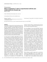
Figure 1 Relative levels of TWIST1 and KRT19 mRNA in BM samples from 267 early breast cancer patients. The levels were calculated using
the 2ΔΔCq method and normalized by dividing by the highest level in the control samples. The horizontal line represents the highest level in the
control samples with the relative value of one. hMAM is not shown in the plot because no expression was found in the normal control samples.
than ICC. It is important to note that BM3 and BM4
have a higher frequency of positive samples because
these samples were only collected from patients with a
positive BM2 sample.
Discussion
This study was undertaken to compare ICC with a MM
RT-qPCR assay for the detection of DTCs in BM after
adjuvant chemotherapy in early breast cancer patients.
Our study revealed a markedly higher frequency of
positive samples by both the MM RT-qPCR assay and
the individual mRNA-assays compared with ICC. Multiple
mRNA markers clearly contributed to a higher number of
positive samples compared to only using single markers.
The relatively high (61%) concordance between ICC and
Table 2 Concordance between ICC and mRNA markers
ICC
Multimarker
KRT19
hMAM
TWIST1
Concordance
Pos
Neg
Pos
12
112
Neg
11
178
Pos
4
42
Neg
19
248
Pos
2
1
Neg
21
289
Pos
9
88
Neg
14
202
Kappa
Value
0.61
0.045
0.81
0.020
RT-qPCR is primarily because a large fraction of the
samples were negative by both methods. Accordingly,
the kappa observer agreement value was only 0.04, suggesting that the apparent concordance was primarily
due to chance. In principle, the ICC assay should stain,
among others, KRT19 positive cells. Thus, we expected
better concordance between the ICC and the KRT19
mRNA results. However, only 4 out of 313 samples were
positive by both methods and as many as 42 ICC-negative
samples were positive for KRT19 mRNA. One possible explanation for this is that the KRT19 mRNA assay is more
sensitive than the ICC assay. The 19 ICC-positive samples
that were not detected by the KRT19 mRNA assay might
be explained by detection of KRT19-negative DTCs that
express other keratins detected by the ICC approach.
The low concentration of DTCs in BM samples may
affect reproducibility in both detection methods. Many
samples had levels near the detection limit for KRT19
mRNA; whereas, the ICC-assay was able to detect only a
single cell in the majority of positive samples. It follows
Table 3 Distribution of BM1-4 with ICC and qPCR data
BM number
0.93
0.14
0.67
0.035
Total
ICC
qPCR
Concordant
313
Positive (%)
Positive (%)
BM results (%)
BM 1
92
7
(7.6)
44
(47.8)
(53)
BM 2
187
11
(5.9)
63
(33.7)
(66)
BM 3
14
2
(14.3)
10
(71.4)
(43)
BM 4
18
3
(16.7)
7
(38.9)
(56)
Gilje et al. BMC Cancer 2014, 14:514
/>
from the Poisson distribution of rare events that there is
a roughly 35% risk that a second sample would be a false
negative. Hence, the reproducibility of DTC detection
might be enhanced by analyzing larger sample volumes.
On the other extreme, it was recently shown that screening a very large volume of peripheral blood by leukapheresis revealed DTCs in 90% of non-metastatic breast
cancer patients [32]. Such high numbers of DTCs does
not correlate with the risk of relapse for this patient
group, and thus implies a dramatic increase in detection
of clinically irrelevant cells.
The hMAM mRNA assay was only positive in a very
small number of samples (3/313); therefore, it might be
of limited value in combination with KRT19 mRNA in
the post-adjuvant treatment setting. Indeed, 2 of the 3
positive hMAM samples were also positive for KRT19
mRNA and by ICC, with convincing DTC-counts of 2
and 46 by ICC. The remaining hMAM positive sample
was TWIST1-positive and KRT19- and ICC-negative.
Thus, it seems that hMAM contributes to the identification of a very small subgroup of patients, possibly those
with a very high risk, consistent with our previous report
on hMAM [4,18].
TWIST1 was shown to add prognostic information to
a DTC MM panel described by Tjensvoll et al. [4]. Interestingly, we noted that a substantially higher number of
patients had elevated TWIST1 mRNA levels in our
present study [4]. The clinical follow-up will ultimately
help determine the relevance of this discrepancy. As
TWIST1 is a proposed epithelial-mesenchymal-transition
(EMT)-marker [33], the higher number of positive samples might indicate that a substantial portion of patients
have DTCs not expressing keratins [34]. The number of
TWIST1 positive samples, however, exceeds the anticipated number of clinical relapses. Thus, our assay might
be too sensitive, or the cut-off level needs refinement to
reveal only clinically relevant information. ROC analysis
in relation to clinical outcome data, when available, may
reveal an optimal cut-off value. However, this will require
confirmation in a validation cohort.
The high number of DTC positive samples by the
RT-qPCR approach is in part explained by the high
number of TWIST1 positive samples. In later years,
there has been much focus on mesenchymal markers
to detect cells that have undergone EMT as part of the
metastatic process. This is thought to be a reversible
process in which the cancer cells gain mesenchymal
properties to be able to infiltrate different tissues and
give rise to micro- and ultimately macro metastases [34].
Yu et al. showed that in circulating tumor cells a shift
towards higher expression of EMT-markers is associated
with tumor progression [35]. Thus, we might speculate
whether cells transiently expressing mesenchymal genes,
like TWIST1, comprise the subgroup of DTCs with
Page 6 of 8
stem-cell properties and, therefore, the proportion of DTCs
that harbor metastasis-generating abilities [36]. The loss of
epithelial characteristics may imply that these cells are difficult to detect by most commonly used ICC DTC assays.
The discrepancy between the RT-qPCR based and
the ICC-based DTC detection is not surprising based on
previous studies. Becker et al. found agreement between
ICC (with the A45-B/B3 mAb) and KRT19 mRNA detection in 73% of the 385 cases, in line with our KRT19 qPCR
results (81% agreement). Although, the results are biased
since the majority of patients were negative by both
methods. In fact, a kappa value of 0.39 can be computed
based on their reported data, confirming this suspicion to
some extent. Moreover, they demonstrated a 35% positive
rate for both ICC and KRT19 mRNA and 49% of the
patients were positive by at least one of the methods. The
time of BM-collection might be an important difference
between their study and the present one. We collected
BM after adjuvant chemotherapy; whereas, Becker et al.
collected the majority of samples prior to surgery and only
a few (n = 63) after surgery and chemotherapy. This may
have contributed to the much lower number of positive
samples based on both ICC and KRT19 mRNA in our
study. Others have reported concordance in the same
range as in our study. Benoy et al. reported concordant
results in 75% of the samples; whereas, Slade et al. found
agreement between the methods in 71% of the samples
[21,22]. Molloy et al. compared a MM RT-qPCR assay
with ICC in a large population of 733 patients and
found both to be significantly predictive of poorer outcome. However, the RT-qPCR assay was applied to blood
samples (circulating tumor cells) and the ICC to BM samples (DTCs). Thus, a direct comparison with the current
study is difficult because the samples were collected from
different body regions in addition to being analyzed by
two different methods [37].
A general issue regarding mRNA-based DTC detection
is the background level of epithelial transcripts in white
blood cells. However, comparison with blood samples from
a normal control cohort may compensate for this issue,
allowing threshold values for pathological marker levels in
blood to be established. The latter strategy was utilized in
the current study to minimize the number of false positives
due to such background expression in leukocytes.
Despite clear evidence that DTCs in BM in early
breast cancer patients predict a poor outcome, a better
understanding is needed for these analyses to be implemented in the routine clinical management of patients.
Braun et al. found, in their large pooled analysis of 4703
patients, a significant prognostic value of BM DTCstatus in all patients including the lymph-node negative subgroup [3]. On the other hand, several smaller
studies, e.g. by Langer et al., did not find any significant
DTC-specific difference in overall and breast cancer
Gilje et al. BMC Cancer 2014, 14:514
/>
specific survival in 411 clinically lymph node negative
patients, a result that may be caused by the low number of
patients in the study [3,38]. However, this result emphasizes
that the prognostic value of BM DTC-status might be
strongest in already defined high-risk patients. In the
current analysis, only intermediate/high-risk patients, i.
e. patients with higher-risk node negative or node positive disease, were included. Thus, this may be a group
where the BM-DTC status may add clinically relevant
prognostic information.
Few studies have investigated the impact of BM-DTCs
after initial therapy. In a pooled analysis, Janni et al. demonstrated that DTCs can be detected several years after
diagnosis [39]. The persistence of BM DTCs after neoadjuvant treatment was also associated with worse prognosis
in a recent study [40], and our previous results showed reduced survival for patients with persistent DTCs, assessed
by our mRNA MM-assay (hMAM, KRT19, and TWIST1),
after surgery [41]. The majority of studies so far have used
ICC for the detection of DTCs in the BM; although, some
studies also used RT-qPCR. The DTC-detection by ICC is
largely based on pan-cytokeratin antibodies, but different
antibody combinations have also been used with varying
results. Effenberger and colleagues found that ICC detection and prognostic relevance were different for the two
most commonly used pan-cytokeratin antibody combinations (A45-B/B3 (A45) and AE1-AE3 (AE)) [12]. The
AE mAb was more prognostic for the lymph node positive patients. Accordingly, the AE mAbs were utilized in
the current SATT-trial, consistent with the inclusion of a
higher risk population.
Conclusions
In conclusion, this study is to our knowledge the largest
comparison between ICC- and RT-qPCR-based DTC
detection methods in BM samples collected after adjuvant
chemotherapy in a defined high-risk early breast cancer
population. We detected more positive BM samples with
RT-qPCR assays, based on KRT19, hMAM, and TWIST1
mRNAs, than with ICC. The clinical implications of these
findings, however, await future clinical follow-up. Due
to a potential shift in DTC phenotype, we included the
mesenchymal TWIST1 mRNA marker in an attempt to
detect the subpopulation of DTCs lacking epithelial characteristics. Hopefully, this might help identify additional
patients with clinically relevant DTCs. The current findings support that the different means of detection could
be complementary and that both RT-qPCR and ICC
should be further studied as methods for DTC detection
in early breast cancer patients.
Abbreviations
DTC: Disseminated tumor cell; BM: Bone marrow; SATT: Secondary Adjuvant
Taxotere Treatment; ICC: Immunocytochemistry; FEC: Fluorouracil, epirubicin,
cyclophosphamide; MM,: Multimarker; KRT19: Keratin 19; hMAM: Mammaglobin
Page 7 of 8
A; RT-qPCR: Reverse transcription quantitative polymerase chain reaction;
TNM: Standard tumor-node-metastasis classification according to AJCC/UICC
2002; ER: Estrogen receptor; PgR: Progesterone receptor; HER2: Human
epidermal growth factor receptor 2; BCR: Breakpoint cluster region;
EMT: Epithelial-mesenchymal-transition.
Competing interests
The authors declare that they have no competing interests.
Authors’ contributions
BG, ON, KT, RS, and BN drafted the manuscript. BG, BN, RS, MS and ON were
responsible for the study design. BG and ON performed the data analysis
and carried out the statistics. EB performed immunocytochemistry detection
of DTCs and BG performed the RT-qPCR-based detection of DTCs. All authors
read and approved the final manuscript.
Acknowledgements
The study was supported by grants from Western Norway Regional Health
Authorities, the Folke Hermansen Foundation and Sanofi.
Author details
1
Department of Hematology and Oncology, Stavanger University Hospital,
Stavanger, Norway. 2Laboratory for Molecular Biology, Stavanger University
Hospital, Stavanger, Norway. 3Division of Surgery and Cancer Medicine,
Department of Pathology, Oslo University Hospital, Oslo, Norway. 4Division of
Surgery, Transplantation and Cancer Medicine, Department of Oncology,
Oslo University Hospital, Oslo, Norway. 5K.G. Jebsen Center for Breast Cancer
Research, Institute for Clinical Medicine, University of Oslo, Oslo, Norway.
Received: 11 April 2014 Accepted: 10 July 2014
Published: 15 July 2014
References
1. Goldhirsch A, Winer EP, Coates AS, Gelber RD, Piccart-Gebhart M, Thurlimann B,
Senn HJ: Personalizing the treatment of women with early breast cancer:
highlights of the St Gallen International Expert Consensus on the Primary
Therapy of Early Breast Cancer 2013. Ann Oncol 2013, 24(9):2206–2223.
2. Braun S, Pantel K, Muller P, Janni W, Hepp F, Kentenich CR, Gastroph S,
Wischnik A, Dimpfl T, Kindermann G, Riethmüller G, Schlimok G:
Cytokeratin-positive cells in the bone marrow and survival of patients
with stage I, II, or III breast cancer. N Engl J Med 2000, 342(8):525–533.
3. Braun S, Vogl FD, Naume B, Janni W, Osborne MP, Coombes RC, Schlimok
G, Diel IJ, Gerber B, Gebauer G, Pierga JY, Marth C, Oruzio D, Wiedswang G,
Solomayer EF, Kundt G, Strobl B, Fehm T, Wong GY, Bliss J, Vincent-Salomon
A, Pantel K: A pooled analysis of bone marrow micrometastasis in breast
cancer. N Engl J Med 2005, 353(8):793–802.
4. Tjensvoll K, Oltedal S, Farmen RK, Shammas FV, Heikkila R, Kvaloy JT, Gilje B,
Smaaland R, Nordgard O: Disseminated tumor cells in bone marrow
assessed by TWIST1, cytokeratin 19, and mammaglobin A mRNA predict
clinical outcome in operable breast cancer patients. Clin Breast Cancer
2010, 10(5):378–384.
5. Wiedswang G, Borgen E, Karesen R, Kvalheim G, Nesland JM, Qvist H,
Schlichting E, Sauer T, Janbu J, Harbitz T, Naume B: Detection of isolated
tumor cells in bone marrow is an independent prognostic factor in
breast cancer. J Clin Oncol 2003, 21(18):3469–3478.
6. Sanger N, Effenberger KE, Riethdorf S, Van Haasteren V, Gauwerky J,
Wiegratz I, Strebhardt K, Kaufmann M, Pantel K: Disseminated tumor cells
in the bone marrow of patients with ductal carcinoma in situ.
Int J Cancer 2011, 129(10):2522–2526.
7. Aft R, Naughton M, Trinkaus K, Watson M, Ylagan L, Chavez-MacGregor M,
Zhai J, Kuo S, Shannon W, Diemer K, Herrmann V, Dietz J, Ali A, Ellis M,
Weiss P, Eberlein T, Ma C, Fracasso PM, Zoberi I, Taylor M, Gillanders W,
Pluard T, Mortimer J, Weilbaecher K: Effect of zoledronic acid on disseminated
tumour cells in women with locally advanced breast cancer: an open label,
randomised, phase 2 trial. Lancet Oncol 2010, 11(5):421–428.
8. Hadji P, Coleman R, Gnant M, Green J: The impact of menopause on bone,
zoledronic acid, and implications for breast cancer growth and
metastasis. Ann Oncol 2012, 23(11):2782–2790.
9. Rack B, Juckstock J, Genss EM, Schoberth A, Schindlbeck C, Strobl B, Heinrigs
M, Rammel G, Zwingers T, Sommer H, Friese K, Janni W: Effect of zoledronate
Gilje et al. BMC Cancer 2014, 14:514
/>
10.
11.
12.
13.
14.
15.
16.
17.
18.
19.
20.
21.
22.
23.
24.
25.
26.
on persisting isolated tumour cells in patients with early breast cancer.
Anticancer Res 2010, 30(5):1807–1813.
Solomayer EF, Gebauer G, Hirnle P, Janni W, Luck HJ, Becker S, Huober J,
Kramer B, Wackwitz B, Wallwiener D, Fehm T: Influence of zoledronic acid
on disseminated tumor cells in primary breast cancer patients. Ann Oncol
2012, 23(9):2271–2277.
Tjensvoll K, Nordgard O, Smaaland R: Circulating tumor cells in pancreatic
cancer patients: methods of detection and clinical implications. Int J
Cancer 2014, 134(1):1–8.
Effenberger KE, Borgen E, Eulenburg CZ, Bartkowiak K, Grosser A,
Synnestvedt M, Kaaresen R, Brandt B, Nesland JM, Pantel K, Naume B:
Detection and clinical relevance of early disseminated breast cancer
cells depend on their cytokeratin expression pattern. Breast Cancer Res
Treat 2011, 125(3):729–738.
Joosse SA, Hannemann J, Spotter J, Bauche A, Andreas A, Muller V, Pantel K:
Changes in keratin expression during metastatic progression of breast
cancer: impact on the detection of circulating tumor cells. Clin Cancer Res
2012, 18(4):993–1003.
Bosma AJ, Weigelt B, Lambrechts AC, Verhagen OJ, Pruntel R, Hart AA,
Rodenhuis S, van ’t Veer LJ: Detection of circulating breast tumor cells by
differential expression of marker genes. Clin Cancer Res 2002, 8(6):1871–1877.
Ignatiadis M, Xenidis N, Perraki M, Apostolaki S, Politaki E, Kafousi M,
Stathopoulos EN, Stathopoulou A, Lianidou E, Chlouverakis G, Sotiriou C,
Georgoulias V, Mavroudis D: Different prognostic value of cytokeratin-19
mRNA positive circulating tumor cells according to estrogen receptor
and HER2 status in early-stage breast cancer. J Clin Oncol 2007,
25(33):5194–5202.
Lacroix M: Significance, detection and markers of disseminated breast
cancer cells. Endocr Relat Cancer 2006, 13(4):1033–1067.
Ooka M, Tamaki Y, Sakita I, Fujiwara Y, Yamamoto H, Miyake Y, Sekimoto M,
Ohue M, Sugita Y, Miyoshi Y, Ikeda N, Noguchi S, Monden M: Bone marrow
micrometastases detected by RT-PCR for mammaglobin can be an
alternative prognostic factor of breast cancer. Breast Cancer Res Treat
2001, 67(2):169–175.
Tjensvoll K, Gilje B, Oltedal S, Shammas VF, Kvaloy JT, Heikkila R, Nordgard O:
A small subgroup of operable breast cancer patients with poor prognosis
identified by quantitative real-time RT-PCR detection of mammaglobin A
and trefoil factor 1 mRNA expression in bone marrow. Breast Cancer Res
Treat 2009, 116(2):329–338.
Zach O, Lutz D: Tumor cell detection in peripheral blood and bone
marrow. Curr Opin Oncol 2006, 18(1):48–56.
Becker S, Becker-Pergola G, Banys M, Krawczyk N, Wallwiener D, Solomayer
E, Schuetz C, Fehm T: Evaluation of a RT-PCR based routine screening tool
for the detection of disseminated epithelial cells in the bone marrow of
breast cancer patients. Breast Cancer Res Treat 2009, 117(2):227–233.
Benoy IH, Elst H, Van der Auwera I, Van Laere S, van Dam P, Van Marck E,
Scharpe S, Vermeulen PB, Dirix LY: Real-time RT-PCR correlates with
immunocytochemistry for the detection of disseminated epithelial cells
in bone marrow aspirates of patients with breast cancer. Br J Cancer
2004, 91(10):1813–1820.
Slade MJ, Singh A, Smith BM, Tripuraneni G, Hall E, Peckitt C, Fox S, Graham
H, Luchtenborg M, Sinnett HD, Cross NC, Coombes RC: Persistence of bone
marrow micrometastases in patients receiving adjuvant therapy for
breast cancer: results at 4 years. Int J Cancer 2005, 114(1):94–100.
Synnestvedt M, Borgen E, Wist E, Wiedswang G, Weyde K, Risberg T, Kersten
C, Mjaaland I, Vindi L, Schirmer C, Nesland JM, Naume B: Disseminated
tumor cells as selection marker and monitoring tool for secondary
adjuvant treatment in early breast cancer. Descriptive results from an
intervention study. BMC Cancer 2012, 12:616.
McShane LM, Altman DG, Sauerbrei W, Taube SE, Gion M, Clark GM:
Reporting recommendations for tumor marker prognostic studies. J Clin
Oncol 2005, 23(36):9067–9072.
Borgen E, Naume B, Nesland JM, Kvalheim G, Beiske K, Fodstad O, Diel I,
Solomayer EF, Theocharous P, Coombes RC, Smith BM, Wunder E, Marolleau
JP, Garcia J, Pantel K: Standardization of the immunocytochemical
detection of cancer cells in BM and blood: I. establishment of objective
criteria for the evaluation of immunostained cells. Cytotherapy 1999,
1(5):377–388.
Fehm T, Braun S, Muller V, Janni W, Gebauer G, Marth C, Schindlbeck C,
Wallwiener D, Borgen E, Naume B, Pantel K, Solomayer E: A concept for the
standardized detection of disseminated tumor cells in bone marrow
Page 8 of 8
27.
28.
29.
30.
31.
32.
33.
34.
35.
36.
37.
38.
39.
40.
41.
from patients with primary breast cancer and its clinical implementation.
Cancer 2006, 107(5):885–892.
Naume B, Wiedswang G, Borgen E, Kvalheim G, Karesen R, Qvist H, Janbu J,
Harbitz T, Nesland JM: The prognostic value of isolated tumor cells in
bone marrow in breast cancer patients: evaluation of morphological
categories and the number of clinically significant cells. Clin Cancer Res
2004, 10(9):3091–3097.
Wiedswang G, Borgen E, Karesen R, Qvist H, Janbu J, Kvalheim G, Nesland
JM, Naume B: Isolated tumor cells in bone marrow three years after
diagnosis in disease-free breast cancer patients predict unfavorable
clinical outcome. Clin Cancer Res 2004, 10(16):5342–5348.
Farmen RK, Nordgard O, Gilje B, Shammas FV, Kvaloy JT, Oltedal S, Heikkila
R: Bone marrow cytokeratin 19 mRNA level is an independent predictor
of relapse-free survival in operable breast cancer patients. Breast Cancer
Res Treat 2008, 108(2):251–258.
Livak KJ, Schmittgen TD: Analysis of relative gene expression data using
real-time quantitative PCR and the 2(-Delta Delta C(T)) Method.
Methods 2001, 25(4):402–408.
Sim J, Wright CC: The kappa statistic in reliability studies: use, interpretation,
and sample size requirements. Phys Ther 2005, 85(3):257–268.
Fischer JC, Niederacher D, Topp SA, Honisch E, Schumacher S, Schmitz N,
Zacarias Fohrding L, Vay C, Hoffmann I, Kasprowicz NS, Hepp PG,
Mohrmann S, Nitz U, Stresemann A, Krahn T, Henze T, Griebsch E, Raba K,
Rox JM, Wenzel F, Sproll C, Janni W, Fehm T, Klein CA, Knoefel WT,
Stoecklein NH: Diagnostic leukapheresis enables reliable detection of
circulating tumor cells of nonmetastatic cancer patients. Proc Natl Acad
Sci U S A 2013, 110(41):16580–16585.
Yang J, Mani SA, Donaher JL, Ramaswamy S, Itzykson RA, Come C, Savagner
P, Gitelman I, Richardson A, Weinberg RA: Twist, a master regulator of
morphogenesis, plays an essential role in tumor metastasis. Cell 2004,
117(7):927–939.
Kalluri R, Weinberg RA: The basics of epithelial-mesenchymal transition.
J Clin Invest 2009, 119(6):1420–1428.
Yu M, Bardia A, Wittner BS, Stott SL, Smas ME, Ting DT, Isakoff SJ, Ciciliano
JC, Wells MN, Shah AM, Concannon KF, Donaldson MC, Sequist LV, Brachtel
E, Sgroi D, Baselga J, Ramaswamy S, Toner M, Haber DA, Maheswaran S:
Circulating breast tumor cells exhibit dynamic changes in epithelial and
mesenchymal composition. Science 2013, 339(6119):580–584.
De Craene B, Berx G: Regulatory networks defining EMT during cancer
initiation and progression. Nat Rev Cancer 2013, 13(2):97–110.
Molloy TJ, Bosma AJ, Baumbusch LO, Synnestvedt M, Borgen E, Russnes HG,
Schlichting E, van’t Veer LJ, Naume B: The prognostic significance of tumour
cell detection in the peripheral blood versus the bone marrow in 733
early-stage breast cancer patients. Breast Cancer Res 2011, 13(3):R61.
Langer I, Guller U, Worni M, Berclaz G, Singer G, Schaer G, Fehr MK, Hess T,
Viehl C, Bronz L, Schnarwyler B, Wight E, Infanger E, Burger D, Koechli OR,
Zuber M: Bone marrow micrometastases do not impact disease-free and
overall survival in early stage sentinel lymph node-negative breast
cancer patients. Ann Surg Oncol 2013, 21(2):401–407.
Janni W, Vogl FD, Wiedswang G, Synnestvedt M, Fehm T, Juckstock J,
Borgen E, Rack B, Braun S, Sommer H, Solomayer E, Pantel K, Nesland J,
Friese K, Naume B: Persistence of disseminated tumor cells in the bone
marrow of breast cancer patients predicts increased risk for relapse–a
European pooled analysis. Clin Cancer Res 2011, 17(9):2967–2976.
Mathiesen RR, Borgen E, Renolen A, Lokkevik E, Nesland JM, Anker G,
Ostenstad B, Lundgren S, Risberg T, Mjaaland I, Kvalheim G, Lonning PE,
Naume B: Persistence of disseminated tumor cells after neoadjuvant
treatment for locally advanced breast cancer predicts poor survival.
Breast Cancer Res 2012, 14(4):R117.
Tjensvoll K, Oltedal S, Heikkila R, Kvaloy JT, Gilje B, Reuben JM, Smaaland R,
Nordgard O: Persistent tumor cells in bone marrow of non-metastatic
breast cancer patients after primary surgery are associated with inferior
outcome. BMC Cancer 2012, 12:190.
doi:10.1186/1471-2407-14-514
Cite this article as: Gilje et al.: Comparison of molecular and
immunocytochemical methods for detection of disseminated tumor cells in
bone marrow from early breast cancer patients. BMC Cancer 2014 14:514.