Incidental advanced-stage Hodgkin lymphoma diagnosed at the time of radical prostatectomy for prostatic cancer: A case report and review of literature
Bạn đang xem bản rút gọn của tài liệu. Xem và tải ngay bản đầy đủ của tài liệu tại đây (2.47 MB, 9 trang )
Di Meglio et al. BMC Cancer 2014, 14:613
/>
CASE REPORT
Open Access
Incidental advanced-stage Hodgkin lymphoma
diagnosed at the time of radical prostatectomy
for prostatic cancer: a case report and review
of literature
Antonio Di Meglio1,2*, Pier Vitale Nuzzo1,2, Francesco Ricci2, Bruno Spina3 and Francesco Boccardo1,2
Abstract
Background: Pelvic lymph nodes removed during radical retropubic prostatectomy for prostatic cancer can be found
on pathological examination to harbor various unexpected pathologies. Among these, hematologic neoplasms are not
infrequent. Given their frequently indolent clinical course, such neoplasms would likely have remained undiagnosed
and non-life threatening. Despite this, the case we are reporting describes a rare association between two aggressive
neoplasms, and it will be helpful to clinicians who encounter similar combinations of pathologies.
Case presentation: We report the challenging case of a 56-year-old, caucasian man in whom pathological assessment
of pelvic lymph nodes removed during radical retropubic prostatectomy for a high-grade prostatic neoplasm revealed
Hodgkin lymphoma, which was subsequently classified as stage IV. There are very few published reports of this
combination of pathologies. This situation required a cautious and expert approach to delivering the most appropriate
treatment with the most appropriate timing for both diseases.
Conclusion: This report describes the multidisciplinary clinical approach we followed at our institution. We have also
presented a review of published reports concerning the incidence, histologic type, and management of such
concurrent malignancies.
Keywords: Prostatic neoplasm, Radical prostatectomy, Hodgkin lymphoma, Hematologic neoplasm, Concurrent
malignancies, CD44, Literature review
Background
Currently, radical retropubic prostatectomy (RRP) is considered the gold standard for local treatment of organconfined prostate cancer (PCa) [1,2]. Recognizing pelvic
lymph node metastases from PCa during pre-operative assessment can be problematic. Because nodal involvement
is often microscopic and therefore undetectable by using
standard imaging techniques and dimensional and morphologic criteria, metastatic involvement of pelvic nodes
can be overlooked preoperatively; only to be discovered
* Correspondence:
1
IRCCS San Martino University Hospital, IST National Cancer Research
Institute, Academic Unit of Medical Oncology, Genoa, Italy
2
Department of Internal Medicine (DiMI), University of Genova School of
Medicine, Genoa, Italy
Full list of author information is available at the end of the article
unexpectedly by pathologists in the resected specimen
[3,4].
Several incidental findings, other than metastases from
PCa, have been reported in pelvic lymph nodes evaluated
at the time of RRP. These have included nodal metastases
from malignancies arising in another primary site and
non-metastatic disease arising directly from lymphoid tissue (i.e., various types of leukemia/lymphoma).
We describe the case of a patient who underwent surgery for a biopsy-proven high-grade PCa and had an incidental diagnosis of Hodgkin lymphoma (HL) involving
pelvic lymph nodes. We then performed a systematic
search of published reports concerning associations between PCa and hematologic malignancies (HM) discovered as a result of surgery for the PCa. Although several
cases of concomitant HM and primary PCa have been
© 2014 Di Meglio et al.; licensee BioMed Central Ltd. This is an Open Access article distributed under the terms of the Creative
Commons Attribution License ( which permits unrestricted use, distribution, and
reproduction in any medium, provided the original work is properly credited. The Creative Commons Public Domain
Dedication waiver ( applies to the data made available in this article,
unless otherwise stated.
Di Meglio et al. BMC Cancer 2014, 14:613
/>
reported, this association is uncommon; no guidelines
for the management of such patients are thus far available. Moreover, the clinical significance and prognostic
impact of these malignancies in the context of PCa remains unclear.
Case presentation
Case description
A 56-year-old man was referred to our unit after undergoing RRP and bilateral pelvic lymphadenectomy at another hospital. Pathological examination had confirmed
the initial diagnosis of high-grade adenocarcinoma,
Gleason score 10 (5 + 5), consistent with the findings on
the biopsies performed preoperatively. Additionally, it
had disclosed disease extension to both lobes of the
gland, apex, and seminal vesicles, and focal involvement
of the resection margins (Figure 1).
None of the 30 lymph nodes removed in the procedure contained metastatic cells from the PCa. Rather and
surprisingly, the larger lymph nodes were found to
Page 2 of 9
contain classic mixed cellularity HL. The malignant
Hodgkin and Reed-Sternberg cells stained positive for
cluster of differentiation (CD) 20, CD30, and CD15.
Additionally, immunohistochemistry was negative for
CD45, CD3, epithelial membrane antigen, and PAX5
(Figure 2).
When the patient was referred to our clinics 1 month
postoperatively, his serum concentration of prostatespecific antigen (PSA) was 0.34 ng/mL (pre-surgical PSA
had been 6.6 ng/mL).
A staging 18-fluoro-deoxyglucose positron emission
tomography (FDG PET) scan showed nodal disease on
both sides of the diaphragm with enhanced metabolic
activity in the spleen and skeleton (Figure 3A). However,
no tumor invasion was detected on bone marrow biopsy.
A whole-body computed tomography (CT) scan confirmed axillary, mediastinal, celiac trunk, and retroperitoneal lymphadenopathies and failed to detect any bone
lesions. Because the PET scan was positive at the bone
level, his HL was classified as stage IV according to the
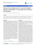
Figure 1 Adenocarcinoma of the prostate, Gleason 10. Hematoxylin and eosin stained photomicrographs (10x magnification) showing: (A)
poorly differentiated adenocarcinoma of the prostate (Gleason score 5 + 5 = 10); (B) disease extension into seminal vesicles; (C) tumor vascular
invasion; and (D) presence of multifocal embolic perineural tumor.
Di Meglio et al. BMC Cancer 2014, 14:613
/>
Page 3 of 9
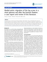
Figure 2 Infiltration of Hodgkin lymphoma within lymph nodes. Photomicrographs of (A) malignant Hodgkin and Reed-Sternberg cell
showing (B) negative staining for CD45; (C) positive staining for CD30; and (D) positive staining for CD15.
Ann Arbor classification, even though bone involvement
from PCa could not be completely excluded. The patient
underwent front-line combination chemotherapy with
the EBVD regimen (epirubicin 35 mg/m2; bleomycin
10 mg/m2; vinblastine 6 mg/m2; dacarbazine 375 mg/
m2). A multidisciplinary team of experts, including hematologists and radiation oncologists, planned and concurred on this approach.
After three cycles of treatment, an interim evaluation
with a FDG PET scan showed no FDG-avid tissue in the
previously positive sites. These data were confirmed by a
whole-body CT scan, which showed shrinkage of previously enlarged lymph nodes. Thus, there was evidence
that the HL had responded well to chemotherapy; however, during this time the PSA concentration had further
increased up to 0.96 ng/mL (PSA doubling time
1.92 months). Therefore, anti-androgen therapy with
bicalutamide, 150 mg per day, was initiated. In addition
to providing evidence of HL response to chemotherapy,
the radiologic images also showed interstitial pneumonia,
which was considered an adverse effect of bleomycin.
Hence, three more cycles of chemotherapy without bleomycin and with the addition of 40 mg of prednisone daily
on days 1–5 of each cycle were scheduled.
After six cycles of chemotherapy, a FDG PET scan
showed no residual disease (Figure 3B); a whole-body
CT scan confirmed complete disappearance of the
lymphoma lesions and resolution of the interstitial pneumonia. PSA was undetectable in his serum. Nevertheless,
because of the adverse prognostic features of his PCa;
namely, the high Gleason score, invasion of seminal vesicles, positive surgical margins, and the increase in PSA
concentrations postoperatively (before commencement
of bicalutamide therapy), on completion of chemotherapy for HL, the patient was also submitted to pelvic irradiation (60 Gy were delivered in 30 fractions to the
whole pelvis followed by an 18-Gy boost to the prostatic
bed, which required the delivery of eight additional daily
fractions). PSA continued to be undetectable in his
serum up until completion of treatment and thereafter.
Di Meglio et al. BMC Cancer 2014, 14:613
/>
Page 4 of 9
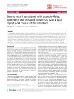
Figure 3 FDG PET scan images before and after treatment. (A) Staging FDG PET scan image showing nodal disease on both sides of the
diaphragm with enhanced metabolic activity in the spleen and skeleton. (B) End of treatment: FDG PET image showing no residual disease.
Bicalutamide single-agent treatment is currently being
continued and the patient is being rigorously followed
up with serum PSA checks 3 monthly and whole body
FDG PET/CT scans 6 monthly. At the time of this report, 30 months after this patient’s referral to our clinics,
there is no evidence of either HL recurrence or of PCa
progression (serum PSA remains undetectable).
Discussion
We performed a systematic search of the PubMed database
using the MeSH keywords “prostatic neoplasms”, “prostatectomy”, “lymphoma”, and “hematologic neoplasms” and
identified retrospective reviews of a total of over 19,000
specimens (most of which had been obtained from patients
who had undergone RRP). We identified seven studies,
Di Meglio et al. BMC Cancer 2014, 14:613
/>
designed ad hoc to assess the frequency and cause of incidental (non-metastatic) lymph node pathology discovered
during RRP that had been performed between 1996 and
2007. The findings of these studies are summarized in
Table 1.
We also identified three case reports of patients who
had been diagnosed with a second malignant hematologic
neoplasm in addition to their PCa. These isolated cases
are also listed in Table 1.
In the evaluated series, the overall incidence of HM
harbored by pelvic lymph nodes removed in the course
of RRP had a range from 0.003% [9] to 1.2% [7]. In the
great majority of these cases, the diagnosis of a HM had
not been suspected preoperatively.
Currently, contrast-enhanced CT scan along with MRI
are the most commonly employed techniques for evaluating nodal disease pre-operatively in patients with PCa.
These imaging techniques are usually reserved for patients with an intermediate or high risk of extraprostatic and/or nodal disease dissemination [16,17].
Evaluation of lymph node metastasis is one of the major
goals of CT scanning in PCa staging. However, such
evaluation is limited by false-positive results and the
paucity of available techniques for identifying lymph
node metastasis [18].
Moreover, unsuspected abnormalities, unrelated to the
known primary PCa, can be revealed during the diagnostic/staging imaging workup. Miller et al. reported discovering a clinically significant coexistent disease by CT
scan in 89/1330 PCa patients (6.7%) who were to
undergo radiation therapy [19].
Elmi et al. retrospectively reviewed 355 initial staging
abdominopelvic CT examinations in patients with PCa
for incidental findings that were unrelated to their primary disease. These “incidentalomas” were classified as
being of low, moderate, or high importance, depending
on the type of medical or surgical management eventually required or on their potential to adversely affect
health. Seventy-five potentially significant findings were
noted in 73 patients (20.6% of all patients): most were
renal masses; these were confirmed to be renal cell malignancies in seven patients (1.97% of all patients). Additionally, lymphadenopathies at sites unlikely to harbor
PCa metastasis were noted in 18 cases, in four of whom
histopathologic examination resulted in a diagnosis of
lymphoma (1.12% of all patients) [20]. Enlarged lymph
nodes were detected in 102 patients; only 18 of these
were in sites uncommonly affected by PCa metastasis
(mainly mesenteric). Accordingly, Coakley et al. suggested
that a diagnosis of lymphoma should be considered in patients with PCa and imaging findings of mesenteric
lymphadenopathies [21].
He et al. reported a <1% incidence of metastases from
PCa in pelvic lymph nodes [11]. This rate of positivity is
Page 5 of 9
unusually low compared with major retrospectively
assessed series reported by Roehl et al. and Daneshmand
et al.: these authors cite an incidence of enlarged lymph
nodes in typical PCa locations in the range of 5.8% [22]
to 12.1% [23] in series of 3478 and 1972 patients, respectively, who had undergone RRP and lymph node
dissection. However, Partin et al. have reported an even
larger series of 5079 cases, considerably more than in either Roehl et al. or Daneshmand et al.’s series. These authors reported pathologically confirmed metastatic
involvement by PCa of lymph nodes in 2% of the 5079
lymph node dissections performed [24].
Winstanley et al. have reported other findings apart
from hematolymphoid pathology in enlarged pelvic
nodes in patients undergoing RRP. Most such lymph
nodes findings did not harbor neoplasms but were affected by other pathologies, including sinus histiocytosis,
non-caseating granulomas, and foreign body reactions.
Therefore, pathologists should be aware of these possibilities, to arrive at the correct diagnosis [9].
In the present case, our patient had not undergone any
pre-surgical staging, probably because of his good general
health and young age. In regard to age, Elmi et al. reported that the overall rate of incidental findings is not
significantly different in patients aged <65 versus >65 years.
However, they reported that patients aged more than
65 years have a higher rate of second neoplasms/synchronous malignancies than younger patients [20]. These
findings are not relevant to our patient, who was aged less
than 65 years.
Preexisting co-morbidities can influence treatment
choices in patients with newly diagnosed PCa [25].
Though possible, incidental discovery of life-threatening
conditions that may force clinicians to delay or modify
the scheduled treatment for PCa is rare: imaging overuse
can lead to over-diagnosis of subclinical conditions that
would never become overt during a patient’s lifetime;
this is a worldwide issue [26].
When discovered incidentally, HM are usually at an
extremely early stage, have limited spread, and are
asymptomatic [7,11]. Although our patient was asymptomatic, he had stage IV HL involving lymph nodal
stations on both sides of the diaphragm, as well as extranodal sites (spleen and skeleton). It is extremely rare to
find such advanced disease incidentally. Of the cases
identified by Eisenberger et al. in over 4000 procedures,
none had diffuse and/or bulky disease [8].
Taking together, only six of 89 reported cases of incidentally discovered HM required an aggressive approach.
Most reported patients with incidentally discovered HM
had low-grade follicular non-Hodgkin lymphoma or small
lymphocytic lymphoma/chronic lymphocytic leukemia.
Considering the indolent nature of these conditions and
the associated expected long-term survival, the decision to
Authors and year
Number of radical prostatectomies
(Number of lymph node dissections
performed)
Total number of concurrent
hematolymphoid malignancies
(Overall incidence)
Encountered Hematolymphoid
malignancy
Treatment required for
hematologic malignancy
Donohue et al. 1996 [5,6]
225 (N.A.)
3 (1.2%)
Lymphoma NOS
N.A.
Terris et al. 1997 [7]
1092 (all patients)
13 (1.2%)
3 HL
Pelvic and abdominal external beam irradiation
1 HCL
Aggressive tp NOS
1 CLL
No tp
6 SLL
No tp
Eisenberger et al. 1999 [8]
4319 (all patients)
10 (0.2%)
8 Lymphocytic lymphoma
Single-agent CT
2 FL
N.A
Winstanley et al. 2002 [9]
1001 (854)
15 (1.8%)
2 SLL/CLL
N.A.
3 neoplastic (0.003%)
1 FL
N.A.
12 Non-neoplastic findings1
N.A.
Weir et al. 2003 [10]
6143 (all patients)
18 (0.3%)
18 SLL/CLL
N.A.
He et al. 2007 [11]
1500 (1150)
13 (1.13%)
9 SLL/CLL
N.A.
Chu et al. 2005 [12]
48313 (N.A.)
29 (0.6%)
3 MZL
N.A.
1 MCL
Aggressive CT NOS
18 incidental cases
N.A.
13 SLL/CLL
N.A.
Di Meglio et al. BMC Cancer 2014, 14:613
/>
Table 1 Published reports of incidental concurrent findings during RRP and lymph node dissections for PCa
3 MZL
1 MCL
11 concurrent known lymphoma2
4 SLL/CLL
4 FL
2 MCL
1 DLBCL
Isolated case reports
Encountered Hematolymphoid malignancy
Treatment required for
hematologic malignancy
Carson H et al. 1996 [13]
1 SLL/CLL
N.A
Mydlo et al. 2001 [14]
1 lymphoma NOS
N.A
Drinis et al. 2009 [15]
1 SLL/CLL
No tp
Page 6 of 9
Abbreviations used: NOS not otherwise specified, tp therapy, N.A. not available, CT chemotherapy, HL Hodgkin lymphom, HCL hairy cell leukemia, CLL chronic lymphocytic leukemia, SLL small lymphocytic lymphoma,
MZL marginal zone lymphoma, MCL mantle cell lymphoma.
1
Including sinus histiocytosis, non-caseating granulomas, foreign body reactions.
2
Prostate and pelvic lymph nodes involved as part of a systemic disease.
3
Specimens were from 3405 biopsies, 266 transurethral resections, and 1160 prostatectomies.
Di Meglio et al. BMC Cancer 2014, 14:613
/>
delay treatment until symptoms developed or diseaserelated complications occurred was made in the majority
of patients reported [27].
No clinical management algorithms have yet been defined for synchronous occurrence of PCa and HL; the
impact of such a double diagnosis on clinical outcome is
unknown. Most authors suggest to treat the more aggressive condition first, thus improving the overall status
of the patient and facilitating a better response of the
second disease to therapy [28]. In our case, treatment
decisions were jointly made by a panel of experts. Because HL appeared to be both the more aggressive of
the two conditions and the disease in which cure was
more likely to be achieved, combination chemotherapy
with the EBVD regimen was initiated as soon as the patient had recovered from his surgery: complete remission of the disease was achieved within a few months.
To avoid any interference with the treatment for his
lymphoma, our panel of experts decided to postpone
pelvic radiotherapy, even though it was robustly indicated in view of the locally advanced stage of PCa and
Page 7 of 9
microscopic residual disease. The decision to postpone
this treatment was supported by the prompt PSA response to the anti-androgen therapy initiated after the
first two chemotherapy cycles. Though no randomized
studies have demonstrated clear superiority for immediate treatment of biochemical recurrence with radiation
or hormonal therapy, several retrospective studies have
shown that anti-androgen therapy prolongs time to metastasis and probably PCa-specific survival [29,30].
Despite the adverse histologic features and high Gleason score of our patient’s PCa, he has had no evidence
of metastatic disease and no increase in PSA since completing pelvic radiotherapy.
As already mentioned, it is not clear yet whether the
co-existence of a malignant lymphoma can alter per se
the natural history of PCa.
Drinis et al. have raised the intriguing possibility that
lymphomas could potentially protect against PCa progression. According to these authors, such protection
could result from the over-expression of circulating transmembrane molecule CD44 in leukemia and lymphoma
Figure 4 Seminal vesicle infiltration by adenocarcinoma of the prostate (Hematoxylin/Eosin and CD44 staining). (A,C) Hematoxylin and
eosin stained photomicrographs showing left seminal vesicle infiltration by poorly differentiated adenocarcinoma of the prostate (10× and 40×
magnification, respectively) (B,D) Photomicrographs showing positive CD44 staining of left seminal vesicle and negative staining CD44 staining of
adenocarcinoma of the prostate (10× and 40× magnification, respectively).
Di Meglio et al. BMC Cancer 2014, 14:613
/>
patients [15]. Some experimental studies support a tumor
suppressor function of CD44 in lymphomas; silencing of
CD44 expression may facilitate lymphoma genesis [31]. In
contrast, circulating concentrations of this protein appear
to be decreased in advanced and metastatic PCa, apparently contributing to tumor progression [32,33].
Gao et al. have also suggested that CD44 is a “metastatic suppressor gene” in PCa. [34-36]. The biological
role of CD44 might not be identical in all organs and tumors. In tissues that do not normally express CD44, its
acquired expression probably correlates with an adverse
outcome, the CD44 having growth- and metastasispromoting actions [37-39].
In light of the above data, we performed CD44 immunostaining on the surgical specimens obtained from our
patient during prostatectomy and lymph node dissection
(Figure 4). The normal prostatic tissue stained positive
for CD44, whereas the PCa tissue did not. The peritumoral stroma, seminal vesicles, and sites of perineural
invasion were mildly CD44 positive. Lymph nodes involved by lymphoma also stained positive for CD44.
These findings support the theory that CD44 is
expressed by normal prostatic epithelium and that capacity for expression is lost during the alterations in
structural differentiation that occur in the course of the
transition to neoplastic tissue. Whether CD44 might be
a prognostic marker indicating the malignant potential
of neoplasm would be difficult to determine because a
standard histologic scoring system that includes CD44
assay would be problematic because of the heterogeneity
and available isoforms of this receptor [40]. Additonally,
the interaction between CD44 expression by lymphoma
cells and PCa cells remains unclear; further investigation
is needed to assign a definite role to this transmembrane
protein [41,42].
Conclusions
Apart from some speculations, we are not able to take a
definitive stance about how the concurrent presence of a
HM may affect or interfere with the natural history of
PCa. What we can confidently state is that, in the
present patient, the concurrent presence of a poor-risk
PCa not only did not hamper treatment of the unexpected and newly diagnosed advanced-stage HL, but did
not even hinder achievement of complete remission of
the latter and long-term relapse-free survival.
Consent
Written informed consent was obtained from the patient
for publication of this case report and the accompanying
images. A copy of the written consent is available for review by the Editor-in-Chief of this journal.
Page 8 of 9
Abbreviations
CD: Cluster of differentiation; FDG PET: 18-fluoro-deoxyglucose positron
emission tomography; HL: Hodgkin lymphoma; HM: Hematologic
malignancy; PCa: Prostatic carcinoma; PSA: Prostate-specific antigen;
RRP: Radical retropubic prostatectomy.
Competing interests
The authors declare that they have no competing interests.
Authors’ contributions
DMA participated in the study conception, acquisition and interpretation of
data, and in drafting the manuscript. NPV participated in the study
conception and made substantial contributions to the acquisition and
interpretation of data. RF participated in updating the patient’s history and
helped in the interpretation of data. SB carried out the
immunohistochemistry analysis and evaluation of staining. BF made
substantial contributions to conception, design, analysis, and interpretation
of data, was also involved in drafting the manuscript and revising it critically
for important intellectual content, and in giving final approval to the
manuscript. All authors have read and approved the final version of the
manuscript.
Acknowledgments
The authors gratefully thank Dr. Bandelloni Roberto (Histopathology Unit,
Hospital Galliera, Genoa, Italy) for the initial pathologic diagnosis and for
providing tissue samples and Drs. Salvi Sandra and Boccardo Simona (IRCCS
San Martino University Hospital - IST National Cancer Research Institute,
Histopathology and Cytology Unit, Genova, Italy) for performing immunostaining
assays on surgical samples.
Author details
1
IRCCS San Martino University Hospital, IST National Cancer Research
Institute, Academic Unit of Medical Oncology, Genoa, Italy. 2Department of
Internal Medicine (DiMI), University of Genova School of Medicine, Genoa,
Italy. 3IRCCS San Martino University Hospital, IST National Cancer Research
Institute, Histopathology and Cytology Unit, Genoa, Italy.
Received: 17 April 2014 Accepted: 31 July 2014
Published: 26 August 2014
References
1. Lu-Yao GL, Yao SL: Population-based study of long-term survival in
patients with clinically localised prostate cancer. Lancet 1997,
349:906–910.
2. Wilt TJ, Brawer MK, Jones KM: Prostate Cancer Intervention versus
Observation Trial (PIVOT) study group. Radical prostatectomy versus
observation for localized prostate cancer. N Engl J Med 2012,
367:203–213.
3. Flanigan RC, McKay TC, Olson M: Limited efficacy of preoperative
computed tomographic scanning for the evaluation of lymph node
metastasis in patients before radical prostatectomy. Urology 1996,
48:428–432.
4. Levran Z, Gonzalez JA, Diokno AC: Are pelvic computed tomography,
bone scan and pelvic lymphadenectomy necessary in the staging of
prostatic cancer? Br J Urol 1995, 75:778–781.
5. Donohue RE: Second malignancies in adenocarcinoma of the prostate.
In Sixth International Prostate Cancer Update. Colorado: Bever Creek; 1996.
6. Donohue RE: Causes of death in a radical prostatectomy series. In Sixth
International Prostate Cancer Update. Colorado: Bever Creek; 1996.
7. Terris MK, Hausdorff J, Freiha FS: Hematolymphoid malignancies
diagnosed at the time of radical prostatectomy. J Urol 1997,
158:1457–1459.
8. Eisenberger CF, Walsh PC, Eisenberger MA: Incidental non-Hodgkin’s
lymphoma in patients with localized prostate cancer. Urology 1999,
53:175–179.
9. Winstanley AM, Sandison A, Bott SR: Incidental findingsin pelvic lymph
nodes at radical prostatectomy. J Clin Pathol 2002, 55:623–626.
10. Weir EG, Epstein JI: Incidental small lymphocytic lymphoma/chronic
lymphocytic leukemia in pelvic lymph nodes excised at radical
prostatectomy. Arch Pathol Lab Med 2003, 127:567–572.
Di Meglio et al. BMC Cancer 2014, 14:613
/>
11. He H, Cheng L, Weiss LM: Clinical outcome of incidental pelvic node
malignant B-cell lymphomas discovered at the time of radical
prostatectomy. Leuk Lymphoma 2007, 48:1976–1980.
12. Chu PG, Huang Q, Weiss LM: Incidental and concurrent malignant
lymphomas discovered at the time of prostatectomy and prostate
biopsy: a study of 29 cases. Am J Surg Pathol 2005, 29:693–699.
13. Carson HJ: Unexpected synchronous non-Hodgkin’s lymphoma
encountered during the treatment of a previously-diagnosed carcinoma:
report of three cases. Leuk Lymphoma 1996, 23:625–629.
14. Mydlo JH, Gerstein M: Patients with urologic cancer and other
nonurologic malignancies: analysis of a sample and review of the
literature. Urology 2001, 58:864–869.
15. Drinis S, Finkelstein MP, Tortorelis DG: Five-year prognosis after radical
prostatectomy in a patient with localized prostate cancer and incidental
non-Hodgkin’s lymphoma. Urol Int 2001, 66:105–107.
16. D’Amico AV, Whittington R, Malkowicz SB: Pretreatmentnomogram for
prostate-specific antigen recurrence after radical prostatectomy or
external-beam radiation therapy for clinically localized prostate cancer.
J Clin Oncol 1999, 17:168–172.
17. D’Amico AV, Whittington R, Malkowicz SB: Biochemical outcome after
radical prostatectomy, external beam radiation therapy, or interstitial
radiation therapy for clinically localized prostate cancer. JAMA 1998,
280:969–974.
18. Tarcan T, Türkeri L, Biren T: The effectiveness ofimaging modalities in
clinical staging of localized prostatic carcinoma. Int Urol Nephrol 1996,
28:773–779.
19. Miller JS, Puckett ML, Johnstone PA: Frequency of coexistent disease at CT
in patients with prostate carcinoma selected for definitive radiation
therapy: is limited treatment-planning CT adequate? Radiology 2000,
215:41–44.
20. Elmi A, Tabatabaei S, Talab SS: Incidental findings at initial imaging
workup of patients with prostate cancer: clinical significance and
outcomes. AJR Am J Roentgenol 2012, 199:1305–1311.
21. Coakley FV, Lin RY, Schwartz LH: Mesenteric adenopathy in patientswith
prostate cancer: frequency and etiology. AJR Am J Roentgenol 2002,
178:125–127.
22. Roehl KA, Han M, Ramos CG: Cancer progression and survival rates
following anatomical radical retropubic prostatectomy in 3,478
consecutive patients: long-term results. J Urol 2004, 172:910–914.
23. Daneshmand S, Quek ML, Stein JP: Prognosis of patients with lymph node
positive prostate cancer following radical prostatectomy: long-term
results. J Urol 2004, 172:2252–2255.
24. Partin AW, Mangold LA, Lamm DM: Contemporaryupdate of prostate
cancer staging nomograms (Partin Tables) for the new millennium.
Urology 2001, 58:843–848.
25. Tewari A, Johnson CC, Divine G: Long-term survival probability in men
with clinically localized prostate cancer:a case–control, propensity
modeling study stratified by race, age, treatment and comorbidities.
J Urol 2004, 171:1513–1519.
26. Heidenreich A, Aus G, Bolla M: European Association of Urology. EAU
guidelines on prostate cancer. Eur Urol 2008, 53:68–80.
27. Young MP, Kirby RS, O’Donoghue EP: Accuracy and cost of intraoperative
lymph node frozen sections at radical prostatectomy. J Clin Pathol 1999,
52:925–927.
28. Mydlo JH, Agins JA, Donohoe J: A review of urologic cancer patients with
multiple primary malignancies. World J Urol 2001, 19:240–243.
29. Sung J, Espiritu JI, Segall GM: Fluorodeoxyglucose positron emission
tomography studies in the diagnosis and staging of clinically advanced
prostate cancer. BJU Int 2003, 92:24–27.
30. Jadvar H: Prostate cancer: PET with 18 F-FDG, 18 F- or 11C-acetate, and
18 F- or 11C-choline. J Nucl Med 2011, 52:81–89.
31. Eberth S, Schneider B, Rosenwald A: Epigenetic regulation of CD44 in
Hodgkin and non-Hodgkin lymphoma. BMC Cancer 2010, 29(10):517.
32. Noordzij MA, van Steenbrugge GJ, Schröder FH: Decreased expression of
CD44 in metastatic prostate cancer. Int J Cancer 1999, 22(84):478–483.
33. Hao J, Madigan MC, Khatri A: In vitro and in vivo prostate cancer
metastasis and chemoresistance can be modulated by expression of
either CD44 or CD147. PLoS One 2012, 7:e40716.
34. Gao AC, Lou W, Dong JT: CD44 is a metastasis suppressor gene for
prostatic cancer located on human chromosome 11p13. Cancer Res 1997,
57:846–849.
Page 9 of 9
35. Gao AC, Lou W, Sleeman JP: Metastasis suppression by the standard
CD44 isoform does not require the binding of prostate cancer cells to
hyaluronate. Cancer Res 1998, 58:2350–2352.
36. Noordzij MA, van Steenbrugge GJ, Verkaik NS: The prognostic value of
CD44 isoforms in prostate cancer patients treated by radical
prostatectomy. Clin Cancer Res 1997, 3:805–815.
37. Negi LM, Talegaonkar S, Jaggi M: Role of CD44 in tumour progression and
strategies for targeting. J Drug Target 2012, 20:561–573.
38. Günthert U, Hofmann M, Rudy W: A new variant of glycoprotein CD44
confers metastatic potential to rat carcinoma cells. Cell 1991, 65:13–24.
39. Kaufmann M, Heider KH, Sinn HP: CD44 variant exon epitopes in primary
breast cancer and length of survival. Lancet 1995, 345:615–619.
40. Ekici S, Ayhan A, Kendi S: Determination of prognosis in patients
withprostate cancer treated with radical prostatectomy: prognostic value
of CD44v6 score. J Urol 2002, 167:2037–2041.
41. Martin P: CD44, intercellular adhesion molecule-1 and vascular cell
adhesion molecule-1: biomarkers in search of validation in lymphomas.
Leuk Lymphoma 2012, 53:1–2.
42. Ghosh SC, NeslihanAlpay S, Klostergaard J: CD44: a validated target for
improved delivery of cancer therapeutics. Expert Opin Ther Targets 2012,
16:635–650.
doi:10.1186/1471-2407-14-613
Cite this article as: Di Meglio et al.: Incidental advanced-stage Hodgkin
lymphoma diagnosed at the time of radical prostatectomy for
prostatic cancer: a case report and review of literature. BMC Cancer
2014 14:613.
Submit your next manuscript to BioMed Central
and take full advantage of:
• Convenient online submission
• Thorough peer review
• No space constraints or color figure charges
• Immediate publication on acceptance
• Inclusion in PubMed, CAS, Scopus and Google Scholar
• Research which is freely available for redistribution
Submit your manuscript at
www.biomedcentral.com/submit