ALDH1A1 overexpression is associated with the progression and prognosis in gastric cancer
Bạn đang xem bản rút gọn của tài liệu. Xem và tải ngay bản đầy đủ của tài liệu tại đây (2.31 MB, 8 trang )
Li et al. BMC Cancer 2014, 14:705
/>
RESEARCH ARTICLE
Open Access
ALDH1A1 overexpression is associated with the
progression and prognosis in gastric cancer
Xiao-shan Li1, Qing Xu2, Xiang-yang Fu1 and Wei-sheng Luo1,3*
Abstract
Background: Aldehyde dehydrogenase 1 family member A1 (ALDH1A1) is a cancer stem cell marker, and its
expression correlates with prognosis in a number of malignancies. The aim of this study is to determine the
relationship of ALDH1A1 expression with clinicopathological parameters and prognosis in gastric cancer.
Methods: ALDH1A1 and matrix metallopeptidase 9 (MMP-9) was evaluated by immunohistochemistry in 216
gastric carcinoma samples. The association between expression of ALDH1A1 and MMP-9, clinicopathological
parameters, and prognosis of gastric cancer was examined.
Results: ALDH1A1 protein expression was significantly associated with depth invasion, lymph node metastasis and
stage of disease (all P < 0.05). Both univariate and multivariate analyses revealed that ALDH1A1 was an independent
prognostic factor for both overall survival (OS) and recurrence-free survival (RFS) (both P < 0.001). Furthermore,
ALDH1A1 overexpression was associated with poor prognosis in patients subgroups stratified by tumor size, depth
invasion and lymph node metastasis. Moreover, ALDH1A1 was significantly correlated with MMP-9 among 216
gastric cancer tissues (P < 0.001). Patients who had ALDH1A1 overexpression, in which tumor cells displayed high
invasiveness, had poor OS and shorter RFS.
Conclusion: ALDH1A1 plays an important role in tumor aggressiveness and prognosis, and may act as a promising
target for prognostic prediction.
Background
Gastric cancer is one of the leading causes of cancerrelated death worldwide due to its frequency, poor
prognosis and limited treatment options [1]. Complete
resection of the tumor and adjacent lymph nodes is the
only effective curative treatment [2]. Unfortunately, after a
complete resection, the 5-year survival rate remains low
[3]. Several studies have shown that various genetic and
epigenetic alterations are involved in the course of carcinogenesis and progression of gastric cancer [4-6]. However,
the molecular mechanism involved in the development of
gastric cancer remains unclear.
Aldehyde dehydrogenases (ALDHs) are a group of
proteins that share highly conserved sequences essential
for function. Each subunit contains a catalytic domain, a
* Correspondence:
1
Department of Gastroenterology, Affiliated Hospital of Guilin Medical
University, Guilin 541004, Guangxi Zhuang Autonomous Region, China
3
Department of Spleen and stomach diseases, The First Affiliated Hospital of
Guangxi University of Chinese Medicine, Nanning 530200, Guangxi Zhuang
Autonomous Region, China
Full list of author information is available at the end of the article
cofactor binding domain, and a bridging domain. Each
subtype’s catalytic pocket has a specificity for a particular substrate [7]. The human ALDH superfamily currently consists of 19 known putatively functional genes
in 11 families and 4 subfamilies with distinct chromosomal locations [7-9]. The ALDH enzymes can be found
in the cytosol, nucleus, mitochrondria, and endoplasmic
reticulum. The function of ALDH is to modulate several
cell functions, including proliferation, differentiation,
and survival, as well as the cellular response to oxidative
stress. It has been reported that the ALDH enzymes that
are involved in normal stem cells as well as cancer stem
cells include the ALDH1 family, ALDH2*2, ALDH3A1,
ALDH4A1 and ALDH7A1 [7]. In particular, ALDH1 has
been used as a marker to identify and isolate normal and
cancer stem cells. It has been known that the ALDH1 subfamily comprises of ALDH1A1, ALDH1A2 and ALDH1A3.
ALDH1A1 is a cytosolic enzyme responsible for oxidizing a variety of intracellular aldehydes to carboxylic
acids [10]. It also plays an important role in the detoxification of peroxidic aldehydes produced by ultraviolet
© 2014 Li et al.; licensee BioMed Central Ltd. This is an Open Access article distributed under the terms of the Creative
Commons Attribution License ( which permits unrestricted use, distribution, and
reproduction in any medium, provided the original work is properly credited. The Creative Commons Public Domain
Dedication waiver ( applies to the data made available in this article,
unless otherwise stated.
Li et al. BMC Cancer 2014, 14:705
/>
light absorption, protecting the lens of the eye. In addition, it exhibits high activity for oxidation of aldophosphamide and has a role in the detoxification of some
commonly used anticancer drugs [11]. Recently, it has
been reported that ALDH1A1 has been related to adverse prognosis in several human malignancies, including
breast cancer, lung cancer, ovarian cancer and esophageal
cancer [12-15]. However, the role of ALDH1A1 on the
prognosis of patients with gastric cancer remains unclear.
In the present study, we assessed the expression of
ALDH1A1 in gastric cancer tissues by immunohistochemistry. Correlation of ALDH1A1 with clinicopathological
parameters and survival of gastric cancer patients were
then analyzed. In addition, it has been reported that
MMP-9 plays an important role in gastric cancer recurrence and prognosis [16]. Therefore, we also investigated
the relationship of ALDH1A1 and MMP-9 protein in
gastric cancer.
Page 2 of 8
Table 1 Clinicopathologic correlation of ALDH1A1
expression in 216 gastric cancer
Characteristics
No. of ALDH1A1 expression (%) P-value
patients
Negative
Positive
Gender
Male
140
64 (45.7%)
76 (54.3%)
Female
76
44 (57.9%)
32 (42.1%)
≤ 60
136
74 (54.4%)
62 (45.6%)
> 60
80
34 (42.5%)
46 (57.5%)
0.087
Age (years)
0.091
Size (cm)
≤ 5.0
139
76 (54.7%)
63 (45.3%)
> 5.0
77
32 (41.6%)
45 (58.4%)
Upper
86
36 (41.9%)
50 (58.1%)
Middle/Lower
130
72 (55.4%)
58 (44.6%)
Well/Moderate
96
41 (42.7%)
55 (57.3%)
Poor
120
67 (55.8%)
53 (44.2%)
T1/T2
81
58 (71.6%)
23 (28.4%)
T3/T4
135
50 (37.0%)
85 (63.0%)
0.065
Tumor site
0.052
Differentiation
Methods
Patients and specimens
The study was approved by the Institutional Review Board
and Human Ethics Committee of Affiliated Hospital of
Guilin Medical University. Written consent for using the
samples for research purposes was obtained from all
patients prior to surgery.
Gastric carcinoma tissues were obtained from gastrectomy specimens of 216 patients from the department of
general surgery, the Affiliated Hospital of Guilin Medical
University (Guilin, China). All the operations were performed between January 2005 and December 2008. The
eligibility criteria of the current study were as follows:
(1) a pathologic examination confirming the presence of
gastric cancer and experienced radical surgery, (2) complete basic clinical data, (3) the absence of any prior treatment for cancer, and (4) no serious complications or other
malignant disease. There were 140 males and 76 females
(mean age, 57.0 years; range, 22–82 years). Relevant
clinical pathologic features (Table 1) were all obtained
from the patients’ files. Tumor stage was classified according to the 7th Union International Cancer Control
(UICC) TNM staging system [17].
Immunohistochemistry staining
A total of 216 gastric carcinoma samples were used in
the immunohistochemistry (IHC) analysis. According to
protocol [18] for IHC on paraffin-embedded tissue sections, paraffin-embedded blocks were sectioned at about
4 μm thickness. Slides were baked at 60°C for 2 h, deparaffinized with xylene and rehydrated using an alcohol
gradient (100% alcohol, 95% alcohol, 80% alcohol, and
70% alcohol). The tissue slides were then treated with
3% hydrogen peroxide in methanol for 30 min to quench
endogenous peroxidase activity, and the antigens were
0.055
Depth of invasion
< 0.001
Lymph node metastasis
Negative
59
46 (78.0%)
13 (22.0%)
Positive
157
62 (39.5%)
95 (60.5%)
I/II
71
52 (73.2%)
19 (26.8%)
III
145
56 (38.6%)
89 (61.4%)
< 0.001
Stages
< 0.001
retrieved in 0.01 M sodium citrate buffer (pH 6.0) using
a microwave oven. After 30 min of preincubation in 10%
normal goat serum to prevent nonspecific staining, the
samples were incubated overnight using a primary
antibody, either anti-ALDH1A1 (Abcam, #ab52492, UK,
dilution 1:200) or anti-MMP-9 (Abcam, #ab38898, UK,
dilution 1:200), in a humidified container at 4°C. The
tissue slides were treated with a non-biotin horseradishperoxidase detection system according to the manufacturer’s instructions (Gene Tech). The IHC results were
evaluated by two independent investigators blinded to
the patients’ identity and clinical status. In discrepant
cases, a pathologist reviewed the cases, and a consensus
was reached.
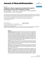
ALDH1A1 and MMP-9 staining intensities were rated
on a scale of 0–3 according to the percentage of positive
tumor (0, < 5% positive cells; 1, 5-10%; 2, 11-50%; or 3, >
50%). The expression is very low for 0, low for 1, moderate
for 2 and high for 3 (Figure 1). ALDH1A1 and MMP-9
Li et al. BMC Cancer 2014, 14:705
/>
Page 3 of 8
Figure 1 Gastric cancer tissue illustrating the range of intensities of ALDH1A1 immunostaining from 0 to 3. The lower panels represent
magnified pictures of boxed area in the corresponding upper panels. The scale bar represents 50 μm.
expression were classified as negative for scores ≤ 1 and
positive for scores ≥ 2.
Follow-up
The follow-up duration was defined as the interval between the date of operation and the date of death or last
follow-up. The study was censored on 30 September
2013. The median follow-up period was 27.0 months
(range, 4–82 months) in 216 patients. All the patients
were followed up every 1–3 months in the first year and
every 3–6 months thereafter. Recurrence were confirmed
by tumor markers levels including CEA, AFP, CA199,
CA125 and CA724, B-type ultrasonic inspection every 3
moths, and computed tomography (CT) or magnetic resonance imaging (MRI) every 6 months after gastrectomy.
The main causes of death were gastric cancer recurrence.
Overall survival (OS) was calculated from the date of
surgery to the date of death or last follow-up. Recurrencefree survival (RFS) was defined as from the date of surgery
until the date of relapse or from the period of resection to
the date of the last observation taken.
Statistical analysis
All statistical analyses were performed using the SPSS
software (version 16.0; Chicago, IL, USA). Interdependence between ALDH1A1 status and clinical data was
calculated using the chi-square test, and displayed in
cross-tables. Correlation of ALDH1A1 with MMP-9 was
calculated by Pearson χ2 test. Survival curves were
plotted using the Kaplan-Meier method and analyzed using
the log-rank test. All reported P values were two-sided
and P < 0.05 was considered statistically significant.
Results
The association of ALDH1A1 with clinicopathological
variables
To elucidate the biological significance of ALDH1A1 in
gastric cancer, we examined the immunohistochemical
expression of ALDH1A1 in gastric cancer tissues (Figure 1).
ALDH1A1 staining mainly located in cytoplasm of tumor
cells. The positive rate of ALDH1A1 was 50.0% (108/216)
in gastric cancer samples.
According to the results of immunohistochemistry, we
correlated ALDH1A1 status in 216 gastric cancer specimens with clinicopathologic parameters (Table 1). Our
analyses showed that the level of ALDH1A1 in gastric
cancer was significantly correlated with depth of invasion (P < 0.001), lymph node metastasis (P < 0.001), and
stage of disease (P < 0.001), but was not associated with
gender, age, tumor size, tumor site and grade of differentiation (P > 0.05) (Table 1). Notably, the correlation of
ALDH1A1 with prominent serosal invasion and lymph
node metastasis positivity suggested a potential role of
ALDH1A1 in increased invasion and metastasis of gastric cancer.
Effect of tumor ALDH1A1 protein level on prognosis
To further determine the effect of ALDH1A1 overexpression on the OS and RFS, we first performed univariate analysis of traditional clinicopathologic variables for
prognosis. The results of the univariate analysis are shown
in Table 2. Overexpression of ALDH1A1 (P < 0.001), larger tumor size (P < 0.001), prominent serosal invasion
(P < 0.001) and lymph node metastasis (P < 0.001) were
significantly associated with the poor OS rate of gastric
cancer patients. In addition, Kaplan-Meier analysis
demonstrated that ALDH1A1 overexpression (P < 0.001),
larger tumor size (P = 0.001), tumor site (P = 0.047),
prominent serosal invasion (P < 0.001) and lymph node
metastasis (P < 0.001) were negative prognostic factors for
RFS in gastric cancer patients (Table 2). Furthermore, to
evaluate the independent impact of ALDH1A1 overexpression on OS and RFS, a multivariate Cox regression
model adjusted for tumor size, tumor site, depth of
invasion, lymph node metastasis and ALDH1A1 expression was performed. Our results showed that ALDH1A1
Li et al. BMC Cancer 2014, 14:705
/>
Page 4 of 8
Table 2 Predictive variables for overall survival and recurrence-free survival of 216 patients with gastric cancer
Variables
No. of patients
OS rate (%)
3y
5y
P-value
RFS rate (%)
3y
5y
41.5
35.4
44.4
41.2
45.3
42.2
38.1
30.6
50.4
45.5
28.3
23.3
34.6
28.6
47.8
43.7
41.2
34.6
43.6
40.1
70.2
65.3
< 0.001
25.6
20.8
82.0
77.5
< 0.001
27.5
22.6
60.5
54.8
23.7
19.2
P-value
Gender
Male
140
46.9
39.6
Female
76
54.0
43.1
≤ 60
136
49.8
43.9
> 60
80
48.9
36.1
≤ 5.0
139
55.6
47.4
> 5.0
77
38.1
28.8
Upper
86
45.9
33.8
Middle/Lower
130
51.8
45.7
Well/Moderate
96
47.8
39.7
Poor
120
50.7
41.6
T1/T2
81
71.4
66.5
T3/T4
135
36.2
25.5
0.308
0.381
Age (years)
0.338
0.391
Size (cm)
0.001
< 0.001
Tumor site
0.074
0.047
Differentiation
0.673
0.822
Depth of invasion
< 0.001
Lymph node metastasis
Negative
59
83.5
79.2
Positive
157
36.5
26.6
Negative
108
69.3
57.1
Positive
108
28.7
23.9
< 0.001
ALDH1A1 protein expression
expression was a poor independent prognostic factor for
OS in gastric cancer patients (hazard ratio, 2.037; 95% CI,
1.407 - 2.950). In addition, positive ALDH1A1 expression
patients were almost 2.0 times more likely to suffer from
relapse than those with negative ALDH1A1 expression
(hazard ratio, 1.945; 95% CI, 1.346 - 2.812). Tumor size,
depth of invasion and lymph node metastasis all had
independent prognostic value in the multivariate analysis
(Table 3).
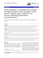
Survival analysis showed that OS and RFS were significant different among 216 patients according to the expression of ALDH1A1 (P < 0.001, P < 0.001) (Figure 2A).
The postoperative median OS and RFS were 27.0 months
and 19.0 months, respectively. The postoperative median
OS times in ALDH1A1-positive (n = 108) and ALDH1A1negative (n = 108) gastric cancer patients subgroup were
12.0 months and 42.0 months, and the median of the RFS
times were 9.0 months and 39.0 months. In addition, the
OS and RFS rates at 5 years were 23.9% and 19.2% for
ALDH1A1-positive patients compared with 57.1% and
< 0.001
< 0.001
54.8% for ALDH1A1-negative patients, respectively (both
P < 0.001; Table 2).
To further evaluate the prognostic value of ALDH1A1
in different subgroups, patients were stratified according to
tumor size (Figure 2B,C), depth of invasion (Figure 2D,E)
and lymph node metastasis (Figure 2F,G). The expression of ALDH1A1 maintained its prognostic value in
predicting shorter OS and RFS in all of these subgroups
for except OS in T1/T2 subgroup (P = 0.054). Therefore, it
appears that ALDH1A1 may serve as a powerful prognostic factor for patients with gastric cancer in different risk
groups.
ALDH1A1 overexpression predict poor prognosis
independent of tumor invasiveness
To better understand the clinical significance of ALDH1A1
on aggressiveness in gastric cancer, we investigated the
relationship of ALDH1A1 and MMP-9 protein expression
in gastric cancer.
Li et al. BMC Cancer 2014, 14:705
/>
Page 5 of 8
Table 3 Cox multivariate analysis of contributory factors to prognosis among 216 gastric cancer patients
after gastrectomy
Variables
β
SE
Hazard ratio (95% CI)
P-valuea
0.410
0.177
1.507 (1.065 ~ 2.133)
0.021
OS
Tumor size
Depth of invasion
0.576
0.255
1.779 (1.079 ~ 2.931)
0.024
Lymph node metastasis
1.018
0.341
2.767 (1.417 ~ 5.403)
0.003
ALDH1A1 protein expression
0.712
0.189
2.037 (1.407 ~ 2.950)
< 0.001
Tumor size
0.411
0.178
1.508 (1.064 ~ 2.138)
0.021
Tumor site
0.043
0.180
1.044 (0.733 ~ 1.486)
0.813
RFS
Depth of invasion
0.649
0.256
1.913 (1.158 ~ 3.162)
0.011
Lymph node metastasis
1.063
0.338
2.894 (1.491 ~ 5.619)
0.002
ALDH1A1 protein expression
0.665
0.188
1.945 (1.346 ~ 2.812)
< 0.001
Abbreviations: ALDH1A1 aldehyde dehydrogenase 1 family member A1, CI confidence interval.
a
Cox proportional hazards regression model.
The positive rates of ALDH1A1 were 63.0% and 60.5%
in the more prominent serosal invasion group (T3/T4)
and more frequent lymph node involvement group (N1-3),
while there were only 28.4% and 22.0% in T1/T2 and N0
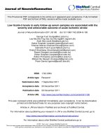
(P < 0.001 and P < 0.001, respectively) (Table 1). In addition, ALDH1A1 was significantly correlated with MMP-9
in 216 gastric carcinoma specimens. Of 108 patients with
low ALDH1A1 expression, 81 patients (75.0%) had low
MMP-9 expression, while 71 of 108 patients (65.7%) with
high ALDH1A1 expression also had high MMP-9 expression (P < 0.001) (Figure 3).
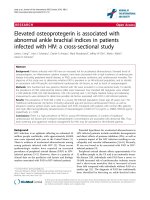
We further explored the influence of tumor invasiveness on the prognostic value of ALDH1A1 expression
in gastric cancer by using MMP-9 as an indicator for
the invasive potential of individual tumor cells. All the
patients were stratified into either a low invasiveness
subgroup (low MMP-9 expression; n = 118) or a high
invasiveness subgroup (high MMP-9 expression; n = 98)
according to the MMP-9 expression index. Kaplan-Meier
survival curves were then plotted to investigate the association between ALDH1A1 status and survival (Figure 4).
In the low invasiveness subgroup, ALDH1A1 overexpression was associated with shorter OS (P < 0.001) and
RFS (P < 0.001) compared with the OS and RFS in
patients with low ALDH1A1 expression (Figure 4A). In
the high tumor invasiveness subgroup (Figure 4B),
patients with ALDH1A1 overexpression were prone to
death (P < 0.001) and relapse (P < 0.001). Furthermore,
the 5-year survival rate was significantly lower in the
low invasiveness subgroup with ALDH1A1 overexpression (23.8%) than that in the high invasiveness
subgroup with low ALDH1A1 expression (53.0%; P =
0.002; data not shown). Therefore, the expression of
ALDH1A1 appears to be a strong postoperative
prognostic parameter for patients with gastric cancer
independent of tumor invasiveness.
Discussion
In the present study, the expression of ALDH1A1 was
investigated in 216 gastric carcinoma tissues by immunohistochemistry. We found that ALDH1A1 was
significantly associated with depth invasion, lymph node
metastasis and stage of disease. In addition, the KaplanMeier survival analysis revealed that the survival times
(OS and RFS) of gastric cancer patients with high
ALDH1A1 expression were significantly shorter than
those with low ALDH1A1 expression. The prognostic
value of ALDH1A1 in different subgroups according to
tumor size, depth of invasion and lymph node metastasis was also estimated, which appears that ALDH1A1
may serve as a powerful prognostic factor for patients
with gastric cancer in different risk groups. Furthermore, the multivariate Cox model analysis indicated
that ALDH1A1 status was an independent factor for
both prognosis indexes (OS and RFS) in gastric cancer.
This finding suggests that ALDH1A1 plays an important role in tumor prognosis, concludes ALDH1A1
could be a potential prognostic factor of gastric cancer.
Our results were consistent with previously reported
results. In several investigations, it has been shown
that the abnormal expression of ALDH1A1 in cancer
cells is associated with tumor progression. Wakamatsu
et al. [19] revealed that ALDH1 was overexpression
and had positively correlated with depth invasion and
TNM stage in gastric cancer, moreover, ALDH1 positivity was significantly higher in diffuse-type lymph node
metastasis than that in the primary tumor. CharafeJauffret et al. [20] reported that the ALDH1A1-positive
Li et al. BMC Cancer 2014, 14:705
/>
Page 6 of 8
Figure 2 Overall survival and recurrence-free survival are shown for patients with gastric cancer. All patients were stratified according to
tumor size, depth of invasion and lymph node metastasis. Kaplan-Meier survival estimates and log-rank tests were used to analyze the prognostic
significance of ALDH1A1 expression in all patients (A) and each subgroup (B-G).
breast cancer cells were able to promote tumor invasion
in vitro and tumor metastasis in mouse xenografts, moreover, expression of ALDH1A1 was an independent predictive factor for early metastasis and decreased survival
in inflammatory breast cancer. Jiang et al. [14] showed
that the ALDH1A1-positive lung cancer cells could
generate tumors in vivo, furthermore, the expression of
ALDH1A1 was positively correlated with the stage and
grade of lung tumors and related to a poor prognosis for
the patients with early-stage lung cancer, which suggested
that ALDH1A1 could be a potential prognostic factor and
therapeutic target for treatment of the patients with lung
cancer. However, Dimou et al. [21] found the contradictory
results that the ALDH1A1-negative expression of lung cancer
patients had shorter survival compared with those with
ALDH1A1-positive expression, which indicated that
Li et al. BMC Cancer 2014, 14:705
/>
Page 7 of 8
Figure 3 ALDH1A1 and MMP-9 levels correlated in 216 gastric cancer tissues. (A, B) IHC staining for ALDH1A1 and MMP-9 was performed
in tumors from 216 gastric cancer patients. Representative examples of ALDH1A1 and MMP-9 staining in serial sections from the same tumor
samples are shown in (A), and percentages of samples displaying low or high ALDH1A1 expression relative to MMP-9 level is shown in (B).
The scale bar represents 200 μm.
ALDH1A1 overexpression was associated with favorable
outcome.
It has been known that degradation of extracellular
matrix (ECM) was a signal for the beginning of invasion
and metastasis, and MMPs are important molecules
involved in ECM degradation during invasion and
metastasis [22]. Chu et al. [16] reported that cancer MMP9 was significantly correlated with depth of invasion and
lymph node metastasis and MMP-9-positive gastric cancer
patients had worse outcomes than those with MMP-9negative tumors. Zhao et al. [23] found that MMP-9
targeted RNA interference was able to successfully suppress MMP-9 expression and inhibit cell growth and invasion of SGC7901 gastric cancer in vitro and in vivo. Our
results demonstrated that the expression of ALDH1A1
and MMP-9 was correlated with each other, indicating
higher invasive and metastasizing activity in ALDH1A1
overexpression cancer cells. In addition, ALDH1A1 was
highly expressed in depth of invasion, especially in T3 and
T4 carcinomas, which was consistent with previously
reported results [19]. As far as lymph node status was
concerned, the patients with lymph node metastasis
tend to show elevated ALDH1A1 expression. Collectively,
ALDH1A1 status in gastric cancer promoting tumor
aggressiveness suggests that ALDH1A1 could be a feasible
target in cancer therapy.
Conclusions
In this study, we demonstrated that ALDH1A1 may play
an important role in tumor invasion, metastasis and
prognosis, and could work as a promising target for
prognostic prediction in gastric cancer. Determination of
ALDH1A1 expression may help to identify high-risk
gastric cancer patients and thus aid the selection of
appropriate therapies. Further investigation is necessary
to clarify the role of ALDH1A1 in the development of
gastric cancer.
Competing interests
The authors declare that they have no competing interests.
Figure 4 Overall survival and recurrence-free survival are
shown for patients with low tumor invasiveness (A) and high
tumor invasiveness (B). Kaplan-Meier survival estimates and
log-rank tests were used to analyze the association between ALDH1A1
expression and overall survival or recurrence-free survival in patients
with low invasiveness (low MMP-9; n = 118) or high invasiveness
(high MMP-9; n = 98).
Authors’ contributions
XSL, WSL, QX participated in the study conception, design, case selection
and experiments. XSL, WSL and XYF carried out the data collection. QX, XYF
and XSL performed the scoring of immunohistochemical staining. XSL and
WSL performed the data analysis and writing of the manuscript. All the
authors read and approved the final manuscript.
Acknowledgements
We gratefully acknowledge the clinical data provided by the pathology
department (Affiliated Hospital of Guilin Medical University).
Li et al. BMC Cancer 2014, 14:705
/>
Author details
1
Department of Gastroenterology, Affiliated Hospital of Guilin Medical
University, Guilin 541004, Guangxi Zhuang Autonomous Region, China.
2
Guilin Medical University, Guilin 541004, Guangxi Zhuang Autonomous
Region, China. 3Department of Spleen and stomach diseases, The First
Affiliated Hospital of Guangxi University of Chinese Medicine, Nanning
530200, Guangxi Zhuang Autonomous Region, China.
Received: 20 June 2014 Accepted: 22 September 2014
Published: 24 September 2014
References
1. Compare D, Rocco A, Nardone G: Risk factors in gastric cancer.
Eur Rev Med Pharmacol Sci 2010, 14:302–308.
2. Dikken JL, van Sandick JW, Maurits Swellengrebel HA, Lind PA, Putter H,
Jansen EP, Boot H, van Grieken NC, van de Velde CJ, Verheij M, Cats A:
Neo-adjuvant chemotherapy followed by surgery and chemotherapy or
by surgery and chemoradiotherapy for patients with resectable gastric
cancer (CRITICS). BMC Cancer 2011, 11:329.
3. Cunningham D, Allum WH, Stenning SP, Thompson JN, Van de Velde CJ,
Nicolson M, Scarffe JH, Lofts FJ, Falk SJ, Iveson TJ, Smith DB, Langley RE,
Verma M, Weeden S, Chua YJ, Participants MT: Perioperative
chemotherapy versus surgery alone for resectable gastroesophageal
cancer. N Engl J Med 2006, 355:11–20.
4. Nobili S, Bruno L, Landini I, Napoli C, Bechi P, Tonelli F, Rubio CA, Mini E,
Nesi G: Genomic and genetic alterations influence the progression of
gastric cancer. World J Gastroenterol 2011, 17:290–299.
5. Yasui W, Sentani K, Sakamoto N, Anami K, Naito Y, Oue N: Molecular
pathology of gastric cancer: research and practice. Pathol Res Pract 2011,
207:608–612.
6. Bornschein J, Rokkas T, Selgrad M, Malfertheiner P: Gastric cancer: clinical
aspects, epidemiology and molecular background. Helicobacter 2011,
16(Suppl 1):45–52.
7. Marchitti SA, Brocker C, Stagos D, Vasiliou V: Non-P450 aldehyde oxidizing
enzymes: the aldehyde dehydrogenase superfamily. Expert Opin Drug
Metab Toxicol 2008, 4:697–720.
8. Vasiliou V, Nebert DW: Analysis and update of the human aldehyde
dehydrogenase (ALDH) gene family. Hum Genomics 2005, 2:138–143.
9. Black WJ, Stagos D, Marchitti SA, Nebert DW, Tipton KF, Bairoch A, Vasiliou V:
Human aldehyde dehydrogenase genes: alternatively spliced transcriptional
variants and their suggested nomenclature. Pharmacogenet Genomics 2009,
19:893–902.
10. Douville J, Beaulieu R, Balicki D: ALDH1 as a functional marker of cancer
stem and progenitor cells. Stem Cells Dev 2009, 18:17–25.
11. Muzio G, Maggiora M, Paiuzzi E, Oraldi M, Canuto RA: Aldehyde
dehydrogenases and cell proliferation. Free Radic Biol Med 2012,
52:735–746.
12. Ginestier C, Hur MH, Charafe-Jauffret E, Monville F, Dutcher J, Brown M,
Jacquemier J, Viens P, Kleer CG, Liu S, Schott A, Hayes D, Birnbaum D,
Wicha MS, Dontu G: ALDH1 is a marker of normal and malignant human
mammary stem cells and a predictor of poor clinical outcome.
Cell Stem Cell 2007, 1:555–567.
13. Deng S, Yang X, Lassus H, Liang S, Kaur S, Ye Q, Li C, Wang LP, Roby KF,
Orsulic S, Connolly DC, Zhang Y, Montone K, Butzow R, Coukos G, Zhang L:
Distinct expression levels and patterns of stem cell marker, aldehyde
dehydrogenase isoform 1 (ALDH1), in human epithelial cancers.
PLoS One 2010, 5:e10277.
14. Jiang F, Qiu Q, Khanna A, Todd NW, Deepak J, Xing L, Wang H, Liu Z,
Su Y, Stass SA, Katz RL: Aldehyde dehydrogenase 1 is a tumor stem
cell-associated marker in lung cancer. Mol Cancer Res 2009, 7:330–338.
15. Wang Y, Zhe H, Gao P, Zhang N, Li G, Qin J: Cancer stem cell marker
ALDH1 expression is associated with lymph node metastasis and poor
survival in esophageal squamous cell carcinoma: a study from high
incidence area of northern China. Dis Esophagus 2012, 25:560–565.
16. Chu D, Zhang Z, Li Y, Zheng J, Dong G, Wang W, Ji G: Matrix
metalloproteinase-9 is associated with disease-free survival and overall
survival in patients with gastric cancer. Int J Cancer 2011, 129:887–895.
17. Edge SB, Compton CC: The American Joint Committee on Cancer:
the 7th edition of the AJCC cancer staging manual and the future of
TNM. Ann Surg Oncol 2010, 17:1471–1474.
Page 8 of 8
18. Wang J, Cui S, Zhang X, Wu Y, Tang H: High expression of heat shock
protein 90 is associated with tumor aggressiveness and poor prognosis
in patients with advanced gastric cancer. PLoS One 2013, 8:e62876.
19. Wakamatsu Y, Sakamoto N, Oo HZ, Naito Y, Uraoka N, Anami K, Sentani K,
Oue N, Yasui W: Expression of cancer stem cell markers ALDH1, CD44
and CD133 in primary tumor and lymph node metastasis of gastric
cancer. Pathol Int 2012, 62:112–119.
20. Charafe-Jauffret E, Ginestier C, Iovino F, Tarpin C, Diebel M, Esterni B,
Houvenaeghel G, Extra JM, Bertucci F, Jacquemier J, Xerri L, Dontu G,
Stassi G, Xiao Y, Barsky SH, Birnbaum D, Viens P, Wicha MS: Aldehyde
dehydrogenase 1-positive cancer stem cells mediate metastasis and
poor clinical outcome in inflammatory breast cancer. Clin Cancer Res
2010, 16:45–55.
21. Dimou A, Neumeister V, Agarwal S, Anagnostou V, Syrigos K, Rimm DL:
Measurement of aldehyde dehydrogenase 1 expression defines a
group with better prognosis in patients with non-small cell lung cancer.
Am J Pathol 2012, 181:1436–1442.
22. Nelson AR, Fingleton B, Rothenberg ML, Matrisian LM: Matrix
metalloproteinases: biologic activity and clinical implications. J Clin Oncol
2000, 18:1135–1149.
23. Zhao F, Zhang Q, Kang C, Cui X, Wang T, Xu P, Zhou X, Liu J, Song X:
Suppression of matrix metalloproteinase-9 expression by RNA interference
inhibits SGC7901 gastric adenocarcinoma cell growth and invasion in vitro
and in vivo. Med Oncol 2010, 27:774–784.
doi:10.1186/1471-2407-14-705
Cite this article as: Li et al.: ALDH1A1 overexpression is associated with
the progression and prognosis in gastric cancer. BMC Cancer
2014 14:705.
Submit your next manuscript to BioMed Central
and take full advantage of:
• Convenient online submission
• Thorough peer review
• No space constraints or color figure charges
• Immediate publication on acceptance
• Inclusion in PubMed, CAS, Scopus and Google Scholar
• Research which is freely available for redistribution
Submit your manuscript at
www.biomedcentral.com/submit