Hypothetical estimate of drug-burden on a diabetic foot ulcer patient and its relevance to microbiological analysis
Bạn đang xem bản rút gọn của tài liệu. Xem và tải ngay bản đầy đủ của tài liệu tại đây (223.09 KB, 15 trang )
Int.J.Curr.Microbiol.App.Sci (2017) 6(6): 1139-1153
International Journal of Current Microbiology and Applied Sciences
ISSN: 2319-7706 Volume 6 Number 6 (2017) pp. 1139-1153
Journal homepage:
Original Research Article
/>
Hypothetical Estimate of Drug-Burden on a Diabetic Foot Ulcer Patient and
its Relevance to Microbiological Analysis
C. Meenakshisundaram1*, J. Uma Rani2, Usha Anand Rao2, V. Mohan3 and R. Vasudevan3
1
Department of Microbiology, Sri Venkateswara Medical College and Research Centre,
Pondicherry-605 102, India
2
Department of Microbiology, Dr. A.L.M. Post-Graduate Institute of Basic Medical Sciences,
Tharamani Campus, University of Madras, Chennai-600 013, India
3
Dr.Mohan’s Diabetic Specialties Research Centre, Gopalapuram, Chennai-600 028, India
*Corresponding author
ABSTRACT
Keywords
Drug burden,
Diabetic foot ulcer
patients
Optimization of
antimicrobial
agents.
Article Info
Accepted:
17 May 2017
Available Online:
10 June 2017
In a retrospective study, conducted in Chennai (India), during 2005, a total of 104 bacterial
isolates, obtained from 75 diabetic foot ulcer patients, revealed the presence of 9-bacterial
species, namely, S. aureus, CONS spp, Streptococcus spp, Corynebacterium spp,
Enterococcus spp, E. coli, Klebsiella spp, Proteus spp, and P. aeruginosa, in different
percentages. The in-vitro antibiotic sensitivity pattern of Pseudomonas aeruginosa, tested
in the retrospective study, and the antibiotic sensitivity patterns of the 8-other pathogens as
adopted from available literature relating to 2-South Indian locations (Kelambakkam and
Bengaluru), and 2-North Indian locations (Chandigarh and New Delhi), were compared
with the data of the multicentre trial studies related to diabetic foot infections, carried out
by Citron, D.M., et al., (2007), in the United States. There was a close agreement among
the AMAs evaluated, in the case of all the 9-pathogens, in the antimicrobial susceptibility
range of 100.0% to 66.7% (bacterial resistance range of 0.0% to 33.3%). A hypothetical
estimate of drug-burden was made, by enlisting the number of AMAs needed to be
administered on a patient, against the 9-pathogens, in order to obtain a cure. A discussion
on the probable adverse reactions caused by AMAs is included. It is suggested that AMAs
such as Ertapenem, Trimethoprim/sulfamethxazole, Tigecycline, Doxycycline, etc, tested
in the United States, can be included in evaluating the antibiotic susceptibility patterns, in
India. Such results, if generated, would be found useful when the currently-used AMAs
happen to become ineffective due to bacterial resistance, in future. More number of
AMAs, if tested, would make it easier to optimize on the choice of drugs.
Introduction
There is an increasing reason for optimism in
offering treatment to diabetic foot ulcers, and
other chronic wounds through enhanced
understanding of pathogenic factors, at the
advent of the latest improvements in
identifying the causative agents versus
suitable antibiotic agents (Cavanagh et al.,
2005; Lipsky et al., in “2012- IDSAGuidelines”). The Infectious Diseases Society
of America (IDSA) classified diabetic foot
infections into four classes, namely, i)
Uninfected, ii) Mild, iii) Moderate to Severe,
and iv) Severe, correlating with the
corresponding clinical symptoms. Obviously,
1139
Int.J.Curr.Microbiol.App.Sci (2017) 6(6): 1139-1153
the treatment approaches could be different in
each class.
The diagnosis must be correctly carried out,
in order to proceed with the treatment option.
Weng et al., (2017) expressed a fear that any
wrong diagnosis of a lower-extremity
infection could lead to unnecessary
medication and hospitalization charges, citing
the estimated number of 50,000 to 130,000
misdiagnosed cases of Cellulitis, during the
period of study, involving a wasteful
expenditure of US$ 195 million to 515
million, in the United States alone. This
would vouch for the importance of the correct
diagnosis, for which the clinical symptoms
and microbiological test results are the
guiding factors.
Gadepalli et al., (2006) suggested that
amputation can be prevented, if the diabetic
foot ulcer can be treated with adequate and
timely care. This optimism gives a lot of hope
for all the stake-holders, pointing towards a
devoted-care needed in the task. However, the
antibiotic resistance exerted by microbial
pathogens against many antimicrobial agents
administered on diabetic foot ulcer patients
continues to pose a problem, often proving as
a challenge to the therapeutic options
preferred by clinicians.
Kruse and Edelman (2006) reported that the
treatment to diabetic foot ulcer must address
all three major concerns, namely, prompt
debridement, offloading procedures, and
infection control, and that antibiotic treatment
must be started after initial culture tests, and
that the treatment must be suitably modified,
as revealed by subsequent culture tests. This
sequence is important for initiating and
enabling a quick-healing process.
Stevens et al., (2005) reported that the
response to initial antibiotic therapy must be
judged by the outcome, namely, reduction in
fever and toxicity, and reduction in the
advancement of infection. Ho Kwong Li et
al., (2015) reported that oral therapy could not
be considered as inferior to intravenous
treatment, in terms of clinical outcomes, and
that the cost of intravenous treatment system
would involve ten times higher cost.
Lipsky et al., (2008) compared the
effectiveness of Pexiganan (a topical cream)
versus Ofloxacin (Oral), in the case of mildlyinfected Diabetic Foot Ulcer (DFU). Lauf et
al., (2014) reported the efficacy and
effectiveness
of
Tigecycline
versus
Ertapenem, in patients, with and without
Osteomyelitis.
In any hospital setting, in India, the
polymicrobial infection could be expected in
the range of around 30.0% of the diabetic foot
infection patients (Sajila et al., 2015).
It was reported that as many as 7-strains of
different bacterial species were present in a
polymicrobial infection in a single diabetic
foot ulcer patient, in India, namely,
Staphylococcus aureus, Enterococcus spp,
Pseudomonas aeruginosa, Pseudomonas
stuzeri, Escherichia coli, Proteus spp, and
Alcaligenes spp. (Shahi et al., 2013).
The polymicrobial infections would often
prove to be severe infections, or limbthreatening infections, requiring to be
covered, in the antibiotic treatment, in the
case of Gram-negative pathogens, as well as
Gram-positive pathogens, among the aerobes
and anaerobes (Grayson et al., 1995; Lipsky
and Berendt et al., 2004; Rao and Lipsky et
al., 2007; Reiber and Lipsky et al., 1998).
In the United States, Lipsky et al., (2005)
compared the effectiveness of two
antimicrobial agents prevalent in diabetic foot
infections, in a major multi centre trial
studies, in two separate groups, namely,
1140
Int.J.Curr.Microbiol.App.Sci (2017) 6(6): 1139-1153
administering Ertapenem @ 1.0 g daily, on
the first-group of 295 diabetic foot infection
patients, for 5-days, and giving Piperacillin/
tazobactam @ 3.375 g every 6-hours, on the
second-group of 291 diabetic foot infection
patients, for 5-days, and thereafter, giving
Amoxicillin/clavulanic acid @ 825/125 mg
every 12-hours, to both groups of patients.
Investigators were given the freedom to
decide on administering Vancomycin to
patients of either group, for the purpose of
giving coverage against the antimicrobial
resistant Enterococcus species, and against
the Methicillin resistant Staphylococcus
aureus (MRSA). Based on clinical and
microbiological outcomes, it was concluded
that the effect of administering Ertapenem
was equivalent to Piperacillin/tazobactam,
and that the nature of adverse effects caused
were similar, in both groups.
In China, Xu et al., (2016) compared the
antibiotic regimens against the pathogens
prevalent in diabetic foot infection of 443
patients, by administering Ertapenem on one
group of 219 patients, and administering
Piperacillin/tazobactam on another group of
224 patients. It was concluded that the
treatment by Ertapenem was non-inferior to
the treatment by Piperacillin/tazobactam, in
respect of clinical outcome, microbiological
outcome, and adverse effects experienced by
the Chinese patients. It was also hinted that
Ertapenem had a lower rate of clinical
resolution in severe diabetic foot infections.
A report by Clinical trials, gov (2010),
compared Tigecycline versus Ertapenem to
check their effectiveness in offering treatment
to diabetic foot infections. Such comparisons
of two antimicrobial agents (AMAs) took into
consideration the various factors, like, the
number of patients getting cured out of the
total number of patients treated, the number
of patients affected with adverse effects such
as blood and lymphatic system disorders,
cardiac disorders, liver-damage, general
disorders such as abdominal pain, renal or
urinary disorders, mental status changes, chest
pain, fever, septic shock, allergic reactions,
metabolism and nutritional disorders, etc.
These factors are related to the choice of
antimicrobial agents, for each pathogen
prevalent in the diabetic foot infection.
Decision to select a particular antimicrobial
agent could be based on rating of Noninferiority or Equivalence margin, such as
5.0% to 10.0%.
In the Indian scenario, fungal pathogens were
reported to be prevalent in chronic wounds of
diabetic patients, in addition to bacterial
pathogens (aerobic and anaerobic), in many
locations in India (Bansal et al., 2008;
Sanniyasi et al., 2015, Chincholikar et al.,
2002). Anaerobic pathogens have been
isolated and treated successfully by Anandi et
al., (2004), with a multi-disciplinary
involvement, in a teaching hospital setting.
In addition to the antimicrobial agents used
for fighting against the infection, in a diabetic
patient, certain oral-hypoglycemic drugs, also,
need to be administered, for maintaining the
desired glycemic control. According to
Armstrong and Lipsky (2004), a diabetic foot
ulcer patient must first be medically
stabilized,
and
secondly,
metabolic
aberrations, if any, must be carefully
addressed. This factor does have relevance to
the “drug-burden” on the diabetic foot ulcer
patient, in the case of patients already
experiencing
system-factors
such
as
hypertension, hyperlipidemia, atherosclerotic
heart disease, obesity or renal insufficiency,
etc. This aspect, therefore, deserves to be
given a due consideration, at the time of
planning the type of treatment to be given to a
diabetic foot ulcer patient, on a case to case
basis (Rowe and Khardori, 2017).
In the case of poly-microbial infections, care
must be taken to optimize the number of
antibiotic classes, so that the adverse reactions
1141
Int.J.Curr.Microbiol.App.Sci (2017) 6(6): 1139-1153
which are characteristically unique to each
class of antimicrobial agents cannot become
additive, in their effects on the same patient.
This effort of optimization of antibiotic
classes would help avoiding the occurrence of
undesirable adverse effect on the diabetic foot
ulcer patient, thereby, reducing the “drugburden” on the patient.
In the selection of antimicrobial agents, the
susceptibility patterns have to be considered,
along
with
the
probable
adverse
reactions/allergies/hyper sensitivity reactions,
etc., which they could cause on the diabetic
foot ulcer patient. Certain details need to be
considered as listed below:
Allergy to Penicillin could vary from 5% to
10 % of hospitalized patients (Green et al.,
1971; Parker, 1972).
Borish et al., (1987) highlighted on the
necessity to look for allergy to penicillin in
patients, before deciding about the
medication.
times
greater
with
Amoxicillin/clavulanate treatment.
this
Joseph and Axler (1990) reported that
combination therapy containing Clindamycin
and Aztreonam or Ciprofloxacin could be
found helpful for diabetic foot infection
patients who are allergic to Beta-lactam
antibiotics, and that less-severe infections can
be treated with a single antimicrobial agent
such as Ticarcillin/clavulanic acid, or
Ampicillin/sulbactam. Cephalosporins with
anaerobic activity (Cefoxitin, or Cefotaxime,
or Ceftizoxime) can be used, in areas where
Enterococcus is not a major problem.
Thus, it becomes necessary to select the
therapy based on the personalized metabolicsystem details of the patient, and also on the
area-specific considerations. This is one
reason as to why bacterial antibiotic
sensitivity has to be assessed in each local
centre, by testing appropriate AMAs against
the pathogenic species.
Indian scenario
Bronze et al., (2017) reported that
Amoxicillin/clavulanate is preferred as an
alternative to patients who are feeling allergic,
or intolerant to Macrolide class of antibiotics
(Erythromycin,
Azithromycin,
and
Clarithomycin) proving effective against
Gram-positive cocci and some intracellular
pathogens.
Bronze et al., (2017) also claimed that
Ertapenem (Invanz) was stable against
hydrolysis by a variety of beta-lactamases,
including Penicillinases, Cephalosporinases,
and Extended beta-lactamases (ESBLs).
Edmonds
(2009)
reported
that
Amoxicillin/clavulanate would be able to kill
the bacteria which would prove resistant to
Amoxicillin (if applied alone), and that the
probability for the occurrence of hepatotoxicity in the patient would be around six-
National
Treatment
Guidelines
for
Antimicrobial Use (2016) have been
prescribed by the National Centre for Disease
Control (NCDC) of the Ministry of Health
and Family Welfare, Government of India,
New Delhi, according to which the following
hints are indicated, relevant to the selection of
antimicrobial agents for treatment against
Skin and Soft Tissue Infections:
For
Vancomycin-resistant
Enterococcal
(VRE)-species, Linezolid has been indicated
to be efficient, although it cannot be used for
a long period of time, as intolerance may
develop in some patients. Its use is not
recommended for patients with impaired renal
function. Daptomycin NOT approved for
treatment of VRE-infection. Its use, as a
Monotherapy, is not recommended.
1142
Int.J.Curr.Microbiol.App.Sci (2017) 6(6): 1139-1153
For the ESBL (Extended Beta-lactamase)producing
Enterobacteriaceae,
the
Carbapenems (Imipenem, Meropenem, or
Ertapenem) have been indicated as the drug of
choice, for serious infections. For mild cases,
Piperacillin/tazobactam or Cefoperazone/
sulbactam could be considered, when
susceptibility in-vitro is favourably indicated.
Citing the CLSI-recommendations, it has
been stated that ESBL-producing isolates
must be considered resistant to all Penicillins,
Cephalosporins (including Cefepime and
Cefpirome) and Aztreonam, irrespective of
the in-vitro test results. (It must be
remembered that the emergence of ESBLproducing Enterobacteriaceae is related to
indiscriminate use of Third Generation
Cephalosporins).
For Carbapenem-resistant Enterobacteriaceae
(CRE)-infections, involving ESBL, or AmpC
and Porin-loss, or Acquired Carbapenemases,
it has been indicated that either Polymixin, or
Colistin, or Tigecycline and Fosfomycin can
be recommended.
In the case of infections involving bones and
joints, treatment must be based on culture of
blood/synovial fluid/bone biopsy, and
necessarily, with orthopaedic consultation.
(These guidelines provide the most
authoritative information applicable to skin &
soft tissue infections. However, there is a
need to generate more data at local and
regional levels, in order to streamline efforts
in the direction of augmenting endeavours to
fight against the ever-increasing menace of
antibiotic resistance. Such data can be made
available to the National Data Bank on
Bacterial antibiotic resistances, in the case of
all diseases, in order to serve the purpose of
justifying policy-formulations, related to
therapeutic
strategy
enhancement
(Meenakshisundaram et al., 2016).
Factors related to glycaemic control
Bansal et al., (2008) reported that about 67%
of the diabetic foot ulcer patients had random
blood sugar (RBS)-levels greater than 200
mg/dL, and a majority of them had HbA1clevels above 7.0%, and that, among the total
of 103-diabetic foot ulcer patients, the HbA1c
(%) varied around 8.15+/-1.75.
In case of Hypoglycemia (blood sugar level
dropping below 70 mg/dL), treatment is given
in the form of 15 to 20 gram of fast-acting
carbohydrates, like a fruit juice, glucosetablet, sugar-cube, etc. But, fats or proteins, if
consumed, can slow down the body’s
absorption of sugar, and may cause an
increase in the blood sugar level (Mayo Clinic
Diseases
and
Conditions:
diabetic
hypoglycemia, 2015).
In maintaining the glycemic control,
Fernando et al., (2016) reported that glycemic
interventions include subcutaneous insulin
administration, continuous insulin infusion,
oral-hypoglycemic
drugs
(anti-diabetes
agents), life-style intervention or a
combination of these interventions. Oral antidiabetes therapy include four classes of
hypoglycemic drugs, namely, Sulfonylureas,
Metformin, Thiazolidinedione’s and Alphaglucosidase inhibitors. The main side-effects
caused by alpha-glucosidase inhibitors are
flatulence (intestinal gas-related problem) and
diarrhea which are usually mild, and not
necessitating the cessation of therapy.
(Thomas Higgins, 2017).
Sawin et al., (2010) reported that Metformin
did not cause hypoglycemia in hospitalized
diabetic patients, and yet the theoretical risk
of Metformin inducing lactic acidosis must be
monitored.
The risk factors relating to the occurrence of
increased lactic acidosis or increased lactate
levels, in case of anti-diabetes drugs, such as
Metformin versus other anti-hypoglycemic
1143
Int.J.Curr.Microbiol.App.Sci (2017) 6(6): 1139-1153
treatments, became a debate, and hence, the
situation must be monitored (Salpter et al.,
2006). Kendall et al., (2006) reported that
persons with diabetes might avoid taking the
antimicrobial agent Gatifloxacin, a thirdgeneration broad spectrum Fluoroquinolone
which has activity against Gram-negative /
Gram-positive
(aerobic/anaerobic)
and
atypical pathogens, as it undergoes minimal
bio-transformation, and is excreted renally
Uckay et al., (2009), and Richard et al.,
(2008) reported that skin commensals, such as
Coagulase negative Staphylococcus (CONS)
spp, Corynebacterium spp, or Bacillus spp,
would require treatment, only when
associated with an infection involving
osteosynthetic material or hardware.
Chloramphenicol (on prolonged use, perhaps)
was associated with anemia (resulting from
decreased production or increased reduction
of red blood cells), according to Smith
Marsch (of the University of Illinois at
Chicago) 2017).
Side effects due to common antimicobial
agents in use
Anderson (2017) reviewed and updated an
exhaustive list of antimicrobial agents,
considering the various side-effects produced
by each class of antibiotics, although it was
concluded that the antimicrobial agents are
generally safe, when used in “appropriate”
doses. The highlights of side-effects are given
below:
Factors related to cross-resistivity
With regard to cross-resistivity between two
antimicrobial agents or alternatives in the
selection of antibiotics, the following points
need to be considered:
Cephalosporins become acceptable to a
majority of patients who were found to be
allergic to Penicillin (Romano et al., 2004).
However, in some cases, fatal ends have been
reported (Pumphrey et al., 1999). Crossresistivity
between
Penicillins
and
Carbapenems were reported to be low
(Romano et al., 2007).
There was no Cross-resistivity between
Penicillins and Monobactam.
In the case of Quinolones, increases of IgEmediated anaphylactic reactions were
reported, perhaps, due to the large scale use of
the Quinolones (Manfredi et al., 2004; Hein,
1997; Sachs et al., 2006; Venturini Diaz et
al., 2007). Aminoglycosides rarely cause
hyper-sensitivity reactions, although some
reports indicated the occurrence of IgEmediated systemic reactions (Solensky et al.,
2010).
Penicillins (Ampicillin, Amc, Pi, Pi/t,
Nafcillin,
Oxacillin)
causing
hypersensitivity, including Anaphylaxis; (in
addition to nausea, vomiting, diarrhea, skinrash, drug-fever, abdominal pain);
Cabapenems (Imipenem/cilastatin identified
with probable hypersensitivity; Meropenem
causing hypersensitivity in penicillin-allergy
patients); in addition to diarrhea, nausea,
vomiting,
liver-toxicity,
eosinophilic
leukocytosis, Aminoglycosides (G, Tob, Ak),
causing oto-toxicity or renal toxicity on longterm use; in addition to dizziness, nausea,
vomiting, nystagmus (rhythmical oscillation
of the eye-balls, either horizontal, rotary or
vertical).
Cephalosporins
(Cefolexin,
Cefaclor,
Cefuroxime, Ceftibuten, Cefdirnir, Cefixime,
Ceftriaxone) causing cross-hyper sensitivity
in penicillin-allergic patients; in addition to
skin-rash,
diarrhea,
nausea,
vomiting
(although rare); serum-sickness (involving
reaction by the immune system)
Glycopeptides (Vancomycin causing Red
man syndrome; Televancin causing Taste-
1144
Int.J.Curr.Microbiol.App.Sci (2017) 6(6): 1139-1153
alteration),
nausea/vomiting,
headache,
dizziness);
Macrolides
(Erythromycin,
Azithromycin,
Clarithromycin)
causing
sometimes high rate of intestinal side effects;
in addition to diarrhea, nausea, vomiting,
taste-alteration, anorexia (aversion for food)
Sulfonamides
(T/S,
Erythromycin/
sulfisoxazole, Sulfasalazine, etc) causing
Stevens Johnson syndrome, Toxic epidermonecrolysis involving necrosis and loosening of
tissues), photosensitivity; in addition to
anorexia, dizziness, diarrhea, nausea,
vomiting, headache, rash, abdominal pain
Tetracyclines (Tet, Doxi, Mino) causing Liver
toxicity, Photosensitivity; in addition to
diarrhea, nausea, vomiting, abdominal pain,
anorexia (diminished appetite)
Quinolones (Cip, Lev, Mxf, Ofl) causing
severe photosensitivity, Insomnia, abdominal
pain, lethargy; in addition to diarrhea, nausea,
vomiting
Metronidazole will cause metallic taste, in
addition to nausea/vomiting, dizziness,
headache; (Alcohol consumption while being
treated with Metronidazole would aggravate
symptoms). Lipsky et al., (2012b), also,
described the relative merits and de-merits of
many antimicrobial agents, to be used in oral
route versus parenteral route.
The above sets of informations derived from
literature, are to be treated as reference
material (or hints) only. It must be inferred
that the local data on bacterial sensitivity
pattern, in the antimicrobial treatment, does
play a major role in deciding the prospects of
healing of the diabetic foot ulcer wound.
Aim and objectives
The present study was conducted with the
following aim and objective:
To estimate the probable drug-burden which
would result on the diabetic foot ulcer patient,
in the treatment process, using the data on the
bacterial pathogens isolated from the wound,
and the data on antibiotic sensitivity, and
To assess whether any other antimicrobial
agents are to be newly tested in India for their
in-vitro susceptibility against each pathogen
found in diabetic foot ulcers, based on hints
acquired from international literature.
Materials and Methods
Pus swabs were collected from 75-diabetic
foot ulcer patients being treated in
Dr.V.Mohan's Diabetes Specialties Center,
Gopalapuram, Chennai-600 028 (South
India), during a period of 5-months, from
May to September, 2005. The 104-pus
samples collected from the patients were
transported to the Laboratory in Carey-Blair
transport medium of Hi-media (India). All the
isolates were identified, adopting the standard
procedures indicated in the NCCLS, 2002
(Meenakshisundaram et al., 2015). Drug
resistance
pattern
of
Pseudomonas
aeruginosa only was evaluated using
antimicrobial agents representing various
classes of standard antimicrobial agents,
namely,
Ampicillin
(10ug),
Amoxicillin/clavulanic acid (20ug + 10ug),
Piperacillin
(100ug),
Imipenem(10ug),
Cefotaxime (30ug), Ceftazidime (30ug),
Ceftriaxone (30ug), Gentamicin(10ug) and
Ciprofloxacin (5ug).
Antimicrobial agents (AMAs), with a
susceptibility range of 100.0% to 66.7%
(facing bacterial resistance of 0.0% to 33.3%
from Pseudomonas aeruginosa) were
evaluated.
Bacterial resistance of other 8-bacterial
species isolated in the diabetic foot ulcer
specimens were not evaluated in the bacterial
sensitivity test, in the retrospective study, due
1145
Int.J.Curr.Microbiol.App.Sci (2017) 6(6): 1139-1153
to limited scope of the study. In order to fill
up this gap, antimicrobial susceptibility
patterns reported in the literature in India,
relating to diabetic foot infections, were used,
choosing 2-other locations in South India,
namely, Kelambakkam (near Chennai,
Tamilnadu State, as reported by Priyadarshini
et al., 2013), and Bengaluru (in Karnataka
State, as reported by Sajila et al., 2015), and
choosing 2-locations in North India, namely,
Chandigarh (in Hariyana State, as reported by
Bansal et al., 2008), and New Delhi (as
reported by Gadepalli et al., 2006).
In all cases, antimicrobial agents (AMAs)
effective against the pathogens in the
susceptibility range of 100.0% to 66.7%, as
evaluated in the 5-cities in India, were
compared with the data relating to similar
data collected in the multicenter trial studies
on diabetic foot ulcers, in the United States,
as reported by Citron et al., (2007).
The overall drug-burden on the individual
diabetic patient will correspond to the
antibiotic agents needed to be administered on
the patient, in order to eradicate all pathogens,
inclusive of aerobic organisms, anaerobic
organisms and fungal species prevalent in the
patient. The drugs have to be optimally
selected, so as to earn a healing of the wound,
with minimum number of drug-types, so that
the side-effects can be minimized. Allergies
and cross-resistances become additional
factors for consideration, in the selection of
the drugs.
Results and Discussion
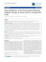
The prevalence of different bacterial species
isolated from the 104-samples in Chennai, in
the retrospective study is presented in table 1.
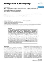
Table 2 presents the details of the most
effective antimicrobial agents (AMAs) whose
susceptibility patterns were in the range of
100.0% to 66.7% against the particular
pathogen, namely, Pseudomonas aeruginosa
(the bacterial resistances varying from 0.0%
to 33.3%).
According to the data shown in table 2, it
becomes clear that any one of the five
antimicrobial agents (AMAs) can be
administered
against
Pseudomonas
aeruginosa found in a diabetic foot ulcer
patient, choosing either Imipenem or
Pipeacillin or Amoxicillin/clavulanic acid, or
Ceftazidime, or Gentamicin.
This result is presented in table 3, in
comparison to the multicentre- trial studies
carried out in the United States by Citron et
al., (2007) who evaluated 7-antimicrobial
agents to be effective against Pseudomonas
aeruginosa, namely, Imipenem, Gentamicin,
Ceftazidime,
Piperacillin/
tazobactam,
Amikacin, Ciprofloxacin, Levofloxacin, and
Moxifloxacin. This implies that there is a
closer agreement between our Chennai-data
of the retrospective study, in comparison to
the multicentre-study data of the United
States.
Being encouraged by this trend, it was
attempted to compare the antimicrobial
sensitivity patterns of the other 8-pathogens,
namely, Staphylococcus aureus (MSSA,
MRSA), CONS spp, Streptococcus spp,
Enterococcus spp, Corynebacterium spp,
Escherichia coli, Klebsiella spp, and Proteus
spp., choosing informations available in the
published literature, as reported for 2-South
Indian
locations
(Kelambakkam
and
Bengaluru), and 2-North Indian locations
(Chandigarh and New Delhi), in contrast to
similar data pertaining to the multicentre trial
study in the United States, as shown in table
3. Several similarities are found among the
antimicrobial agents evaluated in India and
the United States, considering separately, the
Gram-positive and Gram-negative aerobic
bacterial categories. It is found that more
number of antimicrobial agents are used in the
antimicrobial susceptibility tests in India, than
1146
Int.J.Curr.Microbiol.App.Sci (2017) 6(6): 1139-1153
in the United States, perhaps due to the
predominance of Gram-negative bacteria
prevalent in diabetic foot ulcers in India or
factors related to the commercial availability
of the various classes of drugs, in different
geographical locations of India.
In the case of the present study, it was
assumed, hypothetically, that all the 9bacterial pathogens to be present in a single
patient, namely, 5-Gram-positive pathogens
(aerobic) and 4-Gram-negative pathogens
(aerobic). A hypothetical estimate was made
to identify the antimicrobial agents needed to
be used against all the 9-pathogens, in order
to obtain a cure for the patient.
Referring to table 3, the antimicrobial agents
evaluated in the 5-Indian cities, were
compared, for optimally choosing the suitable
antimicrobial agents, as detailed below:
Table.1 Isolation rate of other bacteria
S.no
A.
1.
2.
3.
4.
5.
B.
6.
7.
8.
9.
S.no
1.
2.
3.
4.
5.
Organism
Gram-positive (aerobic): (40.4%)
Staphylococcus aureus
Coagulase Negative Staphylococcus (CONS)
Streptococcus spp.
Corynebacterium spp.
Enterococcus spp.
Gram-negative (aerobic): (59.6 %)
Escherichia coli
Pseudomonas aeruginosa
Klebsiella spp.
Proteus spp.
(Number of isolates=104)
No. of organisms (%)
18 (17.3%)
11 (10.6%)
6 (5.8)
4 (3.8)
3 (2.9
23 (22.2)
18 (17.3)
11 (10.6)
10 (9.6)
Table.2 List of AMAs effective against Pseudomonas aeruginosa
(Total number of P. aeruginosa isolates=n=18)
Antimicrobial agent
No of resistant strains
% Resistence
Imipenem
1
5.5
Piperacillin
2
11.0
Co-amoxyclav
4
22.0
Ceftazidime
6
33.0
Gentamicin
6
33.0
1147
Int.J.Curr.Microbiol.App.Sci (2017) 6(6): 1139-1153
Table.3 Comparison of antibiotic agents effective against pathogens found in diabetic foot ulcers,
in the workable susceptibility range of 100.0% to 66.7%
S.
No
1.
MicroOrganism
P. aeruginosa
2.
S, aureus
2.11
MSSA
2.2.2
MRSA
3.
CONS
Spp.
Streptococcus spp.
4.
5.
6.
Enterococcus
Spp.
Corynebacterium
Spp.
(1) Multi-Centre
Trials (U.S.A).
Ak,G,Cip,I,Pi/t,Lev
Mxf,Caz
Amc,Etp,Pi/t,Lev
Mxf,Cip,Cldm
T/S,Dox, Cfl
Dox
T/S
Amc,Etp,Pi/t,LevMxf,
Cldm,T/S,Cip,Dox,Cfl
Amc,Etp,Lev,Mxf
Cip,Cldm,Pi/t
T/S,Cfl
Amc,Pi/t,Cip,Lev
Mxf,T/S
Amc,Etp,Pi/t
Dox, Cfl
(2) Chennai
(S.India)
Amc,Pi,G.I
Caz
………
……
….
….
(3) KelamBakkam,(S.India)
Ak,Crb,Ci,I
Mer,Pi/t,PmB,Cfs
G,Ntlmc,E,Cldm,
Clrmp,Van,Lin,Rif
Teic,Clxcln,Cfzln Cot
Ntlmc,Cldm,Van
Clrmp,Teic,Lin
Ntlmc,Lin
Van, Teic
Ofl,Van,Teic
Tet,Cldm,,Lin
…
.
….
….
Ak, Etp,I,Pi/t,G,Cip
Lev,Mxf,Dox,T/S,
Caz
….
…..
7.1
EnteroBacteriaceae
7.21
E. coli
……….
8.
Klebsiella
Spp.
…….
….
9.
Proteus
Spp.
Etp,I, Pi/t,Ak,Cip,Lev
Mxf,G,T/S,Dox,Caz
……
Ak,Tet,Tob,G,Cip
Mer,Ofl,Ci,Pi/t
Clrmp,I,PmB,Cot
Cpm, Cfs,Cfrxm
Cftxm
Ak, I, G Pi/t,Clrmp
Ci, PmB,Cfs
Ak, I,Mer,Pi/t, Ci
PmB,Cfs
Cfs, I,Clrmp
Tet, Pi/t
(5) Bengaluru
(S.India)
Ak,G,I,Pi/t,Lev
Mer,Tob
Cldm,Ofl,Oxa,E,G
Lom,Tet,Lin,Van Cpz
…
…..
(7) Chandigarh
(N.India)
Ak,Pi,Tob,I
Ctr,Caz,Cfs
Amp,Amc,Cip G,
E,I,Ctr, Cfrxm,Cfs
….
(8) New Delhi
(N.India)
Ak,I,Mer,Pi/t
Tcc,Cfs
Rif
…..
….
Amc,Cldm,Ofl.Oxa
….
G,Tet,Lin,Van,Cpz,Cpm
(S.pyo):Amc,Cdm,E
…
Ofl,Oxa,Lom,G,Lin
Tet,Van,Cpz,Cpm
Amc,Tet,Lin,Van
(E.faec)Amc,
Caz,Cfs,G
….
Ak, Tet,Cldm
Clrmp,Rif,Cot
….
….
……
……
….
…
……..
Ak, Cip,G,Lev,Pi/t
Mer,Tob,Ntlmc
Ak, G,I,
Lev,Pi/t
Ak,G,I,Lev,Pi/t
Ak, I,G.Cip,Lev,Pi/t
Cfrxm,Cpm,Cpz
Ak,I,Cfs
Caz
(K.oxy)Ak,I,Cfs,
Caz (K.pne)
I,Ctr,Cfs
(P.vul)Ak,G,I,
Caz,Cfs,Cfrxm Ctr
I, Mer,Tcc
Cfs
(K.pne) Amc,Cip
Mer,Pi/t,Tcc,Cfs
Ak,I,Caz,Cftxm
I,Cip,Tcc
Mer,Cfs
Amp=Ampicillin;Amc=Amoxicillin/clavulaninicacid;Ak=Amikacin; Caz=Ceftazidime;Ctr=Ceftriaxone;Cip=Ciprofloxacin;Cftxm=Cefotaxime;
Cfrxm=Cefuroxime; Cfl=Cefalexin; Cfzln=Cefazolin; Cpm=Cefepime; Cpz=Cefoperazone; Cfs=Cefoperazone/sulbactam; Crb=Carbenicillin;
Cot=Cotrimoxazole; Cldm=Clindamycin; Ci=Colistin; Clxcln=Cloxacillin; Clrmp=Chloramphenicol; Dox=Doxycycline; E=Erythromycin; Etp=Ertapenem;
G=Gentamicin; I=Imipenem; Lev=Levofloxacin; Lin=Linezolid; Lom=Lomefloxacin; Mer=Meropenem; Mxf=Moxifloxcin; Ntlmc=Netimycin; Oxa=Oxacillin;
Ofl=Ofloxacin; Pi=Piperacillin; Pi/t=Piperacillin/tazobactam; PmB=PolymixinB; Rif=Rifampicin; Tcc=Ticarcillin/clavulanicacid; Tet=Tetracycline;
Teic=Teicoplanin; Tob=Tobramycin; T/S=Trimethoprim/sulfamethoxazole; Van=Vancomycin
1148
Int.J.Curr.Microbiol.App.Sci (2017) 6(6): 1139-1153
Case-1: For the 4-species of Gram-positive
bacteria present in the diabetic ulcer wound,
namely, Staphylococcus aureus, Coagulase
negative staphylococcus (CONS) species,
Streptococcus species, and Enterococcus
species, the recommended AMAs, would be
Linezolid or Vancomycin, as evaluated in
India. For the Corynebacterium species,
Amoxicillin/clavulanate may be tried as
reported in the multi centre-trial data of the
United States, in the absence of Indian data
for this particular pathogen.
Case-2: For the 4-numbers of Gram-negative
bacterial species present in the same diabetic
foot ulcer wound, namely, Escherichia coli,
Klebsiella species, Proteus species and
Pseudomonas aeruginosa, the recommended
AMAs would be either Piperacillin/
tazobactam, or Cefoperazone/ sulbactam, or
Imipenem (chosen from data reported for the
5-Indian cities).
In addition to the estimates made in Case-1
and Case-2, it is to be said that appropriate
medication must be included for covering the
anaerobic pathogens and fungal pathogens, if
present in any other situation.
Metronidazole has been reported to be
effective against majority of anaerobic
pathogens (Anandi et al., 2004; Chincholikar,
2002). An antifungal cream (such as
Fluconazole) must be included in the list, as a
topical medicine, if fungal pathogens are
present (Sanniyasi et al., 2015). Also, Citron
et al., (2007) reported that Ertapenem,
Piperacillin / tazobactam, Amoxicillin /
clavulanic acid, or Clindamycin could be
effective against the anaerobic pathogens
found in diabetic foot infections.
Thus, all these medicines administered on a
hypothetical patient would represent the
“drug-burden”, on the patient, in addition to
the anti-diabetes (oral-hypoglycemic) drugs to
be consumed by the patient, for the purpose of
maintaining a normal glycemic control.
There is, therefore, a necessity to optimize on
the number of drugs to be administered on the
patient, by choosing the AMAs, in such a way
that the chosen drug would be effective
against more than one pathogen, without
causing any adverse effect.
The choice of drugs to be administered is to
be left to the prerogative decision of the team
of experts who attend on the diabetic foot
ulcer patient. The input from a microbiologist
is needed during the various stages of the
treatment process.
Certain antimicrobial agents such as
Ertapenem,
Tigecycline,
Doxycycline,
Trimethoprim/ sulfamethoxazole, etc., found
useful in the multicentre studies in the United
States, can be included in the in-vitro
susceptibility tests in India, to assess their
effectiveness in offering treatment to diabetic
foot infections.
Acknowledgement
The authors are grateful to the wisdom of all
other investigators whose findings have been
cited.
References
Anandi, C., Alaguraja, D., Natarajan, V.,
Ramanathan, M., Subramaniam, C.S.,
Thulasiram, M., Sumithra, S., 2004,
“Bacteriology of foot infections”. Indian
J. Med. Microbiol., 22: 175- 178.
Anderson, L. 2017. “Antibiotics-Common Side
Effects, Allergies and Reactions”,
Drugs.com, Know More, Be sure,
(Medically reviewed on March 5, 2017.
Armstrong, D.G., and Lipsky, B.A. 2004.
“Diabetic foot infections: stepwise
medical and surgical management”, Int.
1149
Int.J.Curr.Microbiol.App.Sci (2017) 6(6): 1139-1153
Wound J., 1(2):123-32. (doi:10.1111/j.
1742-4801.2004.00035.x).
Bansal, E., Garg, A., Bhatia, A., Attri, A.K.,
Chander, J., 2008. “Spectrum of
Microbial Flora in Diabetic Foot Ulcers”,
Indian J. Pathol. Microbiol., 51: 204-208.
Borish, L., Tamir, R., Rossenwaser, L.J., 1987.
“Intravenous desensitisation to betalactam antibiotics”, The J. Allergy and
Clin. Immunol., 80(3): 314-319.
Bronze, M.S., Khardori, R. 2016. “Diabetic
Foot Infections Medication: Penicillin,
Cephalosporins”,
emedicine.
medscape.com/article/237378-medication
#2. (Updated April 05, 2017).
Cavanagh, P.R., Lipsky, B.A., Bradbury, A.W.,
Botek, G. 2005. “Treatment for Diabetic
Foot Ulcers”. Lancet, 366(9498): 1725175.
Chincholikar, D.A., Pal, R.B. 2002. A study of
fungal and bacteriological infections of
the diabetic foot. Indian J. Pathol.
Microbiol., 45:15-22.
Citron, D.M., Goldstein, E.J.C., Merriam, C.V.,
Lipsky, B.A., Abraham, M.A. 2007.
“Bacteriology of the moderate to severe
diabetic foot infections and the in-vitro
activity of the antimicrobial agents”, J.
Clin. Micobiol., 45(9): 2819-28.
Clinicaltrials.gov: Trial record 13 of 82 for:
drugs for diabetic foot infections: “Study
Evaluating the Safety and Efficacy of a
Once-daily Dose of Tigecycline versus
Ertapenem in Diabetic Foot Infections
(DFI) with a Sub-study in Patients with
Diabetic Foot Infections complicated by
Osteomyelitis”. (Clinical Trials. gov.
identifier: NCT00366249 Last updated
April 2010).
Edmonds, M. 2009. “The treatment of diabetic
foot infection: Focus on Ertapenem”,
Vasc. Health Risk Manag., 5: 949-963.
Fernando, M.E., Seneviratne, R.M., Tan, Y.,
Lazzarini,
P.A.,
Sangla,
K.S.,
Cunningham,
M.,
Buttner,
P.G.,
Golledge, J. 2016. “Intensive versus
conventional glycemic control for treating
diabetic foot ulcers”, Cochrane Database
of
Systematic
Rev.,
doi:
10.1002/14651858.CDO10764.pub2.
Gadapalli, R., Dhawan, B., Sreenivas, V. et al.
2006. “A Clinico-microbiological studyof
diabetic Foot Ulcers in an Indian Tertiary
Care Hospital”, Diabetes Care, 29: 17271732.
Grayson, M.L. 1995. “Diabetic foot infections:
Antibiotic therapy”, Infect. Dis. Clin.
North Am., 9:143-61.
Green, C.R., Rosenblum, A. 1971, “A report of
the penicillin study group”, J. Allergy
Clin. Immunol., 48: 331.
Heine, H. 1997. “Cutaneous adverse reaction to
ciprofloxacin: demonstration of specific
lymphocyte Proliferation and crossreactivity to ofloxacin in vitro”, Acta
Dermato-Venereol., 77: 285-286.
Ho Kwong Li, Mathew Searborough, et al.
2015. “Oral versus intravenous antibiotic
treatment for bone and joint intervention
(OVIV): study protocol for a randomized
controlled trial”, Trials, 16: 583. PMCID:
PMC4687165.
Joseph, W.S., and Axler, D.A. 1990.
Microbiology and antimicrobial therapy
for diabetic foot infections, Clin. Podiatr.
Med. Surg., 7(3): 467-81.
Kendall, C., and Wooltorton, E. 2006. People
with diabetes should avoid antibiotic
Gatifloxacin, Canadian Med. Association
J., 174(8): 1089-1090. PMCID: 1421471.
Kruse, I., and Edelman, S. 2006. “Evaluation
and Treatment of Diabetic Foot Ulcers”,
American Diabetes Association: Clin.
Diabetes,
24(2):
91-93.
( />Lauf, L., Ozsvar, Z., Mitha, I., Regoli-Merei, J.,
Embil, J.M., Coope, A., Sabol, M.B.,
Castaing, N., Dartois, N., Yan, J., Dukart,
G., Maroka, R. 2014. “Phase 3 study of
comparing Tygecycline and Ertapenem in
patients with diabetic foot infection with
and without Osteomyelitis”, Diagn.
Microbiol. Infect. Dis., 78(4): 469-80.
(doi:
10/1016/j.diagmicrobio.2013.12.
007).
1150
Int.J.Curr.Microbiol.App.Sci (2017) 6(6): 1139-1153
Lipsky, B.A., 2009 Diagnosing and Treating
Diabetic Foot Infections, Klimik Dergisi;
22(1): 2-13.
Lauf, L., Ozsvar, Z., Mitha, I., Regoli-Merei, J.,
Embil, J.M., Coope, A., Sabol, M.B.,
Castaing, N., Dartois, N., Maroko, R.
2014. Phase 3 study of comparing
Tygecycline and Ertapenem in patients
with diabetic foot infection with and
without Osteomyelitis, Diagn. Microbial.
Infect. Dis., 78(4): 469-80.
Lipsky, B.A., Armstrong, D.G., Citron, D.M.,
Tice,
A.D.,
Morgenstern,
D.E.,
Abramson, M.A. 2005. Ertapenem versus
Piperacillin/tazobactam
for
diabetic
(SIDESTEP): prospective, randomized,
controlled, double-blinded, multicenter
trial. Lancet, 366(9498): 1695-1703.
Lipsky, B.A. 2007. Empirical therapy for
diabetic foot infections: Are there clinical
clues to guide antibiotic selection? Clin.
Microbiol.
Infect.,
13(4):
3513.PMID:17359317. (doi:10.1111/j.14690691.2007.01697.x).
Lipsky, B.A., Berendt, A.R., Cornia, P.B., Plie,
J.C., Peters, E.J.G., Armstrong, D.G.,
Deery, H.G., Embil, J.M., Joseph, W.S.,
Karchmer, A.W., Pinzur, M.S., and
Senneville, E. 2012. 2012 Infectious
Disease Society of America Clinical
Practice Guidelines for the Diagnosis and
Treatment of Diabetic Foot Infections,
Clin. Infect. Dis., 54(12): 132-137.
Lipsky, B.A., Peters, E.J.C., Sonneville, E.,
Berendt, A.R., Embil, J.M., Lavery, L.A.,
Urbancic-Rovan, V., Jeffcoate, W.J.
2012(b). “Expert Opinion on the
Management of Infections in the Diabetic
foot”, Diabetes Metabolism Res. Rev., 38(
S1): 163-178. doi:10.1002/dmrr.2248,
Lipsky, B.A., Berendt, A.R., Deery, H.G., Emil,
J.M., Joseph, W.S., Karchmer, A.W.,
LeFock, J.L., Lew, D.P., Mader, J.T.,
Norden, C., and Tan, J.S. 2004.
“Diagnosis and Treatment of Diabetic
Foot Infections”, Clin. Infect. Dis., 39(7):
885-910. doi:10.1086/424846.
Lipsky, B.A., Holoyd, K.J., Zasloff, M. 2008.
“Topical versus systemic antimicrobial
therapy for treating mildly infected
diabetic foot ulcers:a randomized,
controlled, double-blinded multi-centre
trial of Pexiganan Cream”, Clin. Infect.
Dis., 47: 1537-45.
Manfredi, M., Severino, M., Testi, S., et al.
2004. “Detection of specific IgE to
quinolones”, J. Allergy Clin. Immunol.,
113: 155-160.
Mayo Clinic: Diseases and Conditions: Diabetic
hypoglycaemia. 2015.
Meenakshisundaram,
C.,
UshaAnandRao,
Rajendran, P., Mohan, V., and
Vasudevan, R. 2015. “Characerisation of
Pseudomonas aeruginosa
and its
association with Diabetic Foot ulcer
isolated from a tertiary care hospital in
Tamilnadu, India”, Int. J. Curr. Microb.
and App. Sci., Vol.4: No.7: pp.122-126.
Meenakshisundaram, C., Uma Rani, J., Usha
Anand Rao, Mohan, V., and Vasudevan,
R. 2016. “Microbial Profiles of Diabetic
Foot Ulcers: A random Comparison
within India”, Int. J. Curr. Microbiol. and
App.
Sci.,
5(12):
835-849.
( />12.092).
National Committee for Clinical Laboratory
Standard: Performance Standard for
Antimicrobial Susceptibility Testing:
Twelfth information standard: M100-S
12, Vol.22,no.1, Villanova, Pa, National
Committee for Clinical Laboratory
Standard, 2002.
National
Treatment
Guidelines
for
Antimicrobials Use in Infectious Diseases
(version 1.0 (2016),National Centre for
Disease Control, Director General of
Health Services, Ministry of Health &
Family Welfare, Government of India,
New Delhi.
Parker, C.W. 1972. “Allergic drug responses –
mechanisms and unsolved problems”,
CRC Crit. Rev. Toxicol., 1: 261.
Priyadarshini, S., Jeya, M., Linda Susan, S.,
2013, “The Bacteriology of Diabetic Foot
Ulcers, with a Special Reference to Multidrug Resistant Strains”, J. Clin. Diagn.
Res., 7(3): 441-445. (Pub Med).
1151
Int.J.Curr.Microbiol.App.Sci (2017) 6(6): 1139-1153
Pumphrey, R.S.H., Dais, S. 1999. “Underreporting of antibiotic anaphylaxis may
put patients at risk”, Lancet, 353: 11571158.
Rao, N., and Lipsky, B.A. 2007. “Optimising
antimicrobial therapy in diabetic foot
infections”, Drugs, 67(2): 195-214.
PMID: 17284084.
Reiber, G., Lipsky, B.A., Gibbons G.W. 1998.
“The burden of diabetic foot ulcers”, The
American J. Surgery, 176(2): Suppl 1:5S10S. doi:10.1016/S0002-9610(98)001810.
Richard, J.L., Sotto, A., Jourdon, N., et al.
2008. “Risk factors and healing impact of
multi-drug resistant bacteria in diabetic
foot ulcers”, Diabetes Metab., 34: 363369.
Romano, A., Gueant-Rodiguez, R.M., Viola,
M., et al. 2004. “Cross-reactivity and
tolerability of cephalosporins in patients
with hypersensitivity to penicillins”, Ann.
Intern. Med., 141: 16-22.
Romano, A., Viola, M., Gueant-Rodriguez,
R.M., et al. 2006. “Imipenem in patients
with immediate hypersensitivity to
penicillins”, N. Eng. J. Med., 354: 28352837.
Romano, A., Viola, M., Gueant-Rodriguez,
R.M., et al. 2007. “Tolerability of
meropenem in patients with IgE-mediated
Hypersensitivity to penicillins”, Ann.
Intern. Med., 146: 266-269.
Rowe, V.L., and Khardori, R., et al., (updated
March 14), 2017. Diabetic Ulcer
Treatment and Management, MedScape,
Drugs
and
Diseases.
(emedicine.medscape.com/article/460282
-treatment).
Sachs, B., Reigel, S., Seebeck, J., et al., 2006.
“Fluoroquinolone-associated anaphylaxis
in spontaneous Adverse drug reaction
reports in Germany: differences in
reporting rates between individual
Fluoroquinolones and occurrence after
first-ever use”, Drug Saf., 29: 1087-1100.
Sajila, N.M., Manjunath, R., Mahesh Desai.
May 2015. “A study of the bacteriology
profile of Diabetic Foot Ulcer, and
Antibiotic Sensitivity Pattern”, J. Evol.
Med. Dent. Sci., 4(35): 6832-6840.
doi:10.14260/jemds/2015/991.
Salpeter, S., Greyber, E., Pastenack, G.,
Salpeter, E. 2006. “Risk of fatal and nonfatal lactic acidosis with Metformin use in
type2 Mellitus”, Ochrane Database Rev.,
2006. CD 002967.
Sanniyasi, S., Balu, J., Narayanan, C.D. 2015.
“Fungal Infection: A hidden enemy in
Diabetic Foot Ulcers”, The J. Foot and
Ankle Surgery (Asia Pacific), July-Dec.,
Sri Ramachandra University, Chennai,
2(2): 74-76. doi: 10.5005/jp-journals10040-1033.
Sawin, G., Shaunghnessy, A.F. 2010. “Glucose
control in hospitalized patients”, Am.
Fam. Physician, 81: 1121-4.
Shahi, S.K., Kumar, A., Gupta, S.K., and Singh,
S.K. 4 Dec., 2013. “Occurrence of
Multiple antibiotic resistance phenotype
and class 1-integron in bacteria isolated
from diabetic foot ulcers”, African J.
Microbiol. Res., 7(48): 5424-5432.
doi:10.5897/AJMR12.979.
Smith Marsch, D.E. 2017. “Severity of Adverse
Drug Reactions: Some serious Adverse
Drug Effects”, in MDS Manual:
Professional/Clinical
pharmacology/
Adverse reactions.
Solensky, R., and Khan, D.A. 2010. “Drug
Allergy: An Updated Practice Parameter“,
Annals of Allergy, Asthma and Immunol.,
(www.jcaai.org) Pp. 273.e9.
Stevens, D.L., Bisno, A.L., Chambers, H.F.,
Everett, E.D., Dellinger, P., Goldstein,
E.J.C., Gorbach, S.L, Hirscmann, J.V.,
Kaplan, E.L., Montoya, J.G., and Wade,
J.C. 2005. “Practice Guidelines for the
diagnosis and Management of Skin and
Soft Tissue Infections”, Clin. Infect. Dis.,
41(10):1373-1406. doi:10.1086/497143.
Thomas Higgins. 2017. Boulder Medical
Centre.
Uckay, I., Pittet, D., Vasudaux, P., Sax, H.,
Lew, D., Walvogel, F. 2009. “Foreign
body infections due to Staphylococcus
epidermidis”, Ann. Med., 41:109-119.
1152
Int.J.Curr.Microbiol.App.Sci (2017) 6(6): 1139-1153
Venturini Diaz, M., Lobera Labairu, T., delPozo
Gil, et al. 2007. “In-vivo diagnostic tests
in adverse reactions to Quinolones”, J.
Investig. Allergol. Clin. Immunol., 17:
393-398.
Weng, Q.Y., Raff, A.B., Cohen, J.M., et al.
2017.
“Costs
and
Consequences
Associated with Misdiagnosed Cellulitis”,
JAMA. Dermatol., 153(2): 141-146. doi:
10.1001/jamadermatol.2016.3816.
Xu, Z.R, Ran, X.W., Xian, Y., Yan, X.D.,
Yuan, G.Y., Mu, S.M., Shen, J.F., Zhang,
B.S., Gan, W.J., Wang, J. 2016.
“Ertapenem
versus
piperacillin/tazobactam for diabetic foot
infections in China: a Phase 3,
muliticentre, randomized, double blind,
active-controlled, non-inferiority trial”, J.
Antimicrob. Chemother., 71(6): 16881696.
How to cite this article:
Meenakshisundaram, C., J. Uma Rani, Usha Anand Rao, V. Mohan and Vasudevan, R. 2017.
Hypothetical Estimate of Drug-Burden on a Diabetic Foot Ulcer Patient, and Its Relevance to
Microbiological Analysis. Int.J.Curr.Microbiol.App.Sci. 6(6): 1139-1153.
doi: />
1153