Báo cáo khoa học: "Dose distribution in the thyroid gland following radiation therapy of breast cancer-a retrospective study" ppsx
Bạn đang xem bản rút gọn của tài liệu. Xem và tải ngay bản đầy đủ của tài liệu tại đây (323.77 KB, 7 trang )
RESEARCH Open Access
Dose distribution in the thyroid gland following
radiation therapy of breast cancer-a retrospective
study
S Johansen
1*
, KV Reinertsen
2,3
, K Knutstad
4
, DR Olsen
5
and SD Fosså
6
Abstract
Purpose: To relate the development of post-treatment hypothyroidism with the dose distribution within the
thyroid gland in breast cancer (BC) patients treated with loco-regional radiotherapy (RT).
Methods and materials: In two groups of BC patients postoperatively irradiated by computer tomography (CT)-
based RT, the individual dose distributions in the thyroid gland were compared with each other; Cases developed
post-treatment hypothyroidism after mul timodal treatment in cluding 4-field RT technique. Matched patients in
Controls remained free for hypothyroidism. Based on each patient’s dose volume histogram (DVH) the volume
percentages of the thyroid absorbing respectively 20, 30, 40 and 50 Gy were then estimated (V20, V30, V40 and
V50) together with the individual mean thyroid dose over the whole gland (MeanTotGy). The mean and median
thyroid dose for the included patients was about 30 Gy, subsequently the total volume of the thyroid gland
(VolTotGy) and the absolute volumes (cm
3
) receiving respectively < 30 Gy and ≥ 30 Gy were calculated (Vol < 30
and Vol ≥ 30) and analyzed.
Results: No statistically significant inter-group differences were found between V20, V30, V40 and V50Gy or the
median of MeanTotGy. The median VolTotGy in Controls was 2.3 times above VolTotGy in Cases (r = 0.003), with
large inter-individual variations in both groups. The volume of the thyroid gland receiving < 30 Gy in Controls was
almost 2.5 times greater than the comparable figure in Cases.
Conclusions: We concluded that in patients with small thyroid glands after loco-radiotherapy of BC, the risk of
post-treatment hypothyroidism depends on the volume of the thyroid gland.
Keywords: Breast cancer, Radiotherapy, hypothyroidism
Introduction
Hypothyroidism has been reported as the most common
thyroid disease following radiotherapy (RT) to the neck
in patients with Hodgkin’ s lymphoma and head and
neck tumors. In such patients the whole or large parts
of the thyroid gland are located within the target
volume and are irradiated at high-dose levels [1-10].
Based on this experience t he adult thyroid gland is
viewed as a relatively radiation-resistant organ though
the range of thyroid-ablative radiation doses seems to be
wide, being 10-80 Gy according to Floo et al. [11]. The
association between RT and hypothyroidism in breast
cancer (BC) patients has been investig ated in only a few
studies [12-16]. On the other hand, radiation exposure
to parts of the thyroid gland seems unavoidable in BC
patients receiving RT to the ipsila teral supraclavicular
fossa. Joensuu et al. [12] demonstrated that 17 of 80
patients (21%) had developed thyroid hypofunction 7
years after postoperative loco-regional RT for BC. Brun-
ing et al. [13] concluded that hypothyroidism was signif-
icantly more frequent in BC patients who had received
irradiation to the supraclavicular lymph nodes compared
to non-irradiated BC patients.
Although the risk of radiation-induced hypothyroidism
in BC patients probably is small, it is of interest to
explore the relationship between radiation exposure and
* Correspondence:
1
Institute for Cancer Research, Oslo University Hospital-Radiumhospitalet, N-
0310 Oslo, Norway
Full list of author information is available at the end of the article
Johansen et al. Radiation Oncology 2011, 6:68
/>© 2011 Johansen et al; licensee BioMed Central Ltd. This is an Open Access article distributed under the terms of the Creative
Commons Attribution License ( which permits unrest ricted use, distribut ion, and
reproduction in any medium, provided the origi nal work is properly cited.
thyroid function in BC patients . Theore tically the devel-
opment of hypothyroidism in these patients would pri-
marily depend on the volume receiving relatively high
radiation doses (≥ 30 Gy) thus with the risk of insuffi-
cient post-radiotherapy hormone p roduction. This
volume may show considerable inter-patient variatio n,
as the size of the thyroid gland may vary from patient to
patient. However, to our knowledge, no study has evalu-
ated the association between the thyroid volume
exposed to high-dose irradiation and the development
of post-RT hypothyroidism in BC.
In the present explorative case-control study, we com-
pared findings from thyroid dose volume histograms
(DVHs) in 16 breast cancer patients with post-radiother-
apy (post-RT) hypothyroidism with 16 similarly treated
patients without this late-effect, all patients being fol-
lowed up after a median of 4 years after their breast
cancer diagnosis. The primary aim was to calculate each
patient’ s absolute volume of the gland receiving a
defined dose and to compare the findings between
Cases and Controls.
Patients and methods
In 2003/2004, 415 women treated with RT at the Nor-
wegian Radi um Hospital during the y ears 1998 and
2002, were invited to take part in a follow-up stu dy
assessing long-term treatment effects [ 14]. All had had
surgery for stage II/III breast cancer (BC) consisting of
modified radical mastectomy (MRM) or lumpectomy
(BCS: breast conserving surgery) and axillary lymph dis-
section, and most patients received chemotherapy and
Tamoxifen.
Women considered for the study were identified by the
hospital’s radiotherapy registry and fulfilled the following
inclusion criteria i ) Adjuvant radiotherapy to the chest
wall and the regional lymph node stations, ii) age ≤ 75
years in 2004, iii) no recurrence of breast cancer, and iv)
noothercancerexceptforbasal cell carcinoma, carci-
noma in situ of the uterine cervix, or prior or simulta-
neous surgery for contralateral breast cancer stage I
treated with surgery only v) no pre-BC hypothyroidism
or nodular goiter. The follow-up study consisted of a
mailed questionnaire and an out-patient examination at
the Norwegian Radium Hospital. Out of 318 patients
who both completed the questionnaire and attended the
out-patient examination, 207 had received RT based on
CT dose planning (CT-RT), and patients included in the
present study were all treated with the same CT-RT.
All BC patients attending the survey had blood sam-
ples drawn for evaluation of thyroid function (TSH, T3
og T4). However, for our sub-study, results from these
tests were not taken into consideration as majority of
the included patients reporting to have hypothyroidism
also received “Thyroxin” . This drug results in
normalization of the thyroidfunctioninbloodtest.
Starting the use of this drug was interprete d as a confir-
mation of hypothyroidism.
Cases were thus women who, according to sel f-report
in their questionnaires and the assumed routinely taken
blood test, had no pre-BC hypothyroidism, but started
their thyroxin replace ment therapy. Controls were iden-
tified among woman part icipating in the survey, con-
sisted of 16 breast cancer patients with no pre-BC
hypothyroidism and without a history of post-treatment
hypothyroidism according to their normal blood test
before survey, self-reported medical history and self
report. For each Case one control was found who as
much as possible matched the Case concerni ng age,
stage at presentation and treatment.
None of the 32 included patients in our study had
ever undergone thyroid surgery.
Radiotherapy
All women were treated with 4-field RT in which the
target volume included the breast (after BCS) or the
chest wall (after MRM), the ipsilateral supra-and infra-
clavicular foss a, ipsilateral lymph nodes along the inter-
nal mammary arte ry and ipsilateral axilla. The RT
planning was based on transverse CT scans covering the
region from the 6th cervical vertebra to the middle part
of the abdomen. CT slice thickness and pitch was 1.0
cm. The clinical target volume, both lungs and the
heart, but not the thyroid gland were routinely deli-
neated in the planning CT images. Treatment planning
and dose calculation were performed using the Helax-
TMS (Version 6.0 or higher) system applying a Pencil
Beam algorithm. The voxel size in the dose calculation
matrix was 0.5 × 0.5 × 0.5 cm
3
.
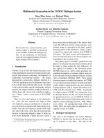
The beam arrangement consisted of 4 half-beams with
two tangential beams covering the caudal part of the
target volume, and one anterior-posterior field (0°) and
one oblique field, typically 110-115°, covering the cranial
part of the chest wall (Figure 1). The beam angles, aper-
tures, weights and dynamic wedges were optimized by
standard (forward) planning. The photon beams energy
was 6 MV using a Varian Clinac (Varian Medical Sys-
tem) linear accelerator. The dose plans were normali zed
to the mean dose to the planning target volume (PTV).
The breast/chest wall should receive a total dose of 50
Gy, and the regional lymph nodes 46-50 Gy. Six of the
women received an additional boost of 10 Gy to the
tumor bed (9 or 12 M eV electrons using a circular field
with a diameter of 5-9 cm, not included in the CT-
based treatment planning).
For the purpose of the current study a radiologist deli-
neated the thyroid gland on the planning CT-images of
the Cases and Controls, and the individual volume of
the gland was calculated (VolTot [cm
3
]). Based on each
Johansen et al. Radiation Oncology 2011, 6:68
/>Page 2 of 7
patient’ s DVH the volume percentages of the thyroid
absorbing respectively 20, 30, 40 and 50 Gy were then
estimated (V20, V30, V40 and V50) together with the
individual mean thyroid dose over the whole gland
(MeanTotGy). Subsequently the absolute volumes (cm
3
)
of the thyroid gland receiving respectively < 30 Gy and
≥ 3 0 Gy were calculated (Vol < 30 and Vol ≥ 30). The
30 Gy dose was taken as the point of influencing the
development of hypothyroidism, as the median of
MeanTotGy was observed to be 31 Gy in both Cases
and Controls in the present study.
Statistics
To assess differences between Cases and Controls, non-
parametric Mann-Whitney test were employed. The
choice of the statistic tests are dependent generally on
whether the data were normally distributed or not. A P-
value < 0.05 was considered to be statistically significant.
Figure 1 Oblique (axillary field) (field 2) and supraclavicular (field 1) fields used in CT-RT. The location of the thyroid gland within the
axillary beam is illustrated. The thyroid gland is pink colored. The same figure with and without isodose lines is shown.
Johansen et al. Radiation Oncology 2011, 6:68
/>Page 3 of 7
Ethics
All patients provided a written consent form to partici-
pate in the study, which was approved by the Ethical
committee of the Health Region South and the Data
Inspectorate of Norway.
Results
Table1confirmsthecomparabilityofthe16Casesand
the 16 Controls as to age, observation time, initial stage,
surgery and systemic treatment as well as the adjuvant
radiotherapy. Median time from BC diagnosis to the
survey was 44 months in both Cases (38-56) and Con-
trols (37-56).
No statistically significant inter-group differences were
found between V20, V30, V40 and V50Gy or the med-
ian of MeanTotGy (Table 2A and 2B, the latter being 31
Gy in both Cases and Controls), if combining both
groups. In contrast, in Controls the median VolTotGy
was 2.3 times above median VolTotGy in Cases (r=
0.003), with large inter-individual variations in both
groups. As a consequence the volume of the thyroid
gland receiving ≥ 30 Gy in Cases was almost 2.2 times
less than the comparable figure in Controls (r =0.001).
Further, among Controls the thyroid volume receiving <
30 Gy was 2.5 times greater than the comparable figure
among Cases (r = 0.000).
Discussion
In this case-control study, breast cancer patients who
developed post-RT hypothyroidism displayed signifi-
cantly smaller thyroid glands volume before the adjuvant
radiotherapy than their controls who had not develope d
post-RT hypothyroidism. This resulted in significantly
smaller absolute thyroid sub-volumes receiving ≥ 30 Gy
in Cases than in Controls. The median of the individual
mean thyroid dose was 31 Gy [22Gy-42Gy] in Cases.
The relatively small volumes with high radiation expo-
sure may be responsible for the post-radiotherapy devel-
opment of hypothyroidism in Cases. Compared to their
Controls, Cases were after radiotherapy left with smaller
thyroid volumes which were enabled to produce suffi-
cient amount of hormone.
When estimating the incidence/prevalence of post-RT
hypothyroidism, it i s important to separate clinical
symptomatic hypothyroidism from biochemical
hypothyroidism. As screening for thyroid function has
not been a routine in breast cancer survivors, we believe
that our BC Cases presented to their family doctor clini-
cal symptoms compatible with decreased thyroid func-
tion which resulted in the diagnosis of hypothyroidism.
In the study of Reinertsen et al. [14] an increased preva-
lence of hypothyroidism (18%) in breast cance r patients
was observed compared to 6 % the prevalence in the
general population in Norway. The difference is related
to a higher incidence after breast cancer treatment.
According to the literature both age and radiation
dose are related to development of post-radiation
hypothyroidism. Radio sensitivity of the thyroid gland is
believed to decrease with increasing age. Bonato and
colleagues [15] showed that 23 of 59 childhood cancer
survivors developed biochemical hypothyroidism after
radiotherapy to the head and neck as well as total body
irradiation. A median thyroid dose in Bonato et al.’ s
study was 42 Gy (inter-quartile range: [27-72Gy]) [15].
After high-dose radiotherapy the 5 years incidence of
Table 1 Individual and Overall
Characteristic Cases Controls
Age
1
(yrs)
<50 3 3
>50 13 13
Median 56 56
Range 44-75 43-73
Obstime
2
(months)
<50 11 12
≥ 50 5 4
Median 44 44
Range 38-56 37-56
Stage
II 13 13
III 3 3
Surgery
3
MRM 12 12
BCS 4 4
Chemo
4
FEC 11 11
other 1 1
No 4 4
Number of Cycles
61111
411
Tamoxifen
yes 15 15
No 1 1
RT (Gy)
50 Gy 13 13
50+10 Gy 3 3
Radiotherapy, surgical and chemotherapy treatment characteristics for cases
(group A) and controls (group B).
1
Age: Age at follow-up
2
Obstime: observation time from breast cancer diagno sis to the outpatient
examination
3
Surgery: MRM = Radical mastectomy, BCS = breast conserving surgery
4
Chemotherapy: FEC = (5Fluoro-Uracil, epirubicin, cyclophosfamide), FEC-
regimen
Other = 4 cycles of epirubicin
No = no chemotherapy
Johansen et al. Radiation Oncology 2011, 6:68
/>Page 4 of 7
Table 2 A: Total thyroid volume (cc) and thyroid dose volume data (%) for dose levels 20, 30, 40 and 50 Gy and for
mean thyroid dose
A: Total thyroid volume (cc) and thyroid dose volume data (%) for dose levels 20, 30, 40 and 50 Gy and for mean thyroid dose.
Case no. VolTot(cm3) V20(%) V30(%) V40(%) V50(%) MeanTotGy Vol ≥ 30(cm3) Vol < 30(cm3)
1 10 95754911 38 7 2
2 8 93 70 37 0 28 5 2
311927143631 8 3
4 7 97 76 47 10 39 6 2
5 9 100 83 53 12 41 7 2
6 3 95 74 43 4 29 2 1
7 7 94 68 33 0 31 5 2
8 5 88 64 31 0 29 4 2
9 8 89 66 34 0 31 6 3
10 8 89 64 30 0 22 5 3
11 2 94 63 19 0 25 1 1
12 3 91 59 13 0 22 2 1
13 5 90 66 33 0 32 3 2
14 6 90 66 33 0 31 4 2
15 5 89 61 24 0 25 3 2
16 7 94 70 37 0 42 5 2
Overall
All: Median: Median: Median: Median: Median: Median: Median: Median:
16 7 93 67 34 0 31 5 2
Range: Range: Range: Range: Range: Range: Range: Range:
1.9-11.4 88-100 59-83 13-53 0-12 22-42 1.2-8.1 0.7-3.3
B: Total thyroid volume (cc) and thyroid dose volume data (%) for dose levels 20, 30, 40 and 50 Gy and for mean thyroid dose.
Control no. VolTot(cm3) V20(%) V30(%) V40(%) V50(%) MeanTotGy Vol ≥ 30(cm3) Vol < 30(cm3)
1 33.4 91.0 72.0 46.0 10.0 33.0 24.0 9.4
2 16.1 95.0 77.0 51.0 16.0 34.9 12.4 3.7
3 40.1 94.0 74.0 46.0 11.0 33.4 29.7 10.4
4 12.8 92.0 70.0 38.0 0.0 32.3 9.0 3.8
5 10.6 90.0 69.0 40.0 4.0 31.6 7.3 3.3
6 6.1 89.0 62.0 26.0 0.0 28.2 3.8 2.3
7 21.8 90.0 66.0 35.0 0.0 28.9 14.4 7.4
8 2.7 92.0 73.0 46.0 9.0 31.1 1.9 0.7
9 18.4 94.0 74.0 46.0 10.0 33.0 13.6 4.8
10 19.2 89.0 66.0 35.0 0.0 29.4 12.7 6.5
11 11.9 91.0 65.0 31.0 0.0 30.3 7.7 4.2
12 16.8 86.0 64.0 33.0 0.0 28.2 10.8 6.0
13 10.6 92.0 72.0 40.0 0.0 37.6 7.6 3.0
14 16.9 90.0 70.0 43.0 8.0 31.3 11.8 5.1
15 12.8 89.0 66.0 34.0 0.0 28.8 8.5 4.3
16 41.0 93.0 70.0 39.0 0.0 32.7 28.7 12.3
Overall
All: Median: Median: Median: Median: Median: Median: Median: Median:
16 16 91 70 40 0 31 11 5
Range: Range: Range: Range: Range: Range: Range: Range:
2.66-41 86-95 62-77 26-51 0-16 28-38 1.9-29.7 0.7-12.3
A. Cases: Individual and Overall. B. Controls: Individual and Overall.
Johansen et al. Radiation Oncology 2011, 6:68
/>Page 5 of 7
biochemical hypothyroidism was 48% in a dults with
head and neck cancer [17]. However, in another study
carried out by Smith et al. [16] the 5 years incidence of
thyroxin requiring hypothyroidism in 38.255 irradiated
and non-irradiated women older than 65 years diag-
nosed with breast cancer and 111.944 cancer-free con-
trols was identified. Their results showed an identical
14% incidence of hypothyroidism development in both
irradiated patient group and non-irradiated [16]. Emami
et al. [18] suggested a tolerance dose of 45 Gy leading
to development of clinical hypothyroidism in 8% of the
individuals followed for 5 years after completion of
radiotherapy with 45 Gy. Yoden et al. [19] have sug-
gested that the percentage volume of the thyroid gland
receiving dose s between 10-60 Gy (V10-V60) would
represent a predictor of hypothyroidism. According to
Yoden et al. [19] V30 Gy had a significant impact on
the peak level of TSH. Other estimations of incidence
after 50 Gy applied to the whole thyroid gland [10-
80Gy] range from 2% - 50% [20,21]. After head and
neck irradiation doses of 10-80 Gy to the thyroid are
reported to lead to dysfunction of the gland [11]. The
diversity of these figures illustrates that the threshold
for thyroid radiation and development of hypothyroid-
ism is not clear. The admittedly small present study
emphasizes the role of the individual thyroid gland
volume for the development of post-radiotherapy
hypothyroidism in BC patients. The sub-volume receiv-
ing ≥ 30 Gy seems to determine whether or not suffi-
cient thyroxin is produced after radiotherapy. Among
Cases the total thyroid volume and the sub-volume
receiving ≥ 30 Gy are sufficiently smaller than in Con-
trols. Interestingly Bonato et al. [15] confirmed in their
study that hypothyroid individuals had smaller glands
than those with normally functioning glands, though it
is not quite clear on the report whether volume mea-
surements have been performed before radiotherapy or
afterwards in connection with the reported survey.
The measurement of the thyroid gland volume repre-
sents the main limitation of this small study. On the
background of the lack of contrast the delineation of the
individual thyroid gland on the CT images remained a
constant difficulty even for an experienced radiologist.
Using ultrasonography, larger volumes have been
described [22] than accomplished in our study. How-
ever, thyroid gland sizes ranging from 3.6-6 cm in
length, 1.5-2 cm in widt h and 1- 2 cm depth are
reported [23], which are more in agreement with our
study. Finally, as our findings are principally based on
the selective differences between the gland volumes in
Cases and Controls, any systematic measurement error
is of less importance, provided that its similar presence
in Cases and Controls.
The impact of adjuvant chemotherapy and hormone
treatment on the risk of hypothyroidism among patients
with head and neck malignancies is investigated by both
Kanti et al. [24] and Sinrad et al. [25]. These authors
found no effect of adjuvant chemotherapy on thyroid
gland function, though chemotherapy for head and neck
cancer differ from that applied in BC p atient group.
Also, Jereczk-Fossa and colleagues [20] have concluded
that the impact of chemothe rapy and endocrine treat-
ment on the risk of hypothyroi dism is still controversial.
The sig nificant difference in thyroid size between Cases
and Controls in the current study and the high similar-
ity of systemic treatment in all Cases and Controls of
our study makes it impossible to analyse the impact of
chemotherapy on post-BC hypothyroidism development.
We concluded that patients with small thyroid glands
are at particular risk to develop hypothyroidism after
radiotherapy for breast cancer, as less tissue with radia-
tion doses less than 30 Gy is available for sufficient thyr-
oxin production. Further investigations in larger cohorts
are required to confirm our results.
Author details
1
Institute for Cancer Research, Oslo University Hospital-Radiumhospitalet, N-
0310 Oslo, Norway.
2
Department of Clincal Cancer Research, Oslo University
Hospital-Radiumhospitalet University Hospital, Norway.
3
The Cancer Center,
Ullevål University Hospital, N-0407 Oslo, Norway.
4
Department of Radiology,
Oslo University Hospital-Radiumhospitalet, Norway.
5
Department of Physics,
University of Bergen, Norway.
6
Faculty of Medicine, University of Oslo, Oslo,
Norway.
Authors’ contributions
All authors read and approved the final manuscript. SJ wrote the paper and
performed the dosimetric and statistical analysis. KVR prepared the patient
material and participated in the discussion of the results. KK delineated the
thyroid gland. DRO participated in the coordination of the study. SDF carried
out the design of the study and edited the manuscript.
Competing interests
The authors declare that they have no competing interests.
Received: 31 January 2011 Accepted: 9 June 2011
Published: 9 June 2011
References
1. Kumpulainen EJ, Hirvikoski PP, Virtaniemi JA, et al: Hypothyroidism after
radiotherapy for laryngeal cancer. Radiother and Oncol 2000, 57:97-101.
2. Kuten A, Lubochitcki R, Fishman G, et al: Postradiotherapy
hypothyroidism: Radiation dose response and chemotherapeutic
radiosensitization at less than 40 Gy. J Surg Oncol 1996, 61:281-283.
3. Bookman MA, Longo DL: Concomitant illness in patients treated for
Hodgkin’s disease. Cancer Treat Rev 1986, 13:77-111.
4. Alterio D, Jereczka-Fossa BA, Franchi B, et al: Thyroid disorders in patients
treated with radiotherapy for head-and-neck cancer: A retrospective
analysis of seventy-three patients. Int J Radiat Oncol Biol Phys 2007,
67:144-150.
5. Ozawa H, Saitou H, Mizutari K, et al: Hypothyroidism after radiotherapy for
patients with head and neck cancer. Am J Otolaryngol 2007, 28:46-49.
6. Tell R, Sjodin H, Lundell G, et al: Hypothyroidism after external
radiotherapy for Head and Neck cancer. Int J Radiat Oncol Biol Phys 1997,
39:303-308.
Johansen et al. Radiation Oncology 2011, 6:68
/>Page 6 of 7
7. Turner SL, Tiver KW, Boyages SC: Thyroid dysfunction following
radiotherapy for head and neck cancer. Int J Radiat Biol Phys 1995,
31:279-283.
8. Nishiyama K, Kozuka T, Higashihara T, et al: Acute radiation Thyroiditis. Int
J Radiat Oncol Biol Phys 1996, 36:1221-1224.
9. Garcia-Serra A, Amdur RJ, Morris CG, et al: Thyroid Function should be
monitored following radiotherapy to the low neck. Am J Clin Oncol 2005,
28:255-258.
10. Hancock SL, McDouGall IR, Onstine LS: Thyroid abonormalities after
therapeutic external radiation. Int J Radiat Oncol Biol Phys 1995,
31:1165-1170.
11. Foo ML, McCullough EC, Foote RL, et al: Doses to radiation sensitive
organs and structures located outside the radiotherapeutic target
volume for four treatment situations. Int J Oncol Biol Phys 1993,
27:403-417.
12. Joensuu H, Viikari J: Thyroid function after postoperative radiation
therapy in patients with breast cancer. Acta Oncol 1986, 25:167-170.
13. Bruning P, Bonfrer J, Jong-Bakker MD, et al: Primary hypothyroidism in
breast cancer patients with irradiated supraclavicular lymph nodes. Br J
Cancer 1985, 51:659-663.
14. Reinertsen KV, Cvancarova M, Wist E, et al: Thyroid function in women
after multimodal treatment for breast cancer stage II/III: comparison
with controls from a population sample. Int J Radiat Oncol Biol Phys 2009,
75(3):764-70.
15. Bonato C, Severino RF, Elnecave RH: Reduced thyroid volume and
hypothyroidism in survivors of childhood cancer treated with
radiotherapy. J Pediatr Endocrinol Metab 2008, 21:943-9.
16. Smith GL, Smith BD, Giordano SH, et al: Risk of hypothyroidism in older
breast cancer patients treated with radiation. Cancer 2008, 112:1371-1379.
17. Mercado G, Adelstein D, Saxton JP, et al: Hypothyroidism: a frequent
event after radiotherapy and after radiotherapy with chemotherapy for
patients with head and neck carcinoma. Cancer 2001, 92(11):2892-7.
18. Emami B, Lyman J, Brown A, et al: Tolerance of normal tissue to
therapeutic irradiation. Int J Radiat Oncol Biol Phys 1991, 21
:109-122.
19. Yoden E, Maruta T, Soejima T, et al: Hypothyroidism after radiotherapy to
the neck. Int Radiat Oncol Biol Phys 2001, S-51:337-338.
20. Jereczek-Fossa BA, Alterio D, Jassem J, et al: Radiotherapy-induced thyroid
disorders. Cancer Treat Rev 2004, 30:369-384.
21. Shakespeare TP, Dwyer M, Mukherjee R, et al: Estimating risk of
radiotherapy complications as part of informed consent: the high
degree of variability between radiation oncologists may be related to
experience. Int Radiat Oncol Biol Phys 2002, 54:647-653.
22. Turken O, Narin Y, Demirbas S, et al: Breast cancer in association with
thyroid disorders. Breast cancer Res 2003, 5:R110-113.
23. Moeller TB, Reif E: Normal findings in CT and MRI. CT: Head and Neck. 1
edition. Yew York: George Thieme Verlag; 2000, 29.
24. Kanti AA, Ranjan DA, Santanu P, et al: Iatrognc hypothyroidism: A
consequence of external beam radiotherapy to the head & neck
malignancies. J Cancer Res Ther 2005, 1:142-146.
25. Sinrad RJ, Tobin EJ, Mazzaferri EL, et al: Hypothyroidism after treatment for
nonthyroid head and neck cancer. Arch Otolaryngol Head Neck Surg 2000,
126:326-328.
doi:10.1186/1748-717X-6-68
Cite this article as: Johansen et al.: Dose distribution in the thyroid
gland following radiation therapy of breast cancer-a retrospective
study. Radiation Oncology 2011 6:68.
Submit your next manuscript to BioMed Central
and take full advantage of:
• Convenient online submission
• Thorough peer review
• No space constraints or color figure charges
• Immediate publication on acceptance
• Inclusion in PubMed, CAS, Scopus and Google Scholar
• Research which is freely available for redistribution
Submit your manuscript at
www.biomedcentral.com/submit
Johansen et al. Radiation Oncology 2011, 6:68
/>Page 7 of 7