Gene expression profiling reveals activation of the FA/BRCA pathway in advanced squamous cervical cancer with intrinsic resistance and therapy failure
Bạn đang xem bản rút gọn của tài liệu. Xem và tải ngay bản đầy đủ của tài liệu tại đây (3.9 MB, 14 trang )
Balacescu et al. BMC Cancer 2014, 14:246
/>
RESEARCH ARTICLE
Open Access
Gene expression profiling reveals activation of
the FA/BRCA pathway in advanced squamous
cervical cancer with intrinsic resistance and
therapy failure
Ovidiu Balacescu1*†, Loredana Balacescu1†, Oana Tudoran1, Nicolae Todor1, Meda Rus1, Rares Buiga1,
Sergiu Susman2, Bogdan Fetica1, Laura Pop2, Laura Maja1, Simona Visan1,3, Claudia Ordeanu1,
Ioana Berindan-Neagoe1,2* and Viorica Nagy1,2
Abstract
Background: Advanced squamous cervical cancer, one of the most commonly diagnosed cancers in women, still
remains a major problem in oncology due to treatment failure and distant metastasis. Antitumor therapy failure is
due to both intrinsic and acquired resistance; intrinsic resistance is often decisive for treatment response. In this
study, we investigated the specific pathways and molecules responsible for baseline therapy failure in locally
advanced squamous cervical cancer.
Methods: Twenty-one patients with locally advanced squamous cell carcinoma were enrolled in this study. Primary
biopsies harvested prior to therapy were analyzed for whole human gene expression (Agilent) based on the
patient’s 6 months clinical response. Ingenuity Pathway Analysis was used to investigate the altered molecular
function and canonical pathways between the responding and non-responding patients. The microarray results
were validated by qRT-PCR and immunohistochemistry. An additional set of 24 formalin-fixed paraffin-embedded
cervical cancer samples was used for independent validation of the proteins of interest.
Results: A 2859-gene signature was identified to distinguish between responder and non-responder patients.
‘DNA Replication, Recombination and Repair’ represented one of the most important mechanisms activated in
non-responsive cervical tumors, and the ‘Role of BRCA1 in DNA Damage Response’ was predicted to be the most
significantly altered canonical pathway involved in intrinsic resistance (p = 1.86E-04, ratio = 0.262). Immunohistological
staining confirmed increased expression of BRCA1, BRIP1, FANCD2 and RAD51 in non-responsive compared with
responsive advanced squamous cervical cancer, both in the initial set of 21 cervical cancer samples and the second set
of 24 samples.
Conclusions: Our findings suggest that FA/BRCA pathway plays an important role in treatment failure in advanced
cervical cancer. The assessment of FANCD2, RAD51, BRCA1 and BRIP1 nuclear proteins could provide important
information about the patients at risk for treatment failure.
Keywords: FANCD2, RAD51, BRCA1, BRIP1, Cervical cancer, Microarray, Treatment response
* Correspondence: ;
†
Equal contributors
1
The Oncology Institute ”Prof Dr. Ion Chiricuta”, 34-36 Republicii street,
400015 Cluj-Napoca, Romania
2
Iuliu Hatieganu, University of Medicine and Pharmacy, 8 Babes street,
400012 Cluj-Napoca, Romania
Full list of author information is available at the end of the article
© 2014 Balacescu et al.; licensee BioMed Central Ltd. This is an Open Access article distributed under the terms of the Creative
Commons Attribution License ( which permits unrestricted use, distribution, and
reproduction in any medium, provided the original work is properly credited.
Balacescu et al. BMC Cancer 2014, 14:246
/>
Background
Cervical cancer, the third most commonly diagnosed cancer in women, with 529,800 cases in 2010 [1], represents a
major problem in oncology due to treatment failure and
distant metastasis. More than 85% of cervical cancers are
diagnosed every year in developing countries, and approximately 90% of overall deaths occur in these countries. If
detected at an early stage, cervical cancer represents one
of the most successfully treated cancers. Unfortunately,
because of the lack of screening programs in developing
countries, cervical cancer is predominantly detected in advanced stages (IIB-IIIB). About half of the patients with
advanced cervical cancer will develop recurrence or metastasis in the first 2 years after completion of therapy.
Although new anticancer drugs are constantly being developed, overcoming drug resistance is still a challenge.
Therefore, there is an urgent need to identify new prognostic factors that could distinguish between patients with
unfavorable prognoses from others with better prognoses.
Almost half of patients present baseline resistance (intrinsic resistance), and a large proportion of the remaining half
will develop resistance during treatment (acquired resistance) [2]. Intrinsic resistance is often complex and occurs
through several mechanisms, depending on the therapy regimen. The treatment for pre-invasive lesions is generally
based on surgery; for invasive cervical cancers, the treatment
is based on surgery and/or radiation and cisplatin-based
chemotherapy [3]. The chemoradiotherapy treatment produces DNA double-strand breaks (DSBs), which is considered to be the most lethal form of DNA damage. DSBs are
caused by radiation and platinum compounds based chemotherapy but also could be produced by endogenous damage,
such as that caused by reactive oxygen species and collapsed
replication forks. DNA damage induces a series of molecular
responses that are responsible for the maintenance of genome integrity [4]. Deficiencies in DSB response and repair
could represent important events for intrinsic resistance.
The diagnosis of baseline resistance in individual patients could improve the cancer treatment by the avoidance of inefficient therapy. Gene expression studies have
been conducted across many tumor types to investigate
the patterns of genes involved in intrinsic resistance. In
cervical cancer, relatively few studies have been focused on
identifying baseline resistance to chemoradiotherapy [5-7].
Therefore, the aim of our study was to investigate the specific pathways and molecules responsible for baseline therapy failure in locally advanced squamous cervical cancer.
Page 2 of 14
ethics committee of The Oncology Institute ‘Prof. Dr. Ion
Chiricuta’. All patients gave informed consent in accordance with the Declaration of Helsinki.
Twenty-one patients with locally advanced squamous cell
carcinoma (FIGO stage IIB-IIIB) were enrolled in the genomics study. A tissue fragment from a primary biopsy and
a cervical lavage specimen were harvested from each patient prior to initiation of the therapy. Tissue samples were
stored in liquid nitrogen until use for RNA extraction.
Corresponding formalin-fixed paraffin-embedded (FFPE)
tissue samples were used for protein validation. Moreover,
an additional set of 24 FFPE samples was used for independent immunohistochemistry validation of the data. All
patients in the validation and study groups had the same
including criteria. The clinical and histopathological characteristics of the patients included in this study are presented
in Table 1.
The therapy schedule
The patients were treated with concomitant chemoradiotherapy (CRT) associated or not with surgery. The radiotherapy protocol includes external beam radiotherapy
(EBRT) to the pelvis delivered by a linear accelerator at
15MV for a dose of 46 Gy/23 fractions and a cervical
boost given by intracavitary high-dose-rate (HDR) brachytherapy (BT) in a dose of 10 Gy/2 fractions. Cisplatin was
administered concomitant with the radiotherapy as a
radiosensitizer. At this dose, patients were evaluated and,
Table 1 Baseline characteristics of the patients in the
genomics study and IHC validation group
Characteristics
Genomics study
group (n = 21)
IHC validation
group (n = 24)
Median age
(range), years
46 (27–73)
52 (28–62)
5 (2–8)
4 (2–7)
12.7 (7.9–14.4)
13.3 (10.2–14.9)
Median tumor
size (range), cm
Median hemoglobin
(range), g/dl
FIGO stage
II B
10
8
III A
5
11
III B
6
5
HPV 16
16
19
Other high-risk*
3
3
Methods
Negative
2
2
Sample collection
Treatment response
Patient samples and clinical data with end points were
obtained from the Departments of Radiotherapy and Pathology of The Oncology Institute ‘Prof. Dr. I. Chiricuta’,
Cluj-Napoca, Romania. This study was approved by the
CR
12
15
NCR
9
9
HPV subtype
*other high-risk in study group: 33,58,73.
other high-risk in validation group: 31,45,58.
Balacescu et al. BMC Cancer 2014, 14:246
/>
according to tumor response, further of CRT (EBRT until
60 Gy concomitant with cisplatin and HDR BT until 20
Gy) or surgery (radical abdominal hysterectomy with
pelvic lymphadenectomy) was decided. In our internal
protocol, surgery was recommended, but not mandatory,
being a patient’s option. The tumor response was clinically
evaluated at 6 months after the end of the CRT treatment
and was defined as complete response (CR) or non-complete
response (NCR) (partial response and stable disease). For the
patients that underwent surgery, the histopathological evaluation confirmed the clinical response.
RNA extraction and purification
Tumor sections with a minimum of 70% tumor cells
were harvested by macrodissection from primary biopsies of cervical cancers. Total RNA was extracted with
TriReagent (Sigma-Aldrich) and purified using an RNeasy
Mini kit (Qiagen) according to the manufacturer’s protocols. Extracted RNA was assessed for quality with a Labon-a-chip Bioanalyzer 2100 (Agilent Technologies). The
RNA Integrity Number (RIN) and rRNA 28S/18S ratio
were used to define the quality of the total RNA. The
RNAs with RINs >7.5 and rRNA 28S/18S ratios >1.8 were
used for further analysis. RNA concentrations were adjusted using a NanoDrop ND-1000 spectrophotometer
(NanoDrop Technologies).
HPV genotyping
Genomic DNA was extracted from 1 ml of cervical lavage using a High Pure DNA extraction kit (Roche). HPV
genotypes, including 37 high- and low-risk genotypes,
were identified with the Linear Array HPV Genotyping
Test (Roche) according to the manufacturer’s protocol.
Oligonucleotide microarray technology
Agilent oligonucleotide technology was used to measure
gene expression changes in the samples of interest. Microarray probes (cRNA-Cy3) were synthesized from 200 ng of
total RNA in two reaction steps using a one-color Agilent
Low Input Quick Amp Labeling Kit according to the manufacturer’s instructions. All labeled cRNAs (Cy3) were purified using an RNeasy Mini kit (Qiagen) and were evaluated
for quality control using a Nanodrop ND-1000 spectrophotometer. cRNAs with minimum yields of 1.65 μg and specific activities of 6 pmol/μl Cy3 per μg cRNA were selected
for further analysis. After fragmentation to an average size
of 60 – 100 nucleotides, each cRNA was hybridized for 17
hours at 65°C to whole-human-genome 4×44K microarray
slides (product G4112F; Agilent) following the manufacturer’s protocol (Agilent Technologies). The slides were
scanned with an Agilent G2505B US45102867 microarray
scanner, and gridding was performed with Feature Extraction Software v.10.5.1.1.
Page 3 of 14
The microarray data have been deposited in the NCBI
Gene Expression Omnibus (GEO) repository under accession number GSE56363.
Microarray data analysis
The microarray data, including median foreground and
background intensities, flags and feature annotations, were
imported into R/Bioconductor. The association between
log2 values of background and foreground intensities
across each array was estimated by computing Pearson
correlation coefficients. Suitable R packages (arrayQualityMetrics, limma, marray) were used for quality control,
normalization, filtering and data summarization. Betweenarray normalization was performed using the quantile
normalization method. The median normalized signals
were used for further data analysis. To reduce the number
of non-informative features, the probes with saturated
and non-uniform signals present in more than 15% of the
samples were removed. Differentially expressed genes/sequences between non-responder and responder samples
were selected using the moderated t-statistic. This method
is an improvement over the standard t-statistic, as it allows
elimination of the influence of random small withingroup variance by sharing information across genes.
The Benjamini and Hochberg method was used to adjust the
p-values for multiple testing (adjusted p-value < 0.05). Only
genes/sequences with at least a 1.5-fold change in expression
between the studied groups were considered differentially
expressed. The hierarchical clustering using Euclidean distances and Ward method was further performed to cluster
the similarities in expression between genes/samples.
Functional analysis
The dataset containing differentially expressed genes was
uploaded into the Ingenuity Pathway Analysis (IPA) software (Ingenuity® Systems, ) and
was associated with the biological functions and canonical
pathways in the Ingenuity Knowledge Base. Fisher’s exact
test (p < 0.05) was used to assess the significance of the associations between genes in the dataset and biological
functions or canonical pathways. In addition, for canonical
pathways, a ratio was computed between the number of
molecules from the dataset and the total number of molecules in that pathway.
Quantitative real-time PCR (qRT-PCR)
The First Strand cDNA Synthesis Kit (Roche) was used to
reverse transcribe 200 ng of total RNA. Five microliters of
1:10 (v/v)-diluted cDNA was amplified in a final volume of
20 μl using a LightCycler 480 (Roche). The amplification
was performed with 1 μM specific primers (Tib Molbiol)
and a 0.2 μM specific hydrolysis probe from the Universal
Probe Library (UPL). The primers and UPL probes were
designed with Roche Applied Science software as follows:
Balacescu et al. BMC Cancer 2014, 14:246
/>
BRCA1 (NM_007294.3): F-ttgttgatgtggaggagcaa, R-ttgttgat
gtggaggagcaa (UPL#11); BRCA2 (NM_000059.3): F-agctta
ctccggccaaaaa, R-ttcctccaatgcttggtaaataa (UPL#50); RAD51
(NM_001164269.1): F-tgagggtacctttaggccaga, R-cactgccaga
gagaccatacc (UPL#66); FANCD2 (NM_033084.3): F-cgacttg
acccaaacttcct, R-tcctccaatctaatagacgacaact (UPL#9); BRIP1
(NM_032043.1): F-aatggcacttcatcaacttgtc, R-tggatgcctgtttc
ttagca (UPL#71); BLM (NM_000057.2): F-gatcagaaagcacca
cccata, R-tcagccatggtgtcacattc (UPL#34); and 18S rRNA
(NR_003286.2): F-gcaattattccccatgaacg, R- gggacttaatcaacgcacgc (UPL#48). Thermal cycling conditions were set as follows: activation at 95°C for 10 minutes; followed by 40
cycles of amplification, including denaturation at 95°C for
15 seconds, annealing at 55°C for 20 seconds and extension
at 72°C for 1 second; followed by a cooling step at 40°C for
30 seconds. The relative expression levels of target genes
(NCR vs. CR) were calculated using the ΔΔCt method [8]
after normalizing to 18S housekeeping gene.
Immunohistochemistry (IHC)
Immunohistochemistry was performed on FFPE 4-μm
thick tissue sections, using a standard protocol. Following
deparaffinization and rehydration of the tissue sections,
antigen retrieval was performed for 20 minutes in 0.01 M
citrate buffer (pH 6.0) using the boiling process (pressure
cooker). Endogenous peroxidase was blocked with H2O2
(3%). Blocking of the nonspecific reactions was performed
using the Novocastra Protein block™ solution. The sections
were incubated 30 minutes with primary antibodies at room
temperature in a humid chamber. The immunohistochemical staining was performed using the following dilutions for
the primary monoclonal antibodies: 1:400 for BRCA1 (BioVision Inc., OH, USA, clone#3364-100), 1:200 for BRCA2
(Covalab, Cambridge, UK, clone pab0457-0), 1:200 for
FANCD2 (Thermo Pierce Biotechnology Inc., IL, USA, clone
PA1-16548), 1:20 for Rad51 (Thermo Pierce Biotechnology
Inc., IL, USA, clone MA5-14416) and 1:300 for BPRIP1
(Abcam, Cambridge, UK, product number ab151509). Sections were sensitized using Post Primary Block™, and then incubated with NovoLink™ polymer containing the secondary
antibody. The peroxidase reaction was developed using
diamino-benzidine tetrachloride (DAB) as chromogen. Sections were counterstained with hematoxylin.
The IHC staining was automatically assessed using the
ImmunoRatio free web-based application [9]. The application is conceived for automated image analysis of immunohistochemical nuclear staining like estrogen receptor (ER),
progesterone receptor (PR), or Ki-67. Briefly, for every case 3
different representative images of immunostained sections
were taken using a CX41 Olympus microscope coupled with
a high resolution video camera AV5100M (MegaVideo IP
camera, Arecont Vision). The application performs the segmentation of brown (DAB-colored), and hematoxylinstained nuclei, than calculates the labeling index as the
Page 4 of 14
percentage of brown stained nuclear area over the total nuclear area. The system also produces a pseudo-colored result image, illustrating the area segmentation. Every
generated image was checked for consistency by two pathologists (BR and SS). Only the correct segmented images
were accepted for further analysis.
Statistical methods
The follow-up endpoint for each patient represents a binary
evaluation of the treatment response at 6 months after the
end of the treatment. All existing factors were compared
when examining the two groups of patients (CR and NCR).
Categorical factors were analyzed using a chi-squared test,
and when reduced numbers of observations were present,
we applied Yates’ correction [10]. A comparison of medians
was performed using the median test and two-tailed unpaired t test was used to evaluate for differences in gene expression between groups of interes (NCR vs CR). The
strengths of the association between genes of interest as well
as between PCR and microarray results were tested with a
Pearson parametric test. The receiver operating characteristic (ROC) curve was used to evaluate the predictive accuracy of genes of interest in the differentiation between
samples with or without complete remission. [11]. The calculation of the area under curve (AUC) and test equality
with a value of 0.5 was performed according to Bamber and
Hanley [12,13]. The point of optimal classification was considered the point nearest to (0.1) of the absolute classification. Unpaired t-test on arcsine-transformed data was used
to determine whether the proportion of stained nuclear protein was different between non-responders and responders
samples, in both genomic and IHC validation groups.
All differences with p < 0.05 were considered statistically significant. The confidence intervals were evaluated
with the level of significance equal to 0.05.
Results
Patient and tumor characteristics
FIGO staging evaluation of the patients included in this
study revealed that approximately 48% of the patients
were in stage II, while the rest of 52% were in stage III.
Among these, 2 patients tested negative for HPV, whereas
HPV-16 subtype has been detected in the majority of the
cases. Based on 6 months treatment outcome evaluation
twelve patients presented complete remission and were
assigned to the CR group, while the rest of 9 patients that
partially responded or had stable disease were assigned to
the NCR group. We observed higher median age value in
the responders group (p < 0.01), however prognostic factors such as tumor size, hemoglobin and FIGO stage were
balanced between the NCR and CR groups (Table 2).
Since almost all the patients presented HPV 16-positive
tumors, the association between HPV subtype and treatment outcome could not be assessed.
Balacescu et al. BMC Cancer 2014, 14:246
/>
Page 5 of 14
Table 2 Association between clinical and
histopathological data and treatment response
Characteristics
Study population
No. of patients CR group NCR group p
n (%)
n
n
21 (100%)
12
9
Age (years)
Median
21
Range
56.5
42
28-73
27-55
< 0.01
Tumor size (cm)
Median
21
Range
4.75
5
2.0-6.5
2.5-8.0
0.054
Hemoglobin (g/dl)
Median
21
Range
12.75
12.2
7.9-14.2
10.1-14.4
0.8
FIGO stage
IIB
10 (47.62%)
4
6
IIIA
5 (23.81%)
4
1
IIIB
6 (28.57%)
4
2
16
16 (76.19%)
10
6
Other high-risk
3 (14.29%)
1
2
Negative
2 (9.52%)
1
1
0.62
HPV subtype
-
Gene expression profiling of cervical cancer samples
Gene expression profiles for NCR and CR samples were
generated using one-color hybridization to whole human genome arrays carrying 43,376 biological sequences. We assessed the quality of the array before
and after normalization and we did not detect batch effects or outlier arrays. We observed a weak correlation
between background and foreground intensities across
each array (r range, 0.06 to 0.2), therefore we did not
perform background correction. To improve data quality, a filtering step was applied. A total number of
40,998 sequences passed the filtering criteria and were
used for further analysis. In class comparison analysis
we identified a signature of 2859 genes whose differential
expression in non-responder compared to responder samples exceeded 1.5-fold at an adjusted p-value < 0.05. Of
these, 1501 genes were up-regulated and 1358 genes were
down-regulated in NCR compared with CR.
To highlight the differences in gene expression a supervised hierarchical clustering was performed on the
set of differentially expressed genes. Based on expression
profiles, non-responder and responder samples were
grouped in two distinct main clusters (Figure 1).
Functional profile assessment
To obtain a global view of the altered biological functions
and canonical pathways that could be responsible for
intrinsic resistance in cervical cancer, we performed functional analysis in IPA. We chose to evaluate the biological
functions and canonical pathways because it provides more
robust results than studying individual genes. Sixty-five significant molecular functions have been predicted in IPA
(p < 0.05) to be mediated by differentially expressed genes
identified in NCR vs. CR samples. ‘Cellular Movement’
(p = 5.30E-08-1.22E-02) was the top biological function
mediated by these genes followed by ‘Cell Cycle’ (p =
7.12E-07-1.22E-02) and ‘DNA Replication, Recombination and Repair’ (p = 7.12E-07-1.18E-02). The dataset
of differentially expressed genes were also integrated in
34 canonical pathways. The ‘Role of BRCA1 in DNA
Damage Response’ was predicted to be the most significantly activated canonical pathway (p = 1.86E-04), which
suggests a baseline intrinsic resistance of non-responding
cervical cancer tumors. The top five molecular and cellular functions and the canonical pathways with associated
p-values are presented in Table 3.
It is known that cancer becomes resistant to therapy by
restoring the DNA repair machinery; therefore, we focused
our attention on the genes involved in ‘DNA Replication,
Recombination and Repair’ molecular mechanisms. In total,
124 genes from our dataset were listed in these mechanisms (Additional file 1). The vast majority of genes (n =
92) were overexpressed with fold change between 1.503
and 2.867 while 32 genes were down-regulated (fold
change: −10.471 to −1.509) in NCR vs. CR cervical samples.
Among these genes, seventeen (RAD51, BRIP1, BLM,
BRCA1, BRCA2, BRCC3, HLTF, FANCD2, FANCI,
FANCM, FANCL, ATF1, E2F4, E2F2, SMARCA2,
SMARCA4 and RFC1) were significantly associated in IPA
with the ‘Role of BRCA1 in DNA Damage Response’ pathway (p = 1.86E-04, ratio = 0.262) (Table 4). The overexpression of BRCA1, BRCA2, RAD51, BRIP1 (BACH1),
FANCD2, BLM and RFC in non-responding versus
responding cervical cancer samples suggests that DNA
repair mechanism activation occurs through cell cycle
arrest and homologous recombination (Figure 2).
qRT-PCR validation of the microarray results
In order to assess the accuracy of microarray results, six
genes including RAD51, BRIP1 (BACH1), BRCA1,
BRCA2, BLM and FANCD2 involved in the ‘Role of
BRCA1 in DNA Damage Response’ pathway were selected for validation by qRT-PCR. The fold changes calculated between NCR vs. CR samples revealed at least
3-fold up-regulation for all genes of interest (Figure 3).
We assessed the correlation between the qRT-PCR and
microarray results by computing Pearson’s correlation
coefficients for each gene. A strong correlation between
the two methods was observed (r = 0.705 - 0.835)
(Table 5).
Balacescu et al. BMC Cancer 2014, 14:246
/>
Page 6 of 14
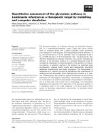
Figure 1 Heatmap of differentially expressed genes between CR (n = 9) and NCR (n = 12) samples obtained from supervised
hierarchical clustering using Euclidean distances and Ward method. The color indicates the level of mRNA expression: red - higher level of
expression, green - lower level of expression, black – no expression changes (each row represents a gene and each column represents a sample).
The CR samples were clustered together and clearly separated from NCR samples.
Assessment of the prognostic significance of genes
involved in ‘Role of BRCA1 in DNA Damage Response’
pathway
We estimated the prognostic significance of the six selected genes by the ROC analysis. We analyzed the ROC
curves for all previously known potential factors, including
age, tumor size, hemoglobin, along with our potential
markers: BRCA1, BRCA2, RAD51, FANCD2, BLM and
BRIP1. If the p-value was not significant (p > 0.05), then the
AUC, sensitivity, specificity and optimal classification point
were omitted. The investigated genes discriminated between the patients in the NCR and CR groups (p < 0.01)
suggesting a superior predictive value compared to classical
factors such as tumor size and hemoglobin. The summary
Balacescu et al. BMC Cancer 2014, 14:246
/>
Page 7 of 14
Table 3 The top significant molecular and cellular
functions identified by IPA
Molecular and cellular functions
p value
No. genes
Cellular movement
5.30E-08-1.22E-02
344
Cell cycle
7.12E-07-1.22E-02
233
DNA replication, recombination
and Rrepair
7.12E-07-1.18E-02
124
Cellular development
1.16E-06-1.22E-02
433
Cellular assembly
and organization
4.97E-06-1.22E-02
322
Canonical pathways
p value
Ratio
Role of BRCA1 in DNA
damage response
1.86E-04
17/65 (0.262)
Primary immunodeficiency
signaling
3.97E-03
11/62 (0.177)
G protein signaling
mediated by Tubby
5.05E-03
9/42 (0.214)
Aryl hydrocarbon
Rreceptor signaling
5.39E-03
25/161 (0.155)
Regulation of actin-based
motility by Rho
7.5E-03
17/89 (0.191)
of the ROC curves (AUC, specificity and sensitivity) for all
six genes is presented in Table 6.
The correlations between the target genes BRCA1,
BRCA2, RAD51, FANCD2, BLM and BRIP1 indicated that
all genes were highly correlated with each other. The correlation coefficients were between 0.69 (BRCA2 vs. BRIP1)
and 0.93 (BRCA1 vs. BRIP1) (Figure 4).
IHC validation of the microarray results
Immunohistochemical staining was performed to obtain
further validation of microarray findings. We assessed the
protein expression of RAD51, BRIP1 (BACH1), BRCA1,
BRCA2, BLM and FANCD2 in all 21 samples used in the
genomic study (Figure 5). For BLM gene we did not identified a specific monoclonal antibody (MoAb), therefore
this gene could not be taken into account for protein validation. An average percentage of nuclear staining on 3
different representative images of every sample was calculated for every protein of interest. We observed a significantly increased protein levels of FANCD2, BRCA1,
RAD51 and BRIP1 in the nuclei of the NCR compared to
the CR cervical tumors. No difference was observed for
nuclear protein expression of BRCA2 in NCR compared
to CR tissues. A ratio between nuclear protein expressions
in NCR and CR groups was calculated (Table 7).
An additional set of 24 FFPE squamous cervical samples
(15 CR and 9 NCR) was used as an independent validation
of the protein data. Increased protein levels of FANCD2,
RAD51, BRCA1, and BRIP 1 (BACH1) in NCR compared
to CR cervical tumors groups were confirmed on the validation set (Table 7).
Discussion
Cervical cancer continues to represent a major health problem for women from developing countries. Cervical cancer
lethality occurs because most patients are first diagnosed in
advanced stages. Even if early stages are successfully treated,
Table 4 Genes involved in the “Role of BRCA1 in DNA Damage Response” pathway with associated p-values obtained
from microarray experiment
Ref seq
Gene symbol
Fold regulation (NCR vs CR)
Adjusted p-value
Description
ATF1
1,747
0,013
activating transcription factor 1
NM_000057
BLM
2,430
0,030
Bloom syndrome, RecQ helicase-like
NM_007300
BRCA1
2,225
0,008
breast cancer 1, early onset
NM_000059
BRCA2
1,842
0,011
breast cancer 2, early onset
NM_001018055
BRCC3
1,621
0,016
BRCA1/BRCA2-containing complex, subunit 3
NM_032043
BRIP1
2,353
0,018
BRCA1 interacting protein C-terminal helicase 1
NM_004091
E2F2
2,137
0,048
E2F transcription factor 2
NM_001950
E2F4
−1,791
0,013
E2F transcription factor 4, p107/p130-binding
NM_005171
NM_001018113
FANCB
2,216
0,010
Fanconi anemia, complementation group B
FANCD2
1,613
0,012
Fanconi anemia, complementation group D2
NM_018062
FANCL
1,701
0,031
Fanconi anemia, complementation group L
NM_020937
FANCM
1,923
0,005
Fanconi anemia, complementation group M
helicase-like transcription factor
NM_033084
NM_139048
HLTF
2,245
0,016
NM_002875
RAD51
2,767
0,010
RAD51 homolog (S. cerevisiae)
NM_002913
RFC1
1,723
0,004
replication factor C (activator 1) 1, 145 kDa
NM_139045
SMARCA2
1,659
0,041
SWI/SNF related, matrix associated, actin dependent
regulator of chromatin, subfamily a, member 2
NM_003072
SMARCA4
−1,519
0,026
SWI/SNF related, matrix associated, actin dependent
regulator of chromatin, subfamily a, member 4
Balacescu et al. BMC Cancer 2014, 14:246
/>
Page 8 of 14
Figure 2 Activation of the “Role of BRCA1 in DNA Damage Response’ pathways in NCR versus CR samples. Genes highlighted in red
were significantly overexpressed in non-responsive compared with responsive cervical cancers.
advanced cervical cancer represents a major problem due
to increased rates of recurrence and distant metastasis. Although knowledge about tumor biology and various mechanisms of resistance has increased in recent years, different
schedules of treatment, including new anticancer drugs,
have not efficiently reduced the occurrence of drug resistance. Intrinsic resistance is often decisive for treatment failure; almost half of patients present with baseline resistance,
rendering classical therapies ineffective.
In an effort to elucidate the patterns of genes involved in
baseline resistance, we performed a genome-wide microarray assay on primary biopsies from patients with advanced
cervical cancers with known clinical and histological responses. All of the patients included in the study received
radiotherapy as the main therapy and cisplatin as a radiosensitizer Based on the microarray analysis, we identified a
supervised gene expression profile that differed dramatically
between the non-responding and responding cervical tumors. ‘DNA Replication, Recombination and Repair’
represents one of the most important molecular patterns
identified as important for intrinsic resistance in cervical
cancer. In our study, the non-responding cervical tumor
cells had more active DNA damage repair machinery than
responding cervical tumor cells, even before starting the
therapy. In total, 92 out of the 124 identified genes involved in ‘DNA replication, recombination and repair’
were overexpressed in the non-responding tumors compared with the responding tumors (Additional file 1).
Balacescu et al. BMC Cancer 2014, 14:246
/>
Page 9 of 14
Figure 3 qRT-PCR validation data for six genes (FANCD2, RAD51, BRCA2, BRCA1, BRIP1/BCH1 and BML) involved in the ‘Role of BRCA1
in DNA Damage Response’ pathway. Fold change was calculated using the ΔΔCt method relative to the CR group.
Cancer cells become resistant to therapy by restoring
DNA repair genes; therefore, we looked for pathways involved in the maintenance of DNA stability. By classifying
the genes according to functional pathways, we identified
the ‘Role of BRCA1 in DNA Damage Response’ as the
most important canonical pathway involved in DNA repair (Table 3). To our knowledge, there are no studies that
describe ‘Role of BRCA1 in DNA Damage Response’ pathway as predictive for treatment outcome in cervical cancer,
even though a conserved pathway for increased DNA repair mediated by BRCA1 was described for other pathologies [14,15]. Among the genes significantly up-regulated in
the BRCA1 canonical pathway, we focused our attention
on a set of six genes that were considered of particular
interest: BRCA1, BRCA2, RAD51, FANCD2, BACH1/
BRIP1/FANCJ and BLM. The expression of these genes
detected by microarray was confirmed by qRT-PCR with
good correlation (Table 5).
Early studies on BRCA1 and BRCA2 have reveled that
both proteins are involved in DSB repair. In this study,
Table 5 Pearson’s correlation coefficients of log2 fold
change values obtained from microarray and PCR
experiments
we showed that BRCA1 and BRCA2 overexpression in patients with advanced cervical cancer is associated with
treatment failure. Several studies have pointed out that
BRCA-deficient cells are inefficient at repairing DNA damage by homologous recombination (HR) [16,17] and are
thus more sensitive to chemotherapeutic drugs. Zhang
et al. [18] reported that the E6 and E7 HPV oncoproteins
interact with BRCA1 and alter its activity in cervical cancer
cells. However, the association between high-risk HPV genotypes and treatment failure could not be evaluated in
our study as our sample set did not comprise a sufficient
number of other high-risk types. Recently, a so-called
BRCAness gene expression profile has also been correlated
with response to chemotherapy and outcome in patients
with epithelial ovarian cancer [19]. BRCA1 is a component
of the BASC complex that is important for efficient DNA
Table 6 ROC analysis for prognostic factors
Nr.crt. Variable AUC Classification Sensitivity Specificity
point
1.
BRCA1
0.81
<0.858565
0.92
0.78
p
<0.01
2.
BRCA2
0.86
<0.602903
0.75
0.89
<0.01
3.
RAD51
0.81
<0.895025
0.91
0.78
<0.01
4.
FANCD2
0.84
<0.673616
0.83
0.78
<0.01
BLM
0.81
>0.871154
0.63
0.99
<0.01
Pearson coefficient
p
5.
BLM
0.835
< 0.0001
6.
BRIP1
0.81
>0.606256
0.78
0.75
<0.01
BRIP1
0.811
< 0.0001
7.
0.86
>46
0.75
0.89
<0.01
BRCA1
0.765
< 0.0001
Age
(years)
BRCA2
0.721
0.0002
RAD51
0.705
0.0005
FANCD2
0.759
< 0.0001
Gene
8.
Tumor 0.65
size (cm)
-
-
-
0.11
(NS)
9.
Hb (g/dl) 0.64
-
-
-
0.39
(NS)
Balacescu et al. BMC Cancer 2014, 14:246
/>
Page 10 of 14
Figure 4 Pearson correlations between fold change values of the target genes.
repair. MSH2/MSH6, PMS2/MLH1, BLM helicase and the
replication factor C (RFC) represent other important members of the BASC complex [20].
Our microarray data pointed out an increased level of
BLM and RFC1 in the non-responding cervical cancers
compared with the responding cancers. Additionally, BRCA1
associates with the SWI/SNF chromatin-remodeling
complex and FANCD2 [21] and plays a role in regulating the cellular localization of BACH1/BRIP1 (BRCA1associated carboxyl-terminal helicase). BRCA2 is also
involved in DNA repair; the protein interacts specifically with RAD51, an essential protein involved in HR
[22]. In our efforts to understand the molecular basis of
treatment response in advanced cervical cancer, besides the
BRCA pathway, we also found the fanconi anemia (FA)
complementation group, FANCD2, FANCL, FANCM,
FANCJ/BRIP1/BACH and FANCI, to be involved in intrinsic resistance to chemo-radiotherapy. These FA proteins
are closely related to the BRCA1 and BRCA2 gene products and their partner proteins and are required for cellular
resistance to agents that cause DNA interstrand cross-links
(ICLs) [23]. The FANCD2 protein colocalizes to nuclear
foci together with BRCA1, BRCA2 and RAD51 and initiates homology-directed DNA repair in a “FA/BRCA
pathway,’ both in response to DNA-damaging agents (cisplatin, ionizing radiation, hydroxyurea, etc.) and in the absence of exogenous DNA damage during the S phase of the
cell cycle [24].
Our results revealed an increased protein level of
FANCD2, RAD51, BRCA1 and BRIP1 in the NCR compared to CR cervical tumor nuclei. These observations
were also confirmed on an independent validation set, emphasizing the role of these four proteins in CRT resistance
(Table 7). Although we observed a 3.8-fold increase in
BRCA2 mRNA in NCR vs. CR cervical samples (qRT-PCR
data), there was no significant difference for BRCA2 protein between NCR and CR groups, which could be due to
either using an inadequate monoclonal antibody clone or
posttranscriptional modifications of the BRCA2 transcript.
A central step in the FA/BRCA pathway is the monoubiquitylation of FANCD2 and its translocation to chromatin
at the site of DNA damage [25]. The ubiquitylation of
FANCD2 is initiated by FANCM and is mediated by the
UBE2T (E2) enzyme and a multisubunit ubiquitin E3 ligase
that consists of eight FA proteins (FANCA/B/C/E/F/G/L/
M) [26]. FANCD2 can also be monoubiquitylated and
chromatin-loaded by the E3 ubiquitin ligase activity of
RAD18 in a FA-independent manner [27].
Balacescu et al. BMC Cancer 2014, 14:246
/>
Page 11 of 14
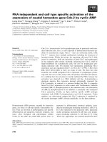
Figure 5 Validation of FANCD2 (A-B), RAD51 (C-D), BRCA1 (E-F), BRCA2 (G-H) and BRIP1 (I-J) protein expression in advanced squamous
cervical tumor cells. Staining for FANCD2, RAD51, BRCA1 and BRIP1 for the NCR cervical samples indicated strongly positive protein expression
compared with the CR cervical samples. The BRCA2 protein expression was comparable between the NCR and CR cervical samples;
(x200 magnification).
Increased expression of FANCM, FANCL, UBE2T and
RAD18 mRNA was observed in NCR compared to CR
cervical tumor samples (Additional file 1). We also observed a slight increase in SLX4/FANCP/BTBD12 mRNA
(fold change = 1.3, p = 0.035) for the NCR vs. CR cervical
samples. SLX4 is a novel member of the FA genes that coordinates multiple DNA repair pathways by acting as a
scaffold for multiple nucleases involved in ICL repair and
the mechanisms involved in HR [28]. Depletion of SLX4
leads to hypersensitivity to cisplatin and reduced efficiency
of HR repair [29]. Narayan et al. [30] showed that advanced
cervical cancer is associated with alterations in the FA/
BRCA pathway by either promoter hypermethylation and/
or deregulated gene expression compared with the normal
cervix. FA inhibitors were recently proposed as important
tools for overcoming cisplatin resistance in tumors [31].
RAD51, one of the key molecules of DNA repair, is another gene we found to be significantly overexpressed in
Balacescu et al. BMC Cancer 2014, 14:246
/>
Page 12 of 14
Table 7 Assessment of the nuclear proteins including
FANCD2, RAD51, BRCA2, BRCA1, BRIP1(BACH1) in NCR
and CR cervical tumors evaluated both for genomics
study set (n = 21) and IHC validation set (n = 24)
Proteins
Cellular
NCR % CR % NCR/CR ratio p-value
localization
Genomics study
set (n = 21)
FANCD2
nucleus
32.53
15.6
2.09
0.032
RAD51
nucleus
BRCA2
nucleus
14.88
7.11
2.09
0.016
21.11
20.81
1.01
BRCA1
0.868
nucleus
27.78
10.69
2.60
0.032
BRIP1 (BACH1)
nucleus
40.44
10.05
4.02
0.001
FANCD2
nucleus
25.34
16.22
1.56
0.011
RAD51
nucleus
16.87
4.86
3.47
0.000
BRCA1
nucleus
27.59
12.97
2.13
0.000
BRIP1 (BACH1)
nucleus
30.73
14.55
2.11
0.011
IHC validation
set (n = 24)
The p-values in the bold format are statistically significant.
the NCR cervical cancers. We observed a 3.4-fold increase
in RAD51 mRNA (qRT-PCR data) and also an increased
protein expression of RAD51 in the nuclei of the NCR vs.
CR cervical cancers. RAD51 has anti-apoptotic activity in
tumor cells [32], and high expression of this protein is correlated with poor prognosis, resistance to ionizing radiation
and drug resistance [33]. RAD51 is essential in the HR
process of DNA repair, its expression being tightly controlled in normal healthy cells to maintain genomic stability [34]. RAD51 co-localizes with BRCA1 and FANCD2 in
S-phase specific nuclear foci and initiates homology-driven
repair activity. Several studies have shown that in cancer
cells, this molecule is overexpressed, leading to radio- and
chemoresistance [35,36]. Elevated levels of RAD51 have been
associated with increased invasiveness in breast cancer patients [37] and have been demonstrated to be an independent prognostic marker of survival in non-small cell lung
cancer patients [38]. A previous microarray study of paired
cervical tumor samples (pre- and post-chemoradiotherapy)
reported down-regulation of RAD51 after treatment [5], supporting the hypothesis that radiation sensitivity is facilitated
by a diminished DNA repair response. Targeting strategies
against this gene have been developed as possible anticancer
treatments; attempts to inhibit RAD51 have proven to
be successful in reducing treatment resistance in tumor
cells [39,40].
In a previous study, it was shown that poly(ADP-ribose)
polymerase (PARP) inhibitors could suppress the expression of BRCA1 and RAD51 [41]. The PARP family, especially PARP1 and PARP2, functions as DNA damage
sensors and recruits a variety of DNA repair proteins to
the site of damaged DNA [42]. In BRCA-positive breast
cancer, PARP inhibitors were found to increase the
cytotoxic effects of radiation and chemotherapy based on
the principle of synthetic lethality [43]. In our microarray
study, we noted a higher level of PARP2 mRNA in the
NCR vs. CR cervical cancer samples (Additional file 1).
Previous studies have shown that prognostic factors
including younger age [44], tumor size [45], anemia
(hemoglobin) and FIGO stage [46], are used to estimate
overall survival, disease-free survival and local control in
cervical cancer. Nevertheless, they not provide information
about the baseline resistance and the tumor heterogeneity.
Our data indicates age as significant factor for treatment
response, however the sample size is limited and no final
conclusions can be drawn. Taking into account our findings and the need to identify new valuable prognostic factors for baseline resistance, we suggest that if our results
are confirmed on a larger study, the assessment of nuclear
expression of FANCD2, RAD51, BRCA1 and BRIP1 proteins could represent a supplementary prognostic factor
that would better tailor the treatment for patients with advanced cervical cancer. These results could be the foundation for the development of new targeting strategies to
improve cervical cancer outcome.
Conclusions
Our data revealed high DNA repair machinery activity even
before starting radio-chemotherapy in NCR patients compared with CR patients. Therefore, our findings demonstrate that baseline FANCD2, RAD51, BRCA1 and BRIP1
nuclear protein expression could have an important role in
treatment failure in advanced squamous cervical cancer. To
our knowledge, this is the first study to demonstrate the
role of the FA/BRCA pathway in baseline resistance and
therapy failure in locally advanced cervical cancer.
Limitations
The limitation of this study is related to the small number
of samples even if we used an independent validation set
for protein data to increased confidence of these findings.
Larger studies have to confirm that the assessment of
these proteins could represent an important prognostic
factor that determines poor response to radiation and
chemotherapy for locally-advanced cervical cancers.
Additional file
Additional file 1: Genes involved in DNA replication,
recombination, and repair mechanisms.
Abbreviations
AUC: Area under curve; BT: Brachytherapy; CR: Complete response;
CRT: Concomitant chemoradiotherapy; Cy3: Cyanine 3; DAB: Diamino-benzidine
tetrachloride; DSBs: DNA double-strand breaks; EBRT: External beam
radiotherapy; FDR: False discovery rate; FIGO: International Federation of
Gynecology and Obstetrics; HDR: High-dose-rate; HPV: Human papillomavirus;
IHC: Immunohistochemistry; IPA: Ingenuity pathway analysis; NCR: Non-complete
response; RIN: RNA integrity number; ROC: Receiver operating characteristic.
Balacescu et al. BMC Cancer 2014, 14:246
/>
Competing interests
The authors declare that there are no competing interests.
Authors’ contributions
OB designed and coordinated the research study, performed the microarray
experiment, interpreted the results and drafted the manuscript; LB performed
bioinformatic analysis of microarray data and drafted the manuscript; OT
performed qRT-PCR analysis and revised the manuscript; NT performed statistical
analysis; MR and SV processed the samples for microarray analysis; RB and SS
assessed the IHC staining; BF performed histopathological evaluation of the
samples; LP and LM processed the tissues for IHC staining; CO enrolled and
followed up patients, IBN interpreted the results and revised the manuscript,
VN enrolled and followed up patients and revised the manuscript. All authors
read and approved the final manuscript.
Acknowledgements
The authors acknowledge the financial support provided by the UEFISCDI
Program - PN-II-PT-PCCA-2011-3.2-1328 (grant no. 96/2012). Additional grants
including the National Grants Program –PNCDI2 (grant no. 42160/2008), the
National Grant Program PN-II- ID - PCE- 2008–2, CNCSIS 1532 and PN-II-RU-PD2011-3-0283, supported parts of this work. The authors thank to all of the
patients who agreed to enter this study.
Author details
1
The Oncology Institute ”Prof Dr. Ion Chiricuta”, 34-36 Republicii street,
400015 Cluj-Napoca, Romania. 2Iuliu Hatieganu, University of Medicine and
Pharmacy, 8 Babes street, 400012 Cluj-Napoca, Romania. 3Faculty of
Veterinary Medicine, University of Agricultural Sciences and Veterinary
Medicine, 3-5 Calea Manastur street, 400372 Cluj-Napoca, Romania.
Received: 12 August 2013 Accepted: 3 April 2014
Published: 8 April 2014
References
1. Jemal A, Bray F, Center MM, Ferlay J, Ward E, Forman D: Global cancer
statistics. CA Cancer J Clin 2011, 61:69–90.
2. Lippert TH, Ruoff HJ, Volm M: Intrinsic and acquired drug resistance in
malignant tumors. The main reason for therapeutic failure.
Arzneimittelforschung 2008, 58:261–264.
3. American Cancer Society: Cancer Facts & Figures 2012. Atlanta: American
Cancer Society; 2012.
4. Branzei D, Foiani M: Regulation of DNA repair throughout the cell cycle.
Nat Rev Mol Cell Biol 2008, 9:297–308.
5. Zempolich K, Fuhrman C, Milash B, Flinner R, Greven K, Ryu J, Forbes A,
Kerlin K, Nichols RC, Gaffney DK: Changes in gene expression induced by
chemoradiation in advanced cervical carcinoma: a microarray study of
RTOG C-0128. Gynecol Oncol 2008, 109:275–279.
6. Wong YF, Selvanayagam ZE, Wei N, Porter J, Vittal R, Hu R, Lin Y, Liao J, Shih JW,
Cheung TH, Lo KW, Yim SF, Yip SK, Ngong DT, Siu N, Chan LK, Chan CS, Kong T,
Kutlina E, McKinnon RD, Denhardt DT, Chin KV, Chung TK: Expression genomics
of cervical cancer: molecular classification and prediction of radiotherapy
response by DNA microarray. Clin Cancer Res 2003, 9:5486–5492.
7. Chao A, Wang TH, Lai CH: Overview of microarray analysis of gene
expression and its applications to cervical cancer investigation. Taiwan J
Obstet Gynecol 2007, 46:363–373.
8. Livak KJ, Schmittgen TD: Analysis of relative gene expression data using
real-time quantitative PCR and the 2(−Delta Delta C(T)) method.
Methods 2001, 25:402–408.
9. Tuominen VJ, Ruotoistenmaki S, Viitanen A, Jumppanen M, Isola J:
ImmunoRatio: a publicly available web application for quantitative
image analysis of estrogen receptor (ER), progesterone receptor (PR),
and Ki-67. Breast Cancer Res 2010, 12:R56.
10. Rosner B: Fundamentals of biostatistics. 6th edition. Pacific Grove, CA:
Duxbury Press; 2006.
11. Zou KH, Liu A, Bandos AI, Ohno-Machado L, Rockette HE: Statistical evaluation of Diagnostic performance topics in ROC Analysis. Boca Raton, Florida:
Chapman & Hall/CRC Press; 2012.
12. Bamber D: The area above the ordinal dominance graph and the area below
the receiver operating characteristics graph. J Math Psychol 1975, 12:387–415.
13. Hanley JA, McNeil BJ: The meaning and use of the area under a receiver
operating characteristic (ROC) curve. Radiol 1982, 143:29–36.
Page 13 of 14
14. Kauffmann A, Rosselli F, Lazar V, Winnepenninckx V, Mansuet-Lupo A,
Dessen P, van den Oord JJ, Spatz A, Sarasin A: High expression of DNA
repair pathways is associated with metastasis in melanoma patients.
Oncogene 2008, 27:565–573.
15. Sanchez-Carbayo M, Socci ND, Lozano J, Saint F, Cordon-Cardo C: Defining
molecular profiles of poor outcome in patients with invasive bladder
cancer using oligonucleotide microarrays. J Clin Oncol 2006, 24:778–789.
16. Rigakos G, Razis E: BRCAness: finding the Achilles heel in ovarian cancer.
Oncologist 2012, 17:956–962.
17. Roy R, Chun J, Powell SN: BRCA1 and BRCA2: different roles in a common
pathway of genome protection. Nat Rev Cancer 2012, 12:68–78.
18. Zhang Y, Fan S, Meng Q, Ma Y, Katiyar P, Schlegel R, Rosen EM: BRCA1
interaction with human papillomavirus oncoproteins. J Biol Chem 2005,
280:33165–33177.
19. Wysham WZ, Mhawech-Fauceglia P, Li H, Hays L, Syriac S, Skrepnik T, Wright J,
Pande N, Hoatlin M, Pejovic T: BRCAness profile of sporadic ovarian cancer
predicts disease recurrence. PLoS One 2012, 7:e30042.
20. Wang Y, Cortez D, Yazdi P, Neff N, Elledge SJ, Qin J: BASC, a super complex
of BRCA1-associated proteins involved in the recognition and repair of
aberrant DNA structures. Genes Dev 2000, 14:927–939.
21. Kerr P, Ashworth A: New complexities for BRCA1 and BRCA2. Curr Biol
2001, 11:R668–R676.
22. Jensen RB, Ozes A, Kim T, Estep A, Kowalczykowski SC: BRCA2 is epistatic
to the RAD51 paralogs in response to DNA damage. DNA Repair (Amst)
2013, 12:306–311.
23. Kim H, D’Andrea AD: Regulation of DNA cross-link repair by the Fanconi
anemia/BRCA pathway. Genes Dev 2012, 26:1393–1408.
24. Wang W: Emergence of a DNA-damage response network consisting of
Fanconi anaemia and BRCA proteins. Nat Rev Genet 2007, 8:735–748.
25. Alpi AF, Patel KJ: Monoubiquitylation in the Fanconi anemia DNA
damage response pathway. DNA Repair (Amst) 2009, 8:430–435.
26. Kelsall IR, Langenick J, MacKay C, Patel KJ, Alpi AF: The Fanconi anaemia
components UBE2T and FANCM are functionally linked to nucleotide
excision repair. PLoS One 2012, 7:e36970.
27. Williams SA, Longerich S, Sung P, Vaziri C, Kupfer GM: The E3 ubiquitin
ligase RAD18 regulates ubiquitylation and chromatin loading of FANCD2
and FANCI. Blood 2011, 117:5078–5087.
28. Svendsen JM, Smogorzewska A, Sowa ME, O’Connell BC, Gygi SP, Elledge SJ,
Harper JW: Mammalian BTBD12/SLX4 assembles a Holliday junction
resolvase and is required for DNA repair. Cell 2009, 138:63–77.
29. Fekairi S, Scaglione S, Chahwan C, Taylor ER, Tissier A, Coulon S, Dong MQ,
Ruse C, Yates JR 3rd, Russell P, Fuchs RP, McGowan CH, Gaillard PH: Human
SLX4 is a Holliday junction resolvase subunit that binds multiple DNA
repair/recombination endonucleases. Cell 2009, 138:78–89.
30. Narayan G, Arias-Pulido H, Nandula SV, Basso K, Sugirtharaj DD, Vargas H,
Mansukhani M, Villella J, Meyer L, Schneider A, Gissmann L, Dürst M,
Pothuri B, Murty VV: Promoter hypermethylation of FANCF: disruption of Fanconi Anemia-BRCA pathway in cervical cancer. Cancer Res 2004, 64:2994–2997.
31. Jacquemont C, Simon JA, D’Andrea AD, Taniguchi T: Non-specific chemical
inhibition of the Fanconi anemia pathway sensitizes cancer cells to
cisplatin. Mol Cancer 2012, 11:26.
32. Henning W, Sturzbecher HW: Homologous recombination and cell cycle
checkpoints: Rad51 in tumour progression and therapy resistance.
Toxicology 2003, 193:91–109.
33. Du LQ, Wang Y, Wang H, Cao J, Liu Q, Fan FY: Knockdown of Rad51
expression induces radiation- and chemo-sensitivity in osteosarcoma
cells. Med Oncol 2011, 28:1481–1487.
34. Richardson C: RAD51, genomic stability, and tumorigenesis. Cancer Lett
2005, 218:127–139.
35. Tennstedt P, Fresow R, Simon R, Marx A, Terracciano L, Petersen C,
Sauter G, Dikomey E, Borgmann K: RAD51 overexpression is a negative
prognostic marker for colorectal adenocarcinoma. Int J Cancer 2013,
132:2118–2126.
36. Hannay JA, Liu J, Zhu QS, Bolshakov SV, Li L, Pisters PW, Lazar AJ, Yu D,
Pollock RE, Lev D: Rad51 overexpression contributes to chemoresistance
in human soft tissue sarcoma cells: a role for p53/activator protein 2
transcriptional regulation. Mol Cancer Ther 2007, 6:1650–1660.
37. Barbano R, Copetti M, Perrone G, Pazienza V, Muscarella LA, Balsamo T,
Storlazzi CT, Ripoli M, Rinaldi M, Valori VM, Latiano TP, Maiello E, Stanziale P,
Carella M, Mangia A, Pellegrini F, Bisceglia M, Muda AO, Altomare V, Murgo
R, Fazio VM, Parrella P: High RAD51 mRNA expression characterize
Balacescu et al. BMC Cancer 2014, 14:246
/>
38.
39.
40.
41.
42.
43.
44.
45.
46.
Page 14 of 14
estrogen receptor-positive/progesteron receptor-negative breast cancer
and is associated with patient's outcome. Int J Cancer 2011, 129:536–545.
Qiao GB, Wu YL, Yang XN, Zhong WZ, Xie D, Guan XY, Fischer D, Kolberg HC,
Kruger S, Stuerzbecher HW: High-level expression of Rad51 is an
independent prognostic marker of survival in non-small-cell lung cancer
patients. Br J Cancer 2005, 93:137–143.
Russell JS, Brady K, Burgan WE, Cerra MA, Oswald KA, Camphausen K, Tofilon PJ:
Gleevec-mediated inhibition of Rad51 expression and enhancement of
tumor cell radiosensitivity. Cancer Res 2003, 63:7377–7383.
Hine CM, Seluanov A, Gorbunova V: Use of the Rad51 promoter for targeted
anti-cancer therapy. Proc Natl Acad Sci U S A 2008, 105:20810–20815.
Yelamos J, Schreiber V, Dantzer F: Toward specific functions of poly(ADPribose) polymerase-2. Trends Mol Med 2008, 14:169–178.
Wang X, Weaver DT: The ups and downs of DNA repair biomarkers for
PARP inhibitor therapies. Am J Cancer Res 2011, 1:301–327.
Sandhu SK, Yap TA, de Bono JS: The emerging role of poly (ADP-Ribose)
polymerase inhibitors in cancer treatment. Curr Drug Targets 2011,
12:2034–2044.
Fyles AW, Pintilie M, Kirkbride P, Levin W, Manchul LA, Rawlings GA:
Prognostic factors in patients with cervix cancer treated by radiation
therapy: results of a multiple regression analysis. Radiother Oncol 1995,
35:107–117.
Narayan K, Fisher R, Bernshaw D: Significance of tumor volume and
corpus uteri invasion in cervical cancer patients treated by radiotherapy.
Int J Gynecol Cancer 2006, 16:623–630.
Grigiene R, Valuckas KP, Aleknavicius E, Kurtinaitis J, Letautiene SR: The
value of prognostic factors for uterine cervical cancer patients treated
with irradiation alone. BMC Cancer 2007, 7:234.
doi:10.1186/1471-2407-14-246
Cite this article as: Balacescu et al.: Gene expression profiling reveals
activation of the FA/BRCA pathway in advanced squamous cervical
cancer with intrinsic resistance and therapy failure. BMC Cancer
2014 14:246.
Submit your next manuscript to BioMed Central
and take full advantage of:
• Convenient online submission
• Thorough peer review
• No space constraints or color figure charges
• Immediate publication on acceptance
• Inclusion in PubMed, CAS, Scopus and Google Scholar
• Research which is freely available for redistribution
Submit your manuscript at
www.biomedcentral.com/submit