Nucleostemin expression in invasive breast cancer
Bạn đang xem bản rút gọn của tài liệu. Xem và tải ngay bản đầy đủ của tài liệu tại đây (799.16 KB, 9 trang )
Kobayashi et al. BMC Cancer 2014, 14:215
/>
RESEARCH ARTICLE
Open Access
Nucleostemin expression in invasive breast
cancer
Takayuki Kobayashi1,2, Kenkichi Masutomi3, Kenji Tamura4, Tomoyuki Moriya5, Tamio Yamasaki5, Yasuhiro Fujiwara4,
Shunji Takahashi2, Junji Yamamoto5 and Hitoshi Tsuda1,6*
Abstract
Background: Recently, the cancer stem cell hypothesis has become widely accepted. Cancer stem cells are
thought to possess the ability to undergo self-renewal and differentiation, similar to normal stem cells.
Nucleostemin (NS), initially cloned from rat neural stem cells, binds to various proteins, including p53, in the nucleus
and is thought to be a key molecule for stemness. NS is expressed in various types of cancers; therefore, its role in
cancer pathogenesis is thought to be important. This study was conducted to clarify the clinicopathological and
prognostic impact of NS in invasive breast cancers.
Method: The correlation between NS immunoreactivity and clinicopathological parameters was examined in 220
consecutive surgically resected invasive breast cancer tissue samples by using tissue microarrays. The presence of
nuclear NS and p53 immunoreactivity in 10% or more of cancer cells was considered as a positive result.
Results: Among the 220 patients, 154 were hormone-receptor (HR)-positive, 22 HER2-positive/HR-negative, and 44
HR-negative/HER2-negative. One hundred and forty-two tumors (64.5%) showed NS positivity, and this positivity
was significantly correlated with estrogen receptor (ER) (P = 0.050), human epidermal growth factor receptor 2
(HER2) (P = 0.021), and p53 (P = 0.031) positivity. The patients with NS-positive tumors showed significantly shorter
disease-free survival than those with NS-negative tumors. Furthermore, the patient group with NS- and p53-positive
tumors showed significantly poorer prognosis than other patient groups. Multivariate analysis showed that NS status
was an independent prognostic indicator.
Conclusions: NS may play a significant role in the determination of breast cancer progression in association with p53
alterations. The NS status of patients with luminal and HER2 type breast cancers may be a useful prognostic marker.
Background
Breast cancer is one of the most prevalent diseases worldwide. While most patients with early breast cancers are
cured with surgically resection followed by appropriate adjuvant drug and radiation therapy, approximately 30% of
these patients experience relapse and develop metastatic
disease [1]. In this metastatic stage, tumor cells frequently
acquire resistance to various drugs during intensive systemic therapies, and eventually their aggressiveness and
growth become uncontrollable. Less than 5% of patients
with distant metastatic tumors live for 5 years [2].
* Correspondence:
1
Department of Basic Pathology, National Defense Medical College, 3-2
Namiki, Tokorozawa, Saitama 359-8513, Japan
6
Department of Pathology and Clinical Laboratories, National Cancer Center
Hospital, 5-1-1 Tsukiji, Chuo-ku, Tokyo 104-0045, Japan
Full list of author information is available at the end of the article
Therefore, identification of potential targets with the
aim of developing interventional drugs is an important
area of research.
The hypothesis that various types of cancers, including
breast cancer, are generated by a limited number of cancer
stem cells has been widely accepted recently [3]. Cancer
stem cells, like normal stem cells, are thought to have
two important characteristics: the ability to undergo selfrenewal and the ability to undergo differentiation into different cell types [4]. Furthermore, these cells are thought
to be inherently resistant to various therapeutic drugs,
making the eradication of tumors containing cancer stem
cells with the use of the current treatment protocols difficult [5]. To overcome these obstacles, the development of
new therapeutic strategies to target cancer stem cells is
essential for the management of breast cancer.
© 2014 Kobayashi et al.; licensee BioMed Central Ltd. This is an Open Access article distributed under the terms of the Creative
Commons Attribution License ( which permits unrestricted use, distribution, and
reproduction in any medium, provided the original work is properly credited. The Creative Commons Public Domain
Dedication waiver ( applies to the data made available in this article,
unless otherwise stated.
Kobayashi et al. BMC Cancer 2014, 14:215
/>
Nucleostemin (NS) is thought to be a key molecule for
maintaining “stemness” [6]. NS was initially cloned from
rat neural stem cells and was found to contain two
GTP-binding motifs and an N-terminal basic domain,
which is essential for binding to p53 [6]. NS accumulates
mainly in the nucleoli and moves to the nucleoplasm
after binding with GTP. Interaction of NS with a multitude of proteins in the nucleoplasm, including p53, may
play a significant role in self-renewal, cell cycle regulation, apoptosis, and cell proliferation [7].
NS is expressed in central nervous system stem cells,
embryonic stem cells, and primitive cells in the bone
marrow and testes [6]. Furthermore, various types of
cancers, including the following, have been reported to
express NS: squamous cell carcinomas of the uterine
cervix; head, neck, esophagus, and renal cell carcinomas;
and prostate cancer [8-13]. Moreover, recent evidence
indicates that NS is involved in maintaining cancer stem
cells [14,15]. These findings suggest that NS may also
play an important role in cancer pathogenesis as well as
in cancer stem cell maintenance. However, no clinical
study has investigated the role of NS in breast cancer.
If NS is expressed in breast cancer stem cells and its expression is correlated with disease progression in breast
cancer, it may serve as a powerful prognostic marker for
clinical use. To test this hypothesis, we investigated the expression of NS in surgically resected invasive breast cancer
specimens from 220 patients by using immunohistochemistry. Furthermore, we examined the prognostic implication of the combination status of NS and p53 and the
significance of NS expression status among the three biological subtypes of breast tumors: (a) hormone-receptor
(HR) positive (luminal type); (b) human epidermal growth
factor receptor 2 (HER2) positive (HER2 type); and (c) HR
negative and HER2 negative (triple negative).
Methods
Patients and tumor specimens
The patient cohort used in the present study was the
same as the cohort reported in our previous study [16].
Briefly, formalin-fixed paraffin-embedded tissue blocks
of invasive breast cancer specimens from 220 consecutive patients were used to construct tissue microarrays
(TMAs). All patients with unilateral invasive breast carcinoma underwent mastectomy or breast-conserving
surgery at the National Defense Medical College (NDMC)
Hospital, Tokorozawa, Japan from 1995 through 1999.
These patients had a median follow-up of 74 months after
surgery (range, 1–151 months), during which 58 patients
experienced relapse. Of the 220 patients, 218 were female
patients and 2 were male patients; 101 (45.9%) patients
had lymph node metastasis and 8 (3.6%) had distant metastasis at the time of breast cancer diagnosis. In most
cases, patients with hormone receptor-positive tumors at
Page 2 of 9
the time of diagnosis were prescribed adjuvant endocrine
therapy (e.g., tamoxifen, toremifene, fadrozole, or LHRH
analogues) for two or more years. The patients with a
large tumor and/or four or more lymph node metastases
received one of the following adjuvant chemotherapy
regimens: cyclophosphamide-epirubicin-5-fluorouracil
(CEF), cyclophosphamide-adriamycin-5-fluorouracil (CAF),
cyclophosphamide-methotrexate-5-fluorouracil (CMF),
and oral fluoropyrimidines. Detailed patient and disease characteristics are documented in Table 1. Clinicopathological data were retrospectively obtained from
medical records [16].
This study was approved by the Medical Ethical
Committee of National Defense Medical College and by
the Institutional Review Board of National Cancer Center.
Tissue microarray construction
We constructed TMA blocks as previously described [16].
Briefly, double tissue cores with a diameter of 2 mm were
obtained from each donor block, and these core specimens were transferred to a recipient block using a Tissue
Microarrayer (Beecher Instruments, Silver Spring, MD,
USA). One TMA block contained a maximum of 26
tumor samples, and 13 TMA sets were used in this study.
Immunohistochemistry
Immunohistochemistry was performed on TMA sections
of 220 patients. The antibodies used were mouse monoclonal anti-human NS (clone BL2858; Bethyl Laboratories, Inc., Montgomery, TX, USA) and mouse monoclonal
anti-human p53 (clone DO-7; Dako, Carpinteria, CA,
USA). Formalin-fixed paraffin-embedded specimens on
the TMA were cut into 4 μm-thick sections. The tissue
sections were deparaffinized twice in xylene for 10 min
and rehydrated through graded ethanol (99%, 90%, 80%,
and 70%) to water. Antigens were retrieved by microwave heating for 30 min in 10 mM sodium citrate
(pH 6.0) for NS and by autoclaving for 15 min in
10 mM Tris–HCl (pH 9.0) for p53. To block endogenous peroxidase activity, the sections were treated with
100% methanol containing 3% hydrogen peroxide for
5 min. Non-specific binding was blocked by incubation
in 2% normal swine serum (Dako) in phosphate-buffered
saline. The slides were incubated with primary antibodies at 4°C overnight and then reacted with a dextran
polymer reagent combined with secondary antibodies
and peroxidase (Envision Plus; Dako) for 30 min at room
temperature. Specific antigen–antibody reactions were
visualized with 0.2% diaminobenzidine tetrahydrochloride and hydrogen peroxide. Counterstaining was performed using Mayer’s hematoxylin. A separate assay was
run using a case of esophageal carcinoma as a positive
control for NS [17]. Reactions without the primary antibodies were used as negative controls.
Kobayashi et al. BMC Cancer 2014, 14:215
/>
Page 3 of 9
Table 1 Correlation between nucleostemin expression
and clinicopathological variables in surgically resected
breast cancers
Table 1 Correlation between nucleostemin expression
and clinicopathological variables in surgically resected
breast cancers (Continued)
Variable
Histological type
Number of cases (%)
Nucleostemin expression
Total
Positive
Negative
(n = 220)
(n = 142)
(n = 78)
P-value
Age
Median (range)
52 (30 ~ 82 y)
≦52
109
71 (65)
38
>52
111
71 (64)
40
<5.0 cm
174
108 (62)
66
≧5.0 cm
42
31 (74)
11
Unknown
4
4(100)
0
Negative
115
70 (61)
45
Positive
101
68 (67)
33
4
4 (100)
0
Negative
209
134 (64)
75
Positive
8
6 (75)
2
Unknown
3
2 (67)
1
I or II
179
111 (61)
68
III or IV
37
28 (76)
9
Unknown
4
3 (75)
1
1, 2
137
86 (63)
51
3
83
56 (67)
27
0.89
0.39
Distant metastasis
0.72
Stage
0.13
Nuclear grade
0.48
ER status
88
50 (57)
38
Positive
132
92 (70)
40
Negative
96
57 (59)
39
Positive
124
85 (69)
39
0.050
0.16
HR (ER/PgR) status
66
40 (61)
26
Positive
154
102 (66)
52
Negative
190
117 (62)
73
Positive
30
25 (83)
5
66
5 (50)
5
Mucinous
6
6 (100)
0
Tubular
5
1 (20)
4
Medullary
3
2 (67)
1
Other
5
3 (60)
2
0.38
NS and p53 expression was assessed according to the
proportion of nuclear staining area. Specimens with 10%
or more immunoreactive tumor cells were considered
positive, and those with less than 10% were considered
negative. Immunohistochemistry results were independently evaluated by two observers (T.K. and H.T.), and
cases with discrepant grades were re-evaluated by discussion until consensus was achieved.
ER, PgR, and HER2 had already been immunohistochemically re-assessed on new sections in our previous
study [16] by using mouse monoclonal anti-human ER
(clone 1D5, Dako), mouse anti-human PgR (clone PgR636,
DAKO), and rabbit polyclonal anti-HER2 antibody
(HercepTest kit, Dako) according to the methods recommended by the manufacturer. ER and PgR were considered positive if the nuclear staining was observed in 10%
or more of tumor cells. Samples were considered hormone
receptor positive if they were ER and/or PgR positive and
hormone receptor negative if they were ER and PgR negative. HER2 results were considered positive if the IHC
score was “3+” or gene amplification was detected by FISH
according to the 2007 ASCO/CAP guideline [18].
Statistical analysis
PgR status
Negative
125 (65)
10
0.21
Lymph node metastasis
Negative
191
Lobular
Abbreviation: ER Estrogen receptor, PgR Progesterone receptor, HR Hormone
receptor.
Tumor size
Unknown
Ductal
0.42
HER2 status
0.02
Comparisons between groups were evaluated using chisquared test or Fisher’s exact test. Disease-free survival (DFS) curves of patients were drawn using the
Kaplan-Meier method and compared using the log-rank
test. Cox multivariate proportional hazards models were
used to explore the association of variables with DFS. For
all tests, P < 0.05 was considered to be statistically significant. All analyses were performed using the software JMP
6.0 for Windows (SAS Institute Inc., Cary, NC, USA).
Results
p53 status
Negative
143
85 (59)
58
Positive
77
57 (74)
20
0.03
Clinicopathological and prognostic implications of NS
expression for the entire patient cohort
Initially, the expression levels of NS were classified as negative (0%), weak (1% to <10%), moderate (10% to <30%), or
strong (30% or more). The number of cases categorized
Kobayashi et al. BMC Cancer 2014, 14:215
/>
into the negative, weak, moderate, and strong groups was
62, 16, 55, and 87, respectively. From these results, we
judged that NS expression showed bimodal distribution
and used a 10% threshold for NS positivity between negative and positive groups.
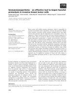
NS protein was frequently detected in the nucleus of
breast cancer cells. Although strong immunoreaction
was observed in both the nucleoli and nucleoplasm of
cancer cells (Figure 1A), nuclear immunoreaction of NS
in some cases was limited to the nucleoli of cancer cells
(Figure 1B). Such cells were also judged as positive for
NS immunoreactivity. Cytoplasmic staining was not observed. These findings are consistent with those of previous reports [6,17,19]. We found that 78 (35.5%) or 142
(64.5%) patients had NS-negative or NS-positive tumors,
respectively (Figure 1C). Unremarkable mammary glands
showed nuclear NS immunoreactivity in almost all luminal epithelial cells (Figure 1D).
Tumors with NS positivity showed a higher frequency
of ER positivity, HER2 positivity, and p53 positivity
(P = 0.050, P = 0.021, and P = 0.031, respectively), whereas
NS expression status was not correlated with tumor size,
lymph node metastasis, distant metastasis, tumor nuclear grade, or PgR positivity. NS expression was detected at 50% or more in all histological types studied
except tubular carcinoma (20%), and the positive rate
was 100% (6 of 6) in mucinous carcinoma. Patients with
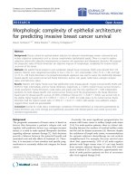
NS-positive tumors showed significantly shorter DFS
Page 4 of 9
time than those with NS-negative tumors (P = 0.020,
Figure 2).
Prognostic implication of the NS and p53 combination
status for the entire patient cohort
Since it has been reported that physical and functional
interaction between NS and p53 appear to be essential
for self-renewal, cell cycle regulation, cell proliferation,
and apoptosis [7], we next examined the prognostic implication of the combination status of NS and p53 for
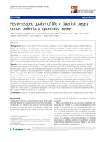
the entire patient cohort. We found that 143 (65%) and
77 (35%) patients had p53-negative and p53-positive tumors, respectively. The patients with p53-positive tumors showed significantly shorter DFS time than those
with p53-negative tumors (P = 0.006, Figure 3A).
A striking stratification of relapse risk was identified
when three different combinations of NS and p53
status were evaluated: 57 cases with a combination of
NS-positive/p53-positive tumors (unfavorable group); 105
cases comprising 20 NS-negative/p53-positive tumors and
85 NS-positive/p53-negative tumors (intermediate group);
and 58 cases with NS-negative/p53-negative tumors
(favorable group). The unfavorable group had a 5-year
DFS rate of 55%, compared with 75% in the intermediate
group and 86% in the favorable group (Figure 3B). The unfavorable group had significantly shorter DFS time than
the intermediate and favorable groups (log-rank test
P = 0.034 and P = 0.0007, respectively).
A
B
C
D
Figure 1 Nucleostemin (NS) expression in human breast cancer tissues. A. A NS-positive tumor. Almost all cancer cells show NS immunoreactivity in both nucleoli and nucleoplasm. B. Another NS-positive tumor, NS immunoreactivity is limited to nucleoli in nuclei of cancer cells. Such
cancer cells are also judged as NS-positive. This case was also classified as NS-positive. C. A NS-negative tumor. D. An unremarkable mammary
gland shows nuclear NS immunoreactivity in almost all luminal epithelial cells.
Kobayashi et al. BMC Cancer 2014, 14:215
/>
Page 5 of 9
Prognostic implication of NS among the three biological
subtypes of breast tumors
1.0
Probability of DFS
Negative NS (n = 78)
Currently, treatment strategies differ between the biological subtypes of breast tumors; therefore, we examined the prognostic implication of NS among three
groups of patients divided based on their biological subtype: 154 patients with luminal-type tumors (HR-positive); 22 patients with HER2-type tumors (HER2-positive
and HR-negative); and 44 patients with triple-negative
tumors (HR and HER2-negative). Eight patients with
HR-positive and HER2-positive tumors were included
and analyzed as luminal-type patients.
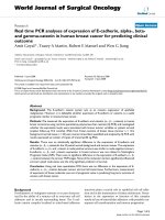
Among the patients with luminal-type tumors, patients with NS-positive tumors showed a significantly
shorter DFS time than those with NS-negative tumors
(P = 0.033, Figure 4A). Among the patients with HER2positive tumors, patients with NS-positive tumors had a
5-year DFS rate of 28% compared with 100% in patients
with NS-negative tumors (Figure 4B). However, the
P-value was not calculated because there was no relapse
0.8
0.6
Positive NS (n = 142)
0.4
0.2
log-rank P = 0.020
0.0
0
20
40
60
80
100
120
140
160
Months
Figure 2 Prognostic impact of NS status in the patients with
primary breast cancer. This figure shows disease-free survival (DFS)
curves for the 142 patients with NS-positive tumors and for the
78 patients with NS-negative tumors. These two curves differ
significantly (P = 0.020).
A
1.0
Probability of DFS
Negative p53 (n = 143)
0.8
0.6
Positive p53 (n = 77)
0.4
0.2
(log-rank P = 0.006)
0.0
0
20
40
60
80
100
120
140
160
Months
B
‘Favorable’ (n = 58)
(Negative NS + Negative p53)
Probability of DFS
1.0
0.8
‘Mixed’ (n =105)
Positive NS + Negative p53
or
Negative NS + Positive p53
0.6
‘Unfavorable’ (n = 57)
(Positive NS + Positive p53)
0.4
0.2
0.0
0
20
40
60
80
100
120
140
160
Months
Figure 3 Prognostic impact of the combination status of NS and p53 in the patients with primary breast cancer. A. Disease-free survival
(DFS) curves for the 77 patients with p53-positive tumors and for the 143 patients with p53-negative tumors. These two curves differ significantly
(P = 0.006). B. This figure shows three disease-free survival (DFS) curves: for the 57 patients with NS-positive and p53-positive tumors (‘unfavorable
group’), for 105 patients comprising 20 NS-negative and p53-positive tumors and 85 NS-positive and p53-negative tumors (‘intermediate’ group),
and for 58 patients with NS-negative and p53-negative tumors (‘favorable’ group). The unfavorable group has significantly shorter DFS time than
the intermediate group or the favorable group (log-rank test P = 0.034 and P = 0.0007, respectively).
Kobayashi et al. BMC Cancer 2014, 14:215
/>
A
HR positive patients (n = 154)
1.0
Probability of DFS
Page 6 of 9
Negative NS (n = 52)
0.8
Positive NS (n = 102)
0.6
0.4
0.2
log-rank P = 0.033
0.0
0
20
40
60
80
100
120
140
160
Figure 4 Prognostic impact of NS status in the three subgroups
of the different biological subtype tumors: luminal-type
tumors, HER2-type tumors, and triple-negative tumors.
A. Subgroup analysis of the 154 patients with luminal-type tumors
(HR-positive tumors). Two disease-free survival (DFS) curves, that for
the 102 patients with NS-positive tumors and that for the 52 patients
with NS negative tumors, differ significantly (P = 0.033). B. Subgroup
analysis of the 22 patients with HER2-type tumors (HER2-positive
and HR-negative tumors). In two disease-free survival (DFS) curves,
that for the 18 patients with NS-positive tumors and that for the 4
patients with NS-negative tumors, five-year DFS rates differ largely
(100% vs 28%). P-value was not available because there was no
relapse in the patients with NS-negative tumors. C. Subgroup
analysis of the 44 patients with triple-negative tumors (HR and HER2
negative tumors). The two curves do not differ significantly (P = 0.41).
Months
in the four patients with NS-negative tumors. Among
the patients with triple-negative tumors, there was no
difference between the survival curves for patients with
NS-positive tumors and those with NS-negative tumors
(P = 0.41, Figure 4C).
HER2 positive patients (n = 22)
B
1.0
Probability of DFS
Negative NS (n = 4)
0.8
0.6
Multivariate analysis of prognostic factors and evaluation
of NS
0.4
0.2
Positive NS (n = 18)
0.0
0
20
40
60
80
100
120
140
Months
HR / HER2 negative patients (n = 44)
C
1.0
Univariate analysis showed that HR, HER2, nuclear
grade, tumor size, nodal status, distant metastasis, and
NS expression were significantly correlated with DFS.
When multivariate analysis was performed using these
seven factors, NS expression status was selected as an
independent prognostic factor (P = 0.036), together with
nuclear grade, tumor size, lymph node status, and distant
metastatic status (P = 0.0008, 0.0007, 0.0038 and <0.0001,
respectively; Table 2).
Probability of DFS
Positive NS (n = 22)
0.8
0.6
Negative NS (n = 22)
0.4
0.2
log-rank P = 0.41
0.0
0
20
40
60
80
Months
100
120
140
160
Discussion
In the present cohort, we found that the NS protein expression status was positively correlated with both ER
and HER2 status and was a powerful prognostic factor.
Patients with NS-positive breast tumors had a significantly shorter DFS time than those with NS-negative tumors (P = 0.020, Figure 2), and multivariate analysis for
DFS showed that NS positivity had an independent impact as a prognostic indicator among breast cancer patients (P = 0.036, Table 2). To our knowledge, this is the
first report to show the clinical implication of NS protein expression in invasive breast cancers.
Although several studies have shown the important
roles of NS in the pathogenesis of various cancer types
[8-13] as well as the maintenance of cancer stem cells
[14,15], no direct evidence is yet available to support
that NS is a marker of cancer stem cells. Currently, molecules such as CD44, CD133, ALDH1, and CXCR4 have
been found to be potential markers of cancer stem cells
[20-25]. Furthermore, the expression of these stem cell
markers has been shown to be a poor prognostic
Kobayashi et al. BMC Cancer 2014, 14:215
/>
Page 7 of 9
Table 2 Prognostic impacts of clinicopathological variables computed by Cox’s univariate and multivariate analyses in
patients with primary breast cancer
Univariate
Hazard ratio
Multivariate
(95% CI)
P-value
Hazard ratio
(95%CI)
P-value
Disease free survival
Nucleostemin
Hormone-receptor
HER2
Nuclear grade
Tumor size
Nodal status
Distant metastasis
Negative
1
Positive
2.06
Positive
1
Negative
1.73
Negative
1
Positive
2.91
1, 2
1
3
3.30
≦5.0 cm
1
>5.0 cm
6.89
Negative
1
Positive
4.51
Negative
1
Positive
71.6
(1.11-3.84)
(1.01-2.95)
(1.59-5.34)
(1.94-5.64)
(3.97-11.9)
(2.45-8.31)
(26.1-196.5)
0.023
0.045
0.0005
<0.0001
<0.0001
<0.0001
<0.0001
1
2.13
(1.05-4.33)
1
0.78
(0.39-1.52)
1
1.65
(0.80-3.41)
1
2.97
(1.57-5.61)
1
2.99
(1.59-5.66)
1
2.73
(1.38-5.38)
1
62.3
(15.5-251.1)
0.036
0.46
0.17
0.0008
0.0007
0.0038
<0.0001
Abbreviation: 95% CI 95% confidence interval.
indicator in several human cancer types [24,26-30].
Based on these observations, our results show that high
NS expression is a powerful indicator of poor outcome,
consistent with the idea that NS may be a breast cancer
stem cell marker.
The limitations of the present study included the
retrospective analyses and the heterogeneity of adjuvant
treatments. Therefore, one should pay careful attention
when interpreting these results. Further studies using a
uniformly treated patient cohort are required to clarify
the role of NS in breast cancer stem cells.
We found that the patient group with tumors coexpressing NS and p53 had shorter DFS times than the patient group with tumors negative for either NS or p53.
GTP binding modulates the movement of NS from the
nucleoli to the nucleoplasm; NS then binds p53 at its
N-terminal basic domain, which results in the suppression of p53 function [6,7]. Because prolongation of the
half-life of most of the mutated p53 protein induces its
nuclear accumulation, it is generally believed that the
p53 pathway does not fully function in tumors with high
p53 nuclear immunoreactivity [31-33]. This evidence
leads to the assumption that p53 function would be profoundly suppressed in tumors coexpressing NS and p53.
Our results show the validity of this concept and that
functional crosstalk between NS and p53 may also occur
in vivo.
Currently, we cannot explain the correlation between
NS expression and p53 expression. Although several
studies have shown that NS modulates the expression of
wild-type p53 [34,35], the role of NS in breast cancers
with mutant p53 has not yet been evaluated. Further research is needed to elucidate the correlation.
We found that the NS expression status was positively
correlated with both ER and HER2 status and also found
a significant prognostic implication of NS expression
for patients with luminal-type tumors and those with
HER2-type tumors, except for those with triple-negative
tumors. NS was first identified as a gene upregulated in
MCF-7 cells upon 17β-estradiol treatment [36]; therefore, our inclusion of subgroup analysis among patients
with luminal-type tumors was reasonable. To our knowledge, this is the first report to demonstrate the possible
association between NS and HER2. Zhang G et al.
showed that NS is required for the expression of EGF
and EGFR in an esophageal squamous carcinoma cell
line [13]. Presumably, NS is required for the expression
of HER2 in a manner similar to that for EGFR. We
found no survival impact of the NS expression status
among patients with triple-negative tumors, who show
higher rates of mutated p53 than patients with luminaltype or HER2-type tumors [37]. NS can function in the
presence of wide-type p53 [7]; therefore, the expression
status of NS may have survival impact only for the
luminal-type and HER2-type tumors.
Conclusions
In summary, our results indicate that the expression status of NS, abundant in stem cells, is a prognostic indicator in breast cancer patients, especially for those with
Kobayashi et al. BMC Cancer 2014, 14:215
/>
luminal-type or HER2-type tumors, and that the coexpression of NS and p53 correlates with poorer prognostic outcomes. Examination of NS expression may be
useful for the stratification and management of breast
cancer patients in future daily practice.
Competing interests
The authors declare no conflicts of interest.
Authors’ contributions
TK and HT conceived of the study, performed experiments, analyzed data
and wrote the manuscript. TM, TY, and JY provided samples, collected
clinical and pathological data. KM, KT, YF, and ST participated in designing
the study and revising the manuscript. HT participated in the overall design
and study coordination and finalized the draft of the manuscript. All authors
read and approved the final manuscript.
Acknowledgments
This work was supported in part by the Foundation for Promotion of
Defense Medicine and by the Cancer Research and Development Fund from
the National Cancer Center, Japan.
Author details
1
Department of Basic Pathology, National Defense Medical College, 3-2
Namiki, Tokorozawa, Saitama 359-8513, Japan. 2Department of Medical
Oncology, Cancer Institute Hospital, 3-8-31 Ariake, Koto-ku, Tokyo 135-8550,
Japan. 3Division of Cancer Stem Cell, National Cancer Center Research
Institute, Tsukiji, Chuo-ku, Tokyo 104-0045, Japan. 4Department of Breast
Oncology and Medical Oncology, National Cancer Center Hospital, 5-1-1
Tsukiji, Chuo-ku, Tokyo 104-0045, Japan. 5Department of Surgery, National
Defense Medical College, 3-2 Namiki, Tokorozawa, Saitama 359-8513, Japan.
6
Department of Pathology and Clinical Laboratories, National Cancer Center
Hospital, 5-1-1 Tsukiji, Chuo-ku, Tokyo 104-0045, Japan.
Received: 19 October 2013 Accepted: 11 March 2014
Published: 21 March 2014
References
1. O’Shaughnessy J: Extending survival with chemotherapy in metastatic
breast cancer. Oncologist 2005, 10(Suppl 3):20–29.
2. Pagani O, Senkus E, Wood W, Colleoni M, Cufer T, Kyriakides S, Costa A,
Winer EP, Cardoso F: International guidelines for management of
metastatic breast cancer: can metastatic breast cancer be cured?
J Natl Cancer Inst 2010, 102(7):456–463.
3. Clarke MF, Fuller M: Stem cells and cancer: two faces of eve. Cell 2006,
124(6):1111–1115.
4. Reya T, Morrison SJ, Clarke MF, Weissman IL: Stem cells, cancer, and cancer
stem cells. Nature 2001, 414(6859):105–111.
5. Dean M, Fojo T, Bates S: Tumour stem cells and drug resistance.
Nat Rev Cancer 2005, 5(4):275–284.
6. Tsai RY, McKay RD: A nucleolar mechanism controlling cell proliferation in
stem cells and cancer cells. Genes Dev 2002, 16(23):2991–3003.
7. Bernardi R, Pandolfi PP: The nucleolus: at the stem of immortality.
Nat Med 2003, 9(1):24–25.
8. Ye F, Zhou C, Cheng Q, Shen J, Chen H: Stem-cell-abundant proteins
Nanog, Nucleostemin and Musashi1 are highly expressed in malignant
cervical epithelial cells. BMC Cancer 2008, 8:108.
9. Fan Y, Liu Z, Zhao S, Lou F, Nilsson S, Ekman P, Xu D, Fang X: Nucleostemin
mRNA is expressed in both normal and malignant renal tissues.
Br J Cancer 2006, 94(11):1658–1662.
10. Liu RL, Zhang ZH, Zhao WM, Wang M, Qi SY, Li J, Zhang Y, Li SZ, Xu Y:
Expression of nucleostemin in prostate cancer and its effect on the
proliferation of PC-3 cells. Chin Med J (Engl) 2008, 121(4):299–304.
11. Cada Z, Boucek J, Dvorankova B, Chovanec M, Plzak J, Kodets R, Betka J,
Pinot GL, Gabius HJ, Smetana K Jr: Nucleostemin expression in squamous
cell carcinoma of the head and neck. Anticancer Res 2007, 27(5A):3279–3284.
12. Malakootian M, Mowla SJ, Saberi H, Asadi MH, Atlasi Y, Shafaroudi AM:
Differential expression of nucleostemin, a stem cell marker, and its
variants in different types of brain tumors. Mol Carcinog 2010,
49(9):818–825.
Page 8 of 9
13. Zhang G, Zhang Q, Yin L, Li S, Cheng K, Zhang Y, Xu H, Wu W: Expression
of nucleostemin, epidermal growth factor and epidermal growth factor
receptor in human esophageal squamous cell carcinoma tissues.
J Cancer Res Clin Oncol 2010, 136(4):587–594.
14. Tamase A, Muraguchi T, Naka K, Tanaka S, Kinoshita M, Hoshii T, Ohmura M,
Shugo H, Ooshio T, Nakada M, Sawamoto K, Onodera M, Matsumoto K,
Oshima M, Asano M, Saya H, Okano H, Suda T, Hamada J, Hirao A:
Identification of tumor-initiating cells in a highly aggressive brain tumor
using promoter activity of nucleostemin. Proc Natl Acad Sci U S A 2009,
106(40):17163–17168.
15. Okamoto N, Yasukawa M, Nguyen C, Kasim V, Maida Y, Possemato R,
Shibata T, Ligon KL, Fukami K, Hahn WC, Masutomi K: Maintenance of
tumor initiating cells of defined genetic composition by nucleostemin.
Proc Natl Acad Sci U S A 2011, 108(51):20388–20393.
16. Kobayashi T, Tsuda H, Moriya T, Yamasaki T, Kikuchi R, Ueda S, Omata J,
Yamamoto J, Matsubara O: Expression pattern of stromal cell-derived factor-1
chemokine in invasive breast cancer is correlated with estrogen receptor
status and patient prognosis. Breast Cancer Res Treat 2010, 123(3):733–745.
17. Nakajima TE, Yoshida H, Okamoto N, Nagashima K, Taniguchi H, Yamada Y,
Shimoda T, Masutomi K: Nucleostemin and TWIST as predictive markers
for recurrence after neoadjuvant chemotherapy for esophageal
carcinoma. Cancer Sci 2012, 103(2):233–238.
18. Wolff AC, Hammond ME, Schwartz JN, Hagerty KL, Allred DC, Cote RJ,
Dowsett M, Fitzgibbons PL, Hanna WM, Langer A, McShane LM, Paik S,
Pegram MD, Perez EA, Press MF, Rhodes A, Sturgeon C, Taube SE, Tubbs R,
Vance GH, van de Vijver M, Wheeler TM, Hayes DF: American Society of
Clinical Oncology/College of American Pathologists guideline
recommendations for human epidermal growth factor receptor 2 testing
in breast cancer. J Clin Oncol 2007, 25(1):118–145.
19. Yoshida R, Fujimoto T, Kudoh S, Nagata M, Nakayama H, Shinohara M, Ito T:
Nucleostemin affects the proliferation but not differentiation of oral
squamous cell carcinoma cells. Cancer Sci 2011, 102(7):1418–1423.
20. Al-Hajj M, Wicha MS, Benito-Hernandez A, Morrison SJ, Clarke MF:
Prospective identification of tumorigenic breast cancer cells. Proc Natl
Acad Sci U S A 2003, 100(7):3983–3988.
21. O’Brien CA, Pollett A, Gallinger S, Dick JE: A human colon cancer cell
capable of initiating tumour growth in immunodeficient mice.
Nature 2007, 445(7123):106–110.
22. Ricci-Vitiani L, Lombardi DG, Pilozzi E, Biffoni M, Todaro M, Peschle C, De
Maria R: Identification and expansion of human colon-cancer-initiating
cells. Nature 2007, 445(7123):111–115.
23. Singh SK, Hawkins C, Clarke ID, Squire JA, Bayani J, Hide T, Henkelman RM,
Cusimano MD, Dirks PB: Identification of human brain tumour initiating
cells. Nature 2004, 432(7015):396–401.
24. Ginestier C, Hur MH, Charafe-Jauffret E, Monville F, Dutcher J, Brown M,
Jacquemier J, Viens P, Kleer CG, Liu S, Schott A, Hayes D, Birnbaum D, Wicha
MS, Dontu G: ALDH1 is a marker of normal and malignant human
mammary stem cells and a predictor of poor clinical outcome.
Cell Stem Cell 2007, 1(5):555–567.
25. Hermann PC, Huber SL, Herrler T, Aicher A, Ellwart JW, Guba M, Bruns CJ,
Heeschen C: Distinct populations of cancer stem cells determine tumor
growth and metastatic activity in human pancreatic cancer. Cell Stem Cell
2007, 1(3):313–323.
26. Joensuu H, Klemi PJ, Toikkanen S, Jalkanen S: Glycoprotein CD44
expression and its association with survival in breast cancer. Am J Pathol
1993, 143(3):867–874.
27. Tempfer C, Losch A, Heinzl H, Hausler G, Hanzal E, Kolbl H, Breitenecker G,
Kainz C: Prognostic value of immunohistochemically detected CD44
isoforms CD44v5, CD44v6 and CD44v7-8 in human breast cancer.
Eur J Cancer 1996, 32A(11):2023–2025.
28. Horst D, Kriegl L, Engel J, Kirchner T, Jung A: CD133 expression is an
independent prognostic marker for low survival in colorectal cancer.
Br J Cancer 2008, 99(8):1285–1289.
29. Maeda S, Shinchi H, Kurahara H, Mataki Y, Maemura K, Sato M, Natsugoe S,
Aikou T, Takao S: CD133 expression is correlated with lymph node
metastasis and vascular endothelial growth factor-C expression in
pancreatic cancer. Br J Cancer 2008, 98(8):1389–1397.
30. Marechal R, Demetter P, Nagy N, Berton A, Decaestecker C, Polus M, Closset
J, Deviere J, Salmon I, Van Laethem JL: High expression of CXCR4 may
predict poor survival in resected pancreatic adenocarcinoma. Br J Cancer
2009, 100(9):1444–1451.
Kobayashi et al. BMC Cancer 2014, 14:215
/>
Page 9 of 9
31. Davidoff AM, Herndon JE 2nd, Glover NS, Kerns BJ, Pence JC, Iglehart JD,
Marks JR: Relation between p53 overexpression and established
prognostic factors in breast cancer. Surgery 1991, 110(2):259–264.
32. Bartek J, Bartkova J, Lukas J, Staskova Z, Vojtesek B, Lane DP:
Immunohistochemical analysis of the p53 oncoprotein on paraffin
sections using a series of novel monoclonal antibodies. J Pathol 1993,
169(1):27–34.
33. Soussi T, Legros Y, Lubin R, Ory K, Schlichtholz B: Multifactorial analysis of
p53 alteration in human cancer: a review. Int J Cancer 1994, 57(1):1–9.
34. Ma H, Pederson T: Depletion of the nucleolar protein nucleostemin
causes G1 cell cycle arrest via the p53 pathway. Mol Biol Cell 2007,
18(7):2630–2635.
35. Dai MS, Sun XX, Lu H: Aberrant expression of nucleostemin activates p53
and induces cell cycle arrest via inhibition of MDM2. Mol Cell Biol 2008,
28(13):4365–4376.
36. Charpentier AH, Bednarek AK, Daniel RL, Hawkins KA, Laflin KJ, Gaddis S,
MacLeod MC, Aldaz CM: Effects of estrogen on global gene expression:
identification of novel targets of estrogen action. Cancer Res 2000,
60(21):5977–5983.
37. Sorlie T, Perou CM, Tibshirani R, Aas T, Geisler S, Johnsen H, Hastie T, Eisen
MB, van de Rijn M, Jeffrey SS, Thorsen T, Quist H, Matese JC, Brown PO,
Botstein D, Eystein Lonning P, Borresen-Dale AL: Gene expression patterns
of breast carcinomas distinguish tumor subclasses with clinical
implications. Proc Natl Acad Sci U S A 2001, 98(19):10869–10874.
doi:10.1186/1471-2407-14-215
Cite this article as: Kobayashi et al.: Nucleostemin expression in invasive
breast cancer. BMC Cancer 2014 14:215.
Submit your next manuscript to BioMed Central
and take full advantage of:
• Convenient online submission
• Thorough peer review
• No space constraints or color figure charges
• Immediate publication on acceptance
• Inclusion in PubMed, CAS, Scopus and Google Scholar
• Research which is freely available for redistribution
Submit your manuscript at
www.biomedcentral.com/submit