Optimisation of an immunohistochemistry method for the determination of androgen receptor expression levels in circulating tumour cells
Bạn đang xem bản rút gọn của tài liệu. Xem và tải ngay bản đầy đủ của tài liệu tại đây (486.56 KB, 8 trang )
Cummings et al. BMC Cancer 2014, 14:226
/>
TECHNICAL ADVANCE
Open Access
Optimisation of an immunohistochemistry method
for the determination of androgen receptor
expression levels in circulating tumour cells
Jeffrey Cummings1*†, Robert Sloane1†, Karen Morris1, Cong Zhou1, Matt Lancashire1, David Moore1, Tony Elliot2,
Noel Clarke3 and Caroline Dive1
Abstract
Background: AZD3514 inhibits and down regulates the androgen receptor (AR) and has undergone clinical trials in
prostate cancer. To provide proof-of-mechanism (POM) in patients, an immunohistochemistry (IHC) method for
determination of AR in circulating tumour cells (CTC) was developed and validated.
Methods: After an assessment of specificity validation focused on intra- and inter-operator reproducibility utilising a
novel modification of incurred sample reanalysis (ISR). β-Content γ-confidence tolerance intervals (BCTI) and Cohen’s
Kappa (κ) were employed in statistical analysis of results.
Results: In a first set of IHC reproducibility experiments, almost perfect agreement was recorded (κ=0.94) when two
different operators scored CTC as overall positive or negative for AR. However, BCTI analysis identified a specific bias
in scoring staining intensity, where one operator favoured moderate over strong assignments, whereas the reverse
was the case with the second operator. After a period of additional training involving deployment of a panel of
standardised images, a second set of validation experiments were conducted. These showed correction of the
inter-operator bias by BCTI with κ for scoring intensity increasing from 0.59 to 0.81, indicative of almost perfect
agreement.
Conclusions: By application of BCTI to the validation of IHC, operator bias and therefore poor reproducibility
can be identified, characterised and corrected to achieve a level of error normally associated with a quantitative
biomarker assay, such as an ELISA. The methodological approach described herein can be applied to any
generic IHC technique.
Keywords: AZD3514, Immunohistochemistry, Method validation, Incurred sample reanalysis, Cohen’s Kappa,
β-Content γ-confidence tolerance intervals
Background
The androgen receptor (AR) axis is a major effector in
the development and progression of prostate cancer and
an important target in the rational drug design of new
anticancer agents [1]. Prior to interaction with ligand (principally 5α-dihydrotestosterone, testosterone and androstenedione) the AR is localised in the cytoplasm bound to
heat shock proteins and remains pre-dominantly inactive
* Correspondence:
†
Equal contributors
1
Clinical and Experimental Pharmacology Group, Cancer Research UK
Manchester Institute, University of Manchester, Manchester Cancer Research
Centre, Manchester M20 4BX, UK
Full list of author information is available at the end of the article
[2,3]. Upon binding of an androgen the receptor dissociates
from heat shock proteins and translocates to the nucleus
where it binds to androgen response elements located in
the promoter and enhancer regions of target genes, resulting eventually in the formation of an active transcription
complex after recruitment of co-regulatory proteins [2,3].
AZD3514 [6-(4-{4-[2-(4-acetylpiperazin-1-yl)ethoxy]
phenyl}piperidin-1-yl)-3-(trifluoromethyl)-7,8-dihydro
[1,2,4]triazolo[4,3-b]pyridazine] emerged as the preferred clinical candidate from an extensive programme of
rational drug design aimed at identifying small molecule inhibitors of the AR [4,5]. The drug has been shown to work
by binding to the AR with high affinity, preventing nuclear
© 2014 Cummings et al.; licensee BioMed Central Ltd. This is an Open Access article distributed under the terms of the
Creative Commons Attribution License ( which permits unrestricted use,
distribution, and reproduction in any medium, provided the original work is properly credited. The Creative Commons Public
Domain Dedication waiver ( applies to the data made available in this
article, unless otherwise stated.
Cummings et al. BMC Cancer 2014, 14:226
/>
translocation of the protein and inhibiting liganddependent and independent transcriptional activity
[4,6]. Unique to AZD3514 as a pharmacological modulator is its ability to down regulate the protein both
in vitro and in vivo [6] and its anti-proliferative activity
against Dunning R3327H prostate tumours in rats
correlated to a reduction in AR levels in tumour tissue
[6]. Also, activity of this class of drug is significantly
enhanced in animal models by castration. Therefore,
AZD3514 entered phase I trials as an oral agent in patients with castration-resistant prostate cancer (CRPC) [7].
During the phase I trial of AZD3514 (EudraCT 2010020232-19, NCT01162395), detection and enumeration
of circulating tumour cells (CTC) pre- and postadministration by the CellSearch System [8] was the
major focus of the pharmacodynamic assessment of
the drug [7]. Several previous studies have demonstrated that pre-dose CTC numbers around a cut-off
value of 5 per 7.5 ml of blood are prognostic of outcome in CRPC (where ≥ 5 predicts for bad prognosis)
[9-11]. Thus, CTC were incorporated primarily as
a surrogate marker of the anti-tumour activity of
AZD3514.
In the present study an immunohistochemistry (IHC)
method has been developed for the determination of AR
expression in CTC and subject to assay validation for potential application to the clinical evaluation of AZD3514.
CTC were harvested from blood samples collected from
the Astra Zeneca Sponsored Clinical Study (D1330N00013)
by an approach termed Isolation by Size of Epithelial
Tumour Cells (ISET), which lends itself more readily to
IHC than the CellSearch System [12]. After an initial evaluation of specificity, validation studies focused on reproducibility [13,14], where advanced statistical techniques, as
recently applied to the validation of CTC enumeration by
CellSearch, were employed in the analysis of data [15].
Methods
Collection of patient samples
Whole blood samples (minimum of 10 ml) were collected from 8 different patients entered into the Astra
Zeneca sponsored clinical study D1330N00013: A Methodology Study to Assess the Variability of and Effect of
Hormone Therapy on (i) Putative Androgen Regulated
Gene Expression in Hair Samples and (ii) Circulating
Tumour Cell Numbers and Androgen Receptor Expression in Patients with Prostate Cancer. The male subjects
were from either Group 1 with localised prostate cancer
with no hormonal treatment or Group 2 with locally advanced/metastatic prostate cancer on castration treatment and their characteristics are reported in Table 1.
Written informed consent was obtained from all subjects
and the studies were ethically approved by the North West
6 Research Ethics Committee (REC) - Greater Manchester
Page 2 of 8
South (Northwest Centre for Research Ethics Committees,
3rd Floor - Barlow House, 4 Minshull Street, Manchester
M1 3DZ, UK) and the Declaration of Helsinki Principles
was followed. The REC reference number for the study was
10/H1013/13. Specimens were obtained by venipuncture
into EDTA tubes, stored at 4°C and processed within 4 hr
by ISET as described below.
Isolation by size of epithelial tumour cells
Isolation of CTC from whole blood by the ISET technique
was performed according to the manufacturer’s instructions
(Rarecells SAS, Paris, France; formerly Metagenex) [12].
Prior to filtration red blood cells were lysed in MetaBuffer
(containing 0.8% formaldehyde, Rarecells, 1:10 v/v). Filtration was then conducted through polycarbonate membranes containing 8 μm pores utilising a 10 place vacuum
system (Metablock, Rarecells). Thus, each 10 ml sample
yielded 10 individual filter spots, which were stored at
−20°C prior to staining for the AR by IHC.
Immunohistochemistry of the androgen receptor in
circulating tumour cells
In order to determine the level of AR expression in
CTC, ISET filters were analysed by IHC as briefly detailed below. Each individual ISET filter was attached by
a paper clip to a glass slide for further handling. Filters
were rehydrated in Tris-buffered saline (TBS). All steps
were conducted at room temperature unless otherwise
stated. Antigen retrieval of samples was conducted in a
water bath at 99°C for 40 min in the presence of 250 ml
antigen retrieval solution (catalogue number S1699, DAKO,
Cambridge, UK). Next, the filters were washed in TBS and
incubated with permeabilisation buffer (0.2% triton in TBS)
for 10 min. Spots were then placed on a clean side and incubated with peroxidase block (3% hydrogen peroxidase in
methanol) for 30 min, after which they were washed in
water. Spots were again transferred to a clean slide and incubated overnight with anti-androgen receptor antibody
(clone AR441, DAKO at 1:400 in DAKO antibody diluent)
in a humidity chamber at 4°C. After incubation with antibody, filters were washed twice in TBS followed by a wash
in water. Filters were then placed on a clean slide, AntiMouse EnVision + Dual Link System-HRP (DAKO) was
added to each filter, which was then incubated for 1 hr after
which they were washed twice in TBS. Filters were stained
for 10 min with DAKO Liquid DAB + Substrate Chromogen System for 10 min after which they were washed in
water. Filters were counter stained with CytoBlue (Innovex
Biosciences, Richom, CA, USA) and finally mounted using
Faramount (DAKO).
Filters were scanned on the Bioview Allegro™ Plus
Scanner (Bioview, Rehovot, Israel) in brightfield mode,
covering the entire area of the 0.6 cm diameter spot
(scan area was set at 0.7 cm diameter). This scan area
Cummings et al. BMC Cancer 2014, 14:226
/>
Page 3 of 8
Table 1 Characteristics of subjects entered into the Astra Zeneca sponsored clinical study D1330N00013 whose blood
samples where utilised in the present study
Patient
Age (Median)
Performance status (Who)
Stage
Grade (Gleason)
Baseline PSA (Median, ng/ml)
Group I
71
Normal-Restricted Activity
Localised
Intermediate (5–7)
7.0
Group II
74
Normal-Restricted Activity
Locally Advanced/ Metastatic
High (8–10)
23.2
was then presented digitally as multiple image frames, from
which CTCs could be selected. The brown staining intensity representing the level of AR expression was graded by
the analyst as follows: negative, 1+ − weak, 2++ − moderate,
3+++ − strong.
Characterisation of the specificity of the ISET/
immunohistochemistry methodology for the androgen
receptor utilising cells lines treated with AZD3514
AZD3514 was received as a kind gift from Astra Zeneca
(Oncology iMED, Alderley Park, Macclesfield, UK) and
used as received. LNCaP and PC3 human prostate cancer cell lines were obtained from the American Type
Culture Collection (ATCC, LGC Standards, Teddington,
UK) and cultured according to ATCC recommendations
in RPMI medium 1640 containing foetal bovine serum.
Prior to drug treatment, LNCaP cells were cultured for
24 hr in phenol-red free RPMI (Life Technologies Ltd,
Paisley, UK) with 10% charcoal stripped foetal bovine
serum (Life Technologies). The cells were then incubated for 24 hr with 10 μM AZD3514 in 1% DMSO vehicle or vehicle alone as a control. After drug treatment
the cells were harvested. Whole blood for spiking with
cell lines was donated by healthy human volunteers according to an ethically approved protocol (North West 6
Research Ethics Committee). Blood was spiked with either LNCaP cells prior to incubation with AZD3514
(positive control), LNCaP cells post drug treatment or
PC3 cells as a negative control and were then processed
by ISET and stained for AR by IHC, as described above.
Statistical methods
Cohen's kappa coefficient (K)
In order to evaluate the degree of inter-operator agreement in the assignment of a staining intensity to each
CTC Cohen's kappa coefficient was calculated using
GraphPad QuickCalcs (San Diego, CA, USA) based on
formula 1 below [16].
κ¼
PrðaÞ− PrðeÞ
1− PrðeÞ
ð1Þ
Here, Pr(a) is the relative observed agreement among
operators, and Pr(e) is the hypothetical probability of
chance agreement, obtained using the observed data to
calculate the probabilities of each observer randomly
assigning each category.
β-Content γ-confidence tolerance intervals
Agreement in the staining intensity assigned by different
operators was also calculated as a measure of incurred
sample reanalysis (ISR) utilising β-content γ-confidence
tolerance intervals (BCTI). This statistic yields an upper
and lower interval where a specified (β) proportion of
measurements will lie with a specified (γ) level of confidence and was calculated as previously reported [17]. In
our adaptation of this methodology, where normally a
single operator assays the same samples twice, data from
a pair of operators who assayed the same samples a single time were substituted into the calculations, as described in full detail recently [15]. Calculation of BCTI
was performed utilising MATLAB (Version R2009a,
MathWorks, Natick, MA, USA) at β = 67% and 95%
[18]. A plot of BCTI (y-axis) against IHC score (x-axis)
represents a modified form of the ‘accuracy profile’.
Results
Specificity of the ISET/IHC technique for the AR in CTC
was investigated employing, as controls, human prostate
cancer cell lines of known AR status, together with treatment of cells with AZD3514 in order to modulate the levels
of the protein. The positive control AR expressing cell line
was LNCaP while the negative control was PC3. AR status
was confirmed in these cell lines by Western blot analysis
utilising the same antibody (AR441) as that employed in
the IHC method through the presence of a band at 110
kDa in LNCaP and the absence of a band in PC3 (Inset to
Figure 1), in keeping with previously published studies [19].
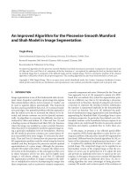
Figure 1 illustrates the results obtained from a typical
ISET/IHC experiment, where blood from healthy volunteers was spiked with either untreated LNCaP cells, PC3
cells or LNCaP cells incubated with AZD3514 at a dose
shown to reduce AR protein expression in this cell line [6].
Untreated LNCaP cells - the positive control - demonstrated strong nuclear brown staining for (translocated)
AR, whereas the PC3 cells - the negative control - displayed
a complete absence of brown staining. The almost
complete absence of cytoplasmic staining in LNCaP cells
may be due to constitutive autocrine stimulation of the AR
signaling pathway [20]. In addition, in LNCaP cells pretreated with AZD3514 prior to spiking into blood there
was a marked reduction in the level of nuclear staining.
These data indicate that the ISET/IHC method described
herein can distinguish between AR positive and AR negative cancer cells in the blood of human subjects and is also
Cummings et al. BMC Cancer 2014, 14:226
/>
Inset
PC3
LNCaP
Page 4 of 8
110kDa
LNCAP Cells Untreated
LNCAP Cells Treated
PC3 Cells
Figure 1 Characterisation of the specificity of the ISET/immunohistochemistry methodology for the androgen receptor utilising cells
lines as controls. Specificity was investigated by employing human prostate cancer cell lines of known AR status, together with treatment of
cells with AZD3514 in order to modulate the levels of the protein, to spike volunteer blood samples prior to processing by ISET and analysis by
IHC. Untreated LNCaP cells were the positive control and demonstrated strong brown nuclear staining for AR. PC3 cells were the negative control
and displayed a complete absence of brown staining. LNCaP cells pre-treated for 24 hours with 10 μM AZD3514 prior to spiking into blood
demonstrated a marked reduction in the level of nuclear staining, compared to untreated LNCaP cells. Inset: Western blot analysis of LNCaP and
PC3 cells for androgen receptor protein expression. Loading was normalised by the addition of 20 μg of protein to each lane and the blot was
run according the standard western blot protocols, utilising anti-androgen receptor antibody clone AR441 as the primary antibody and enhanced
chemiluminescence detection for visualisation of bands.
sufficiently sensitive to detect a drug induced (pharmacodynamic) knockdown in protein levels.
The major focus of the present study was to characterise between-operator and within-operator variability,
employing multiple blood samples collected from either
different patients or at different time points in the same
patient. The number of CTCs captured in the different
ISET filter spots ranged from 0 to 30 per ml of blood,
within the range of CTC previously reported in prostate
cancer patients [21].
In the first validation experiment 4 spots from 8 different
patient blood samples were stained by IHC for AR expression and presented blindly to two different operators to
both enumerate the CTC and score the staining intensity of
each cell. The degree of inter-operator agreement was
assessed both as a percentage and as κ and is presented in
Table 2. Although Cohen's kappa coefficient is a statistical
measure of inter-operator agreement for qualitative (categorical) items the κ statistic is not a test of significance.
Nonetheless, it is a robust measure since it takes into account random agreement occurring by chance. Guidelines
have been published to aid in the interpretation of κ [22],
and these have been previously applied to the enumeration
of CTC by CellSearch [23]. Significantly, almost perfect
agreement was observed when the two different operators
scored CTC as overall positive or negative for AR, with a κ
value of 0.94. However, when the scores produced by different operators for staining intensity were analysed there was
a large reduction in κ from 0.94 to 0.59, indicating a significant degree of disagreement.
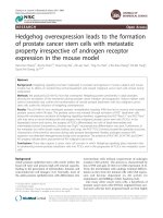
β-Content γ-confidence tolerance intervals (BCTI) reports on ISR both in the form of precision/imprecision
and trueness/bias. Figure 2 illustrates the accuracy profiles at both 67% and 95% probability for the staining intensity assignments of two operators. These demonstrate
a relatively large degree of imprecision, as might be expected with categorical data. However, they also highlight a significant bias, where operator 1 favoured a
score of 2++ (moderate staining intensity) over 3+++
(strong), whereas operator 2 favoured 3+++ over 2++.
Such a bias would not be evident by κ alone.
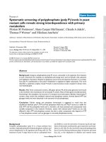
After the first validation experiment a programme of
staff training was embarked upon. A gallery of 20 IHC
images of CTC harvested by ISET from a number of different patients was constructed in order to facilitate a
supervised training workshop. Here analysts were presented with 5 different sets of images where each set included an example of a patient CTC expressing AR at
weak, moderate, strong and negative staining levels. One
such set of 4 graded images taken from the gallery is
Table 2 Degree of inter-operator agreement in scoring of
CTC for expression of the androgen receptor by
immunohistochemistry
1st Run
Evaluation
Positive v Negative
Staining intensity3
1
2
2nd Run
Kappa1
% Agreement
Kappa
% Agreement
0.94
97.2
0.98
98.9
0.59
75.0
0.81
87.9
Kappa values where calculated as described in Methods where the
agreement between operators is defined as follows: < 0 none, 0–0.20 slight,
0.21–0.40 fair, 0.41–0.60 moderate, 0.61–0.80 substantial, and 0.81–1 almost
perfect [22].
2
This table entry refers to an overall assessment of the level of positive versus
negative staining regardless of the staining intensity.
3
Staining intensity was graded into 4 categories as negative, weak (1 +),
moderate (2 ++) or strong (3 +++).
Cummings et al. BMC Cancer 2014, 14:226
/>
Page 5 of 8
2.5
95% Probability
2
67% Probability
1.5
Relative Error
1
0.5
0
-0.5
-1
-1.5
-2
1+
2 ++
3 +++
Scoring Category
Figure 2 Characterisation of inter-operator variability in AR staining intensity by IHC in CTC harvested from patient samples by ISET.
ISET membrane spots obtained after filtration of a number of different patient blood samples were stained by IHC for AR expression and
presented blindly to two different analysts to both enumerate the CTC and score the staining intensity. Results were then analysed by a
modification of ISR where the staining intensities obtained by each operator were substituted into the calculations. β-Content γ-confidence
tolerance intervals (±) were calculated at β = 95% and 67% and the resulting accuracy profiles plotted. These revealed a systematic bias
characterised by one operator favouring a score of 2 ++ over another favouring 3 +++.
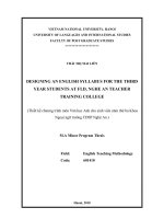
illustrated in Figure 3. In the second validation experiment, the bias between operators observed in experiment 1 was completely eliminated (see Figure 4), and
the value of κ for inter-operator agreement increased
from 0.59 to 0.81 (Table 1), the latter being in the category of almost perfect agreement [22].
Since the samples proffered to the analysts in the
second validation experiment were identical to those
proffered in the first, this allowed for a conventional assessment of ISR [17] (see Figure 5). These data highlight
the effect of the training programme, where analyst 1
showed a greater difference in scoring objects as 2++
between experiments , while analyst 2, as anticipated,
showed a greater difference in scoring objects in the
3+++ category. It is also clear that the major effect of
Negative
Weak
Moderate
Negative
1+
2 ++
Strong
3 +++
Figure 3 Typical example of the training set of images used to
standardise AR receptor staining intensity in CTC isolated by
ISET. After identifying significant operator bias in assigning staining
intensities (see Figure 2), a standard gallery of 20 different images was
produced as an aid to staff training. The figure contains a typical set of
images from the gallery each containing a single patient derived CTC
isolated on an ISET membrane and stained for AR expression by IHC to
illustrate the different levels of staining intensity observed and the
scoring system utilised in the present study.
the training programme was more manifest on operator 2, while the degree of ISR achieved by operator 1
approached 30%.
Discussion
Fit-for-purpose biomarker method validation defines 5
categories of assay based on readout ranging from absolute quantitation to a nominal positive/negative result
[13,14,24,25]. Along this spectrum, IHC in ocular microscopy mode is identified as an ordinal qualitative
assay that yields categorical data presented as discrete
scoring scales. Many of the performance characteristics
normally associated with bioanalytical methods are not
relevant to IHC, for example accuracy [26,27]. However,
the main parameter of relevance to a qualitative assay is
reproducibility: defined by the ICH as the “precision of
repeated measurements between (operators and) laboratories” [14]. Or put more simply, “the property of receiving
consistent results from following a specific procedure” [26].
Although there are many technical variables that could impact on the reproducibility of an IHC method, such as processing and embedding tissue and selection of section
thickness, [27] the major source of error is recognised as
that which is introduced by the reader [28,29].
In the present paper, method validation was performed
on an IHC method for the determination of AR in CTC.
While the focus was obviously reproducibility in addition
to an assessment of specificity, the issue of inter-operator
variability was addressed in a novel manner, through a
modification of ISR [30,31]. Thus, in addition to conducting
ISR in the conventional format (where typically a single
Cummings et al. BMC Cancer 2014, 14:226
/>
Page 6 of 8
2.5
2
95% Probability
1.5
67% Probability
Relative Error
1
0.5
0
-0.5
-1
-1.5
-2
1+
2 ++
3 +++
Scoring Category
Figure 4 Further characterisation of inter-operator variability in AR staining intensity by IHC in CTC harvested from patient samples by ISET.
After additional training utilising the standard gallery of images (see Figure 3), the same two analysts were invited to blindly score AR staining
intensity in CTC harvested from patient blood samples by ISET. Again, results were analysed by a modification of ISR, β-content γ-confidence
tolerance intervals (±) were calculated at β = 95% and 67% and the resulting accuracy profiles constructed. In this case, the bias observed in the
first validation experiment (see Figure 2) appeared to be effectively eliminated.
operator analyses the same set of samples twice), in our
modification two different operators each analysed the
same set of samples once and the results were then subjected to statistical analysis by BCTI. We have previously
demonstrated that in the case of CTC enumeration by CellSearch this modified approach to ISR was exquisitely sensitive in differentiating between both systematic (bias) and
random (imprecision) errors [15]. Our results confirm that
even in the relatively less complex scenario of a single CTC
sitting on a filter, there is still considerable betweenoperator variability in the assignment of a scoring intensity.
However, since BCTI identifies the nature of the interoperator error, it also allowed for its correction through
additional staff training. In addition, by adoption of this approach it has been demonstrated that a highly trained analyst is capable of achieving scores in the repeat analysis of
8
95% Probability
Operator 2
7
95% Probability
Operator 1
6
Relative Error
5
4
3
2
1
0
-1
-2
1+
2 ++
3 +++
Negative
Scoring Category
Figure 5 Incurred sample reanalysis in AR staining intensity determined by β-content γ-confidence tolerance intervals. Due to the fact
that the set of samples analysed blindly by two different operators in the first (Figure 2) and second (Figure 4) validation experiments were
identical, this also allowed a conventional analysis of results by ISR utilising β-content γ-confidence tolerance intervals (±) at β = 95%. The resulting
accuracy profiles clearly demonstrated that the training programme impacted almost exclusively on operator 2, correcting the between-operator
bias in the process (Figure 2). They also highlight that the degree of ISR achievable in this analysis by operator 1 approached 30%, which is the
accepted benchmark for total error for a typical quantitative biomarker assay.
Cummings et al. BMC Cancer 2014, 14:226
/>
samples that varied by 30%, which is within the accepted benchmark for total error of a typical quantitative biomarker/pharmacodynamic assay such as an
ELISA [14,25,32].
As a categorical assay, IHC has a dynamic range restricted to a limited number (normally 3 to 4) of band
widths of staining intensity. Nonetheless, in preclinical
studies with AZD3514, and utilising the same scoring
structure as the present method (0+, 1+, 2+ and 3+),
IHC was able to demonstrate a dose dependent reduction on AR in tumour tissue, at doses of drug that produced only a modest inhibition of tumour growth [6]. In
the same report a comparative evaluation was conducted
between ocular microscopy and image analysis using the
Aperio system (ePathology Solutions, Oxford, UK). Reassuringly, both approaches reported a similar level of
knockdown in AR. Among current IHC techniques approved by the FDA as diagnostic, prognostic or predicative biomarkers, no claims are made that image analysis
is any more accurate than visual assessment by a trained
pathologist [27].
The ISET technique is an example of a tumour-markerindependent technology based on filtration through 8 μm
pores, unlike the FDA cleared CellSearch System which relies critically on the presence of epithelial markers (EpCam)
and antibody directed capture [21,33]. However, the ISET
technique may have an advantage over CellSearch since it
is now believed that a significant proportion of malignant
CTCs lose their “epithelial markers” in preference to mesenchymal antigens, in a process known as epithelial to mesenchymal transition [34-36]. As a consequence the ISET
technique invariably harvests larger populations of CTC
from patient blood than the CellSearch system [37]. Nonetheless, while the ISET technique is claimed to effectively
remove the majority of hematologic cells - red blood cells
by lysis and peripheral blood leukocytes by filtration [12] rare hematologic cells (megakaryocytes or large monocytes)
or mesenchymal (endothelial) cells may be difficult to distinguish from epithelial tumour cells by cytopathologic or
immunochemical analysis [38]. Indeed, it has been demonstrated in a number of different disease types, that ISET
harvests cells of a non-malignant morphology from a small
but significant group of subjects that could potentially result in a false-positive diagnosis [38]. These results advise
caution when relying on a single technique to isolate CTC
from patients.
Conclusions
A novel procedure is presented for the fit-for-purpose
evaluation of the reproducibility of an IHC method for
the determination of AR receptor expression levels in
CTC isolated from patients as a biomarker assay utilising a modification of ISR and BCTI for statistical analysis of results. The procedure was employed to identify
Page 7 of 8
and correct inter-operator bias in the assignment of a
scoring intensity and poor reproducibility, resulting potentially in a reduction of measurement error to a level
normally associated with a quantitative biomarker assay,
such as an ELISA. The methodological approach could
be applied theoretically to any generic IHC method.
Abbreviations
AR: Androgen receptor; IHC: Immunohistochemistry; CTC: Circulating tumour
cells; BCTI: β-content γ-confidence tolerance intervals; K: Cohen’s Kappa;
CRPC: Castration-resistant prostate cancer; POM: Proof-of-mechanism;
ISET: Isolation by size of epithelial tumour cells; ISR: Incurred sample
reanalysis; REC: Research ethics committee.
Competing interests
The authors declare that they have no competing interests.
Authors’ contributions
JC, KM, CZ, RS and CD were the main authors of the manuscript. CZ
developed and validated all programming code utilised in statistical analysis.
JC conducted the statistical analysis and interpretation of data. RS and ML
performed all the laboratory analysis of samples. NC and TE collected blood
samples and clinical data from patients. All authors have read and approved
the final version of the manuscript.
Acknowledgements
The present study was supported with funding from the following: Cancer
Research UK (C147/A12328), the Experimental Cancer Medicine Centre
Network (ECMC) and Astra Zeneca. The authors would like to extend their
gratitude to the staff of the Prostate Cancer Clinic at Salford Royal NHS
Foundation Trust (Stott Lane, Greater Manchester, M6 8HD) from their
contribution to the present study.
Author details
1
Clinical and Experimental Pharmacology Group, Cancer Research UK
Manchester Institute, University of Manchester, Manchester Cancer Research
Centre, Manchester M20 4BX, UK. 2Department of Clinical Oncology, Christie
NHS Foundation Trust, Wilmslow Road, Manchester M20 4BX, UK. 3Urology,
Christie NHS Foundation Trust, Wilmslow Road, Manchester M20 4BX, UK.
Received: 28 October 2013 Accepted: 10 March 2014
Published: 28 March 2014
References
1. Mateo J, Smith A, Ong M, de Bono JS: Novel drugs targeting the
androgen receptor pathway in prostate cancer. Cancer metastasis reviews
2014. doi:10.1007/s10555-013-9472-2.
2. Taplin ME: Drug insight: role of the androgen receptor in the
development and progression of prostate cancer. Nature Clin Pract
Oncology 2007, 4(4):236–244.
3. Knudsen KE, Penning TM: Partners in crime: deregulation of AR activity
and androgen synthesis in prostate cancer. Trends Endocrinol Metab 2010,
21(5):315–324.
4. Bradbury RH, Acton DG, Broadbent NL, Brooks AN, Carr GR, Hatter G, Hayter BR,
Hill KJ, Howe NJ, Jones RD, Jude D, Lamont SG, Loddick SA, McFarland HL,
Parveen Z, Rabow AA, Sharma-Singh G, Stratton NC, Thomason AG, Trueman D,
Walker GE, Wells SL, Wilson J, Wood JM: Discovery of AZD3514, a smallmolecule androgen receptor downregulator for treatment of advanced prostate cancer. Bioorg Med Chem Lett 2013, 23(7):1945–1948.
5. Bradbury RH, Hales NJ, Rabow AA, Walker GE, Acton DG, Andrews DM,
Ballard P, Brooks NA, Colclough N, Girdwood A, Hancox UJ, Jones O, Jude
D, Loddick SA, Mortlock AA: Small-molecule androgen receptor
downregulators as an approach to treatment of advanced prostate
cancer. Bioorg Med Chem Lett 2011, 21(18):5442–5445.
6. Loddick SA, Ross SJ, Thomason AG, Robinson DM, Walker GE, Dunkley TP,
Brave SR, Broadbent N, Stratton NC, Trueman D, Mouchet E, Shaheen FS,
Jacobs VN, Cumberbatch M, Wilson J, Jones RD, Bradbury RH, Rabow A,
Gaughan L, Womack C, Barry ST, Robson CN, Critchlow SE, Wedge SR,
Brooks AN: AZD3514: a small molecule that modulates androgen
Cummings et al. BMC Cancer 2014, 14:226
/>
7.
8.
9.
10.
11.
12.
13.
14.
15.
16.
17.
18.
19.
20.
21.
22.
23.
24.
25.
26.
receptor signaling and function in vitro and in vivo. Mol Cancer Ther
2013, 12(9):1715–1727.
Omlin AG, Jones RJ, van der Noll R, Graham J, Ong M, Finkelman RD,
Schellens JH, Zivi A, Crespo M, Clack G, Alumkal JJ, Dymond A, Dickinson A,
Ranson M, Malone M, De Bono JS, Elliott T: A first-in-human study of the
oral selective androgen receptor down-regulating drug (SARD) AZD3514
in patients with castration-resistant prostate cancer (CRPC). J Clin Oncol
2013, 31(Supplement):4511.
Tibbe AG, de Grooth BG, Greve J, Dolan GJ, Terstappen LW: Imaging technique
implemented in Cell Tracks system. Cytometry 2002, 47(4):248–255.
de Bono JS, Scher HI, Montgomery RB, Parker C, Miller MC, Tissing H, Doyle
GV, Terstappen LW, Pienta KJ, Raghavan D: Circulating tumor cells predict
survival benefit from treatment in metastatic castration-resistant
prostate cancer. Clin Cancer Res 2008, 14(19):6302–6309.
Miller MC, Doyle GV, Terstappen LW: Significance of Circulating Tumor
Cells Detected by the Cell Search System in Patients with Metastatic
Breast Colorectal and Prostate Cancer. J Oncol 2010, 2010:617421.
Scher HI, Jia X, de Bono JS, Fleisher M, Pienta KJ, Raghavan D, Heller G:
Circulating tumour cells as prognostic markers in progressive, castrationresistant prostate cancer: a reanalysis of IMMC38 trial data. Lancet Oncol
2009, 10(3):233–239.
Vona G, Sabile A, Louha M, Sitruk V, Romana S, Schutze K, Capron F, Franco
D, Pazzagli M, Vekemans M, Lacour B, Brechot C, Paterlini-Brechot P: Isolation by size of epithelial tumor cells : a new method for the immunomorphological and molecular characterization of circulatingtumor cells.
Am J Pathol 2000, 156(1):57–63.
Cummings J, Raynaud F, Jones L, Sugar R, Dive C: Fit-for-purpose
biomarker method validation for application in clinical trials of
anticancer drugs. Br J Cancer 2010, 103(9):1313–1317.
Cummings J, Ward TH, Greystoke A, Ranson M, Dive C: Biomarker method
validation in anticancer drug development. Br J Pharmacol 2008,
153(4):646–656.
Cummings J, Morris K, Zhou C, Sloane R, Lancashire M, Morris D, Bramley S,
Krebs M, Khoja L, Dive C: Method validation of circulating tumour cell
enumeration at low cell counts. BMC Cancer 2013, 13(1):415–423.
Cohen J: A coefficient of agreement for nominal scales. Educ Psychol
Menaurement 1960, 20(1):37–46.
Hoffman D: Statistical considerations for assessment of bioanalytical
incurred sample reproducibility. Aaps J 2009, 11(3):570–580.
Cummings J, Zhou C, Dive C: Application of the beta-expectation tolerance
interval to method validation of the M30 and M65 ELISA cell death
biomarker assays. J Chromatogr B Analyt Technol Biomed Life Sci 2011,
879(13–14):887–893.
Tai S, Sun Y, Squires JM, Zhang H, Oh WK, Liang CZ, Huang J: PC3 is a cell
line characteristic of prostatic small cell carcinoma. The Prostate 2011,
71(15):1668–1679.
Gnanapragasam VJ, McCahy PJ, Neal DE, Robson CN: Insulin-like growth
factor II and androgen receptor expression in the prostate. BJU
international 2000, 86(6):731–735.
Allard WJ, Matera J, Miller MC, Repollet M, Connelly MC, Rao C, Tibbe AG,
Uhr JW, Terstappen LW: Tumor cells circulate in the peripheral blood of
all major carcinomas but not in healthy subjects or patients with
nonmalignant diseases. Clin Cancer Res 2004, 10(20):6897–6904.
Landis JR, Koch GG: The measurement of observer agreement for
categorical data. Biometrics 1977, 33(1):159–174.
Kraan J, Sleijfer S, Strijbos MH, Ignatiadis M, Peeters D, Pierga JY, Farace F,
Riethdorf S, Fehm T, Zorzino L, Tibbe AG, Maestro M, Gisbert-Criado R,
Denton G, de Bono JS, Dive C, Foekens JA, Gratama JW: External quality
assurance of circulating tumor cell enumeration using the Cell Search
((R)) system: a feasibility study. Cytometry B Clin Cytom 2011, 80(2):112–118.
Cummings J, Ward TH, Dive C: Fit-for-purpose biomarker method
validation in anticancer drug development. Drug Discov Today 2010,
15(19–20):816–825.
Lee JW, Devanarayan V, Barrett YC, Weiner R, Allinson J, Fountain S, Keller S,
Weinryb I, Green M, Duan L, Rogers JA, Millham R, O'Brien PJ, Sailstad J,
Khan M, Ray C, Wagner JA: Fit-for-purpose method development and
validation for successful biomarker measurement. Pharm Res 2006,
23(2):312–328.
Jennings L, Van Deerlin VM, Gulley ML: Recommended principles and
practices for validating clinical molecular pathology tests. Arch Pathol Lab
Med 2009, 133(5):743–755.
Page 8 of 8
27. Dunstan RW, Wharton KA Jr, Quigley C, Lowe A: The use of
immunohistochemistry for biomarker assessment–can it compete with
other technologies? Toxicol Pathol 2011, 39(6):988–1002.
28. Fandel TM, Pfnur M, Schafer SC, Bacchetti P, Mast FW, Corinth C, Ansorge M,
Melchior SW, Thuroff JW, Kirkpatrick CJ, Lehr HA: Do we truly see what we
think we see? The role of cognitive bias in pathological interpretation.
J Pathol 2008, 216(2):193–200.
29. Hamilton PW, van Diest PJ, Williams R, Gallagher AG: Do we see what we
think we see? The complexities of morphological assessment. J Pathol
2009, 218(3):285–291.
30. Fast DM, Kelley M, Viswanathan CT, O'Shaughnessy J, King SP, Chaudhary A,
Weiner R, DeStefano AJ, Tang D: Workshop report and follow-up–AAPS
Workshop on current topics in GLP bioanalysis: Assay reproducibility for
incurred samples–implications of Crystal City recommendations. Aaps J
2009, 11(2):238–241.
31. Viswanathan CT, Bansal S, Booth B, Destefano AJ, Rose MJ, Sailstad J, Shah
VP, Skelly JP, Swann PG, Weiner R: Quantitative Bioanalytical Methods
Validation and Implementation: Best Practices for Chromatographic and
Ligand Binding Assays. Pharm Res 2007, 24(10):1962–1973.
32. Miller KJ, Bowsher RR, Celniker A, Gibbons J, Gupta S, Lee JW, Swanson SJ,
Smith WC, Weiner RS: Workshop on bioanalytical methods validation for
macromolecules: summary report. Pharm Res 2001, 18(9):1373–1383.
33. Tibbe AG, Miller MC, Terstappen LW: Statistical considerations for
enumeration of circulating tumor cells. Cytometry A 2007, 71(3):154–162.
34. Lecharpentier A, Vielh P, Perez-Moreno P, Planchard D, Soria JC, Farace F:
Detection of circulating tumour cells with a hybrid (epithelial/mesenchymal)
phenotype in patients with metastatic non-small cell lung cancer. Br J Cancer
2011, 105(9):1338–1341.
35. Ma YC, Wang L, Yu FL: Recent Advances and Prospects in the Isolation by
Size of Epithelial Tumor Cells (ISET) Methodology. Technol Cancer Res
Treat 2013, 12(4):295–309.
36. Weinberg RA: Twisted epithelial-mesenchymal transition blocks senescence. Nature Cell Biology 2008, 10(9):1021–1023.
37. Khoja L, Backen A, Sloane R, Menasce L, Ryder D, Krebs M, Board R, Clack G,
Hughes A, Blackhall F, Valle JW, Dive C: A pilot study to explore circulating
tumour cells in pancreatic cancer as a novel biomarker. Br J Cancer 2012,
106(3):508–516.
38. Hofman VJ, Ilie MI, Bonnetaud C, Selva E, Long E, Molina T, Vignaud JM,
Flejou JF, Lantuejoul S, Piaton E, Butori C, Mourad N, Poudenx M, Bahadoran
P, Sibon S, Guevara N, Santini J, Venissac N, Mouroux J, Vielh P, Hofman PM:
Cytopathologic detection of circulating tumor cells using the isolation
by size of epithelial tumor cell method: promises and pitfalls. Am J Clin
Pathol 2011, 135(1):146–156.
doi:10.1186/1471-2407-14-226
Cite this article as: Cummings et al.: Optimisation of an
immunohistochemistry method for the determination of androgen receptor
expression levels in circulating tumour cells. BMC Cancer 2014 14:226.
Submit your next manuscript to BioMed Central
and take full advantage of:
• Convenient online submission
• Thorough peer review
• No space constraints or color figure charges
• Immediate publication on acceptance
• Inclusion in PubMed, CAS, Scopus and Google Scholar
• Research which is freely available for redistribution
Submit your manuscript at
www.biomedcentral.com/submit