Loss of the NKX3.1 tumorsuppressor promotes the TMPRSS2-ERG fusion gene expression in prostate cancer
Bạn đang xem bản rút gọn của tài liệu. Xem và tải ngay bản đầy đủ của tài liệu tại đây (1.59 MB, 12 trang )
Thangapazham et al. BMC Cancer 2014, 14:16
/>
RESEARCH ARTICLE
Open Access
Loss of the NKX3.1 tumorsuppressor promotes
the TMPRSS2-ERG fusion gene expression in
prostate cancer
Rajesh Thangapazham, Francisco Saenz, Shilpa Katta, Ahmed A Mohamed, Shyh-Han Tan, Gyorgy Petrovics,
Shiv Srivastava and Albert Dobi*
Abstract
Background: In normal prostate epithelium the TMPRSS2 gene encoding a type II serine protease is directly
regulated by male hormones through the androgen receptor. In prostate cancer ERG protooncogene frequently
gains hormonal control by seizing gene regulatory elements of TMPRSS2 through genomic fusion events. Although,
the androgenic activation of TMPRSS2 gene has been established, little is known about other elements that may
interact with TMPRSS2 promoter sequences to modulate ERG expression in TMPRSS2-ERG gene fusion context.
Methods: Comparative genomic analyses of the TMPRSS2 promoter upstream sequences and pathway analyses
were performed by the Genomatix Software. NKX3.1 and ERG genes expressions were evaluated by immunoblot or
by quantitative Real-Time PCR (qRT-PCR) assays in response to siRNA knockdown or heterologous expression.
QRT-PCR assay was used for monitoring the gene expression levels of NKX3.1-regulated genes. Transcriptional
regulatory function of NKX3.1 was assessed by luciferase assay. Recruitment of NKX3.1 to its cognate elements was
monitored by Chromatin Immunoprecipitation assay.
Results: Comparative analysis of the TMPRSS2 promoter upstream sequences among different species revealed the
conservation of binding sites for the androgen inducible NKX3.1 tumor suppressor. Defects of NKX3.1, such as, allelic
loss, haploinsufficiency, attenuated expression or decreased protein stability represent established pathways in
prostate tumorigenesis. We found that NKX3.1 directly binds to TMPRSS2 upstream sequences and negatively
regulates the expression of the ERG protooncogene through the TMPRSS2-ERG gene fusion.
Conclusions: These observations imply that the frequently noted loss-of-function of NKX3.1 cooperates with the
activation of TMPRSS2-ERG fusions in prostate tumorigenesis.
Keywords: Tumor suppressor, NKX3.1, Prostate, ERG, NFкB, Oncogene
Background
Activation of the ERG oncogene [1] represents an early
event in pre-neoplastic to neoplastic transition during
prostate tumorigenesis [2-4]. Rearrangements between
the androgen regulated TMPRSS2 gene promoter and
the ETS-related ERG gene result in TMPRSS2-ERG fusion transcripts that have been found in approximately
half of prostate cancer cases in the Western world [5].
Fusion of other androgen regulated genes, such as, the
prostein coding SLC45A3, prostate specific antigen
* Correspondence:
Center for Prostate Disease Research, Uniform Services University of the
Health Sciences, 1530 East Jefferson Street, Rockville, Maryland 20852, USA
homologue kallikrein 2 (KLK2) or the N-MYC downstream regulated gene 1 (NDRG1) contribute to ERG activation with lower frequencies [6]. At protein levels
ERG is detected as a nearly uniformly overexpressed
protein in over 60% of prostate cancer patients as revealed by the diagnostic evaluation of ERG oncoprotein
detection in prostatic carcinoma [7,8].
Much has been learned about the androgenic regulation of TMPRSS2 promoter [9-13] in prostate cancer. In
contrast, other control elements of the TMPRSS2 promoter are largely unexplored both in the wild type, as
well as, in the TMPRSS2-ERG fusion genomic context.
In the current study comparative analysis of TMPRSS2
© 2014 Thangapazham et al.; licensee BioMed Central Ltd. This is an Open Access article distributed under the terms of the
Creative Commons Attribution License ( which permits unrestricted use,
distribution, and reproduction in any medium, provided the original work is properly cited. The Creative Commons Public
Domain Dedication waiver ( applies to the data made available in this
article, unless otherwise stated.
Thangapazham et al. BMC Cancer 2014, 14:16
/>
Page 2 of 12
Results
region of the TMPRSS2 gene on chromosome 21 (NCBI
build 36.3) for further analyses. This genomic region encompasses upstream regulatory elements (−13.5 kb)
shown to control cancer-associated expression of the
ERG oncogene [30].
Comparative analysis of modular regulatory sequences
of various species is a powerful approach for pinpointing
functionally relevant regulatory elements [31-33]. We applied a computational approach (FrameWoker software,
release 5.4.3.3) that has been shown to identify conserved
orientation, relative position and relative distance of binding motif (matrix) clusters [34,35] also known as the
“motif grammar” [36] using the Matrix Family Library
Version 7.1. We have examined the −15,000;+78 bp regions of human, rhesus monkey, rat and mouse TMPRSS2
gene promoter upstream sequences for the conservation
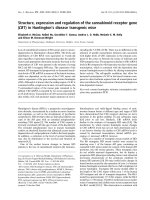
of composite regulatory elements. Striking conservation of
a composite model was noted in this analysis that was
mapped to the human TMPRSS2 -2350; -2258 sequences
relative to the TSS. Within the composite model we have
identified the vertebrate NKX3.1 matrix (V$NKXH) as the
prostate-specific component of the model and putative
binding site was termed as NKX3.1 binding site 1 (NBS1)
(Figure 1B).
Identification of an NKX3.1 binding site within the
TMPRSS2 gene promoter upstream sequences
NFкB-centered network of NKX3.1 target gene signatures
Within the TMPRSS2 gene locus promoter downstream
sequences beyond the +78 position of the first non-coding
exon (NM_005656) frequently participate in genomic rearrangement events. These genomic rearrangements are
characterized by the recurrent TMPRSS2 (first noncoding exon:+78) [26] to ERG (exon 8 or Exon 9)
[1,27,28] fusion junctions also known as fusion type “A”
or “C”, respectively [11]. In this gene fusion event the
TMPRSS2 promoter-proximal and promoter upstream
sequences are retained. Towards the bioinformatic analysis of TMPRSS2-ERG regulatory elements we mapped
the transcription start sites (TSS) of TMPRSS2 gene in
TMPRSS2-ERG fusion harboring human prostate tumors. From a carefully characterized RNA pool of ERG
expressing and TMPRSS2-ERG fusion harboring prostate tumors obtained from six radical prostatectomy
specimens [29], cDNA molecules were generated and
amplified using 5’ cap-specific forward primers and
ERG-specific reverse primers. Amplicons were isolated
and cloned. Individual clones (n = 20) were analyzed by
DNA sequencing and the frequency of cap-tags were
plotted on the transcription start region (TSR_200587)
of the TMPRSS2 gene (Figure 1A). The DNA sequence
analysis revealed that the most frequent (50%) transcription start of TMPRSS2-ERG fusion transcripts is at
+5, relative to the wild type TMPRSS2 promoter +1 position. By confirming the TSS position we focused our investigation on the +78 to15,000 upstream regulatory
Utilizing this highly conserved model the entire human
genome was searched for model matches (ModelInspector
Release 5.6) to define gene loci potentially targeted by
NKX3.1. After filtering for non-redundant, intronic, exonic and promoter model matches within gene loci of annotated genes, knowledge-based pathway analysis was
performed using functional co-citation settings. The analysis revealed a network with NFкB in the central regulatory node (Additional file 1: Figure S1). As expected,
searching of the entire human genome for this composite
model precisely identified the TMPRSS2 gene upstream
−2350; -2258 sequences. In contrast, search of the dog,
bovine, opossum and zebra fish genome failed to identify
model matches within the Tmprss2 loci of these species.
In a meta-analysis approach we compared the comparative
genome analysis-derived network to the signature of
Nkx3.1-targeted genes defined by in vivo ChIP assay in a
mouse model (Additional file 1: Figure S2) [21]. Strikingly
similar NFкB-centered regulatory network was revealed
by the analysis (Figure 2). NKX3.1 target genes within the
compared datasets were enriched in functionally related
genes. Moreover, the analysis highlighted orthologues of
TMPRSS2, JARID2 and the NFкB genes. The apparent
similarity between these datasets has prompted us to
examine the disease association of NKX3.1 target genes by
gene ontology analyses. Enrichment of chromosome aberrations, inversion, breakage and associated diseases was
revealed by the analysis (Table 1).
promoter upstream elements among different species revealed the presence of a conserved NKX3.1 binding site.
NKX3.1 is a bona fide tumor suppressor gene with
prostate-restricted expression [14]. Loss or decreases in
NKX3.1 levels has been frequently observed in prostatic
intraepithelial neoplasia and at the pre-neoplastic to
neoplastic transformation stages of prostate cancer
[15,16]. Loss of Nkx3.1 cooperates with loss of Pten in
engineered mouse models of prostate tumorigenesis
[17,18]. Furthermore, Nkx3.1 defects cooperate with
Pten-Akt pathways [19] and disrupt cellular response to
DNA damage [20]. Nkx3.1 was also shown to oppose
the transcription regulatory function of C-Myc [21] in
mouse models. In prostate cancer cells C-MYC is activated by ERG [22-24]. A recent study has shown that
ERG is a repressor of NKX3.1 raising the possibility of a
feed-forward circuit in prostate tumorigenesis [25]. Our
observation of conserved NKX3.1 binding elements in
the TMPRSS2 promoter prompted us to examine the
hypothesis that NKX3.1 is a repressor of ERG in the
TMPRSS2-ERG fusion genomic context in prostate
cancer.
Thangapazham et al. BMC Cancer 2014, 14:16
/>
Page 3 of 12
Figure 1 Defining a conserved composite model for NKX3.1 binding within the TMPRSS2 gene promoter upstream sequences.
(A) Frequency of TMPRSS2-ERG transcript initiation sites within the TMPRSS2 promoter transcriptional start region (TSR). (B) NKX3.1 model match
within the human TMPRSS2 promoter upstream region with conserved distance, positions and orientations (arrows) of transcription factor
binding sites.
Figure 2 Summary of NFкB centered NKX3.1 target gene signatures from in silico (left panel) and from the meta-analysis of in vivo
data (right panel). Experimentally validated human genes and their orthologues in mouse are highlighted in yellow. Secondary nodes
representing genes with four or more functional connections are stemming from the central regulatory node (green boxes). Nodes with four or
more functional connections are outlined by red. Connected genes are marked with white background color.
Thangapazham et al. BMC Cancer 2014, 14:16
/>
Page 4 of 12
Table 1 Disease association analysis of predicted NKX3.1
targeted genes within the human genome reveals the
enrichment of chromosome aberrations, inversion,
breakage gene ontology categories
MeSH Disease/input n = 464
Genes
P-value
Expected
Observed
Chromosome inversion
1.67e-04
120
152
Chromosome aberration
2.06e-04
13
27
Angelman Syndrome
2.99e-04
3
10
Chromosome breakage
3.45e-04
20
36
Uniparental disomy
3.95e-04
4
12
Prader-Willi Syndrome
8.64e-04
5
13
Translocation, genetic
9.83e-04
59
82
Altered expression of predicted downstream target genes
in response to NKX3.1 depletion
To evaluate NKX3.1 in TMPRSS2-ERG fusion harboring
prostate cancer cells we utilized the siRNA depletion
strategy. Consistent with a negative regulatory function
of NKX3.1, the transcripts of endogenous TMPRSS2ERG fusion allele, as well as, the wild type TMPRSS2
showed elevated expression along with HDAC9, RUNX1,
NFкB and JARID2 genes in response to NKX3.1 inhibition (Figure 3A). In line with previous reports we also
noted the reduction of CFTR expression in response
NKX3.1si. This finding suggests that CFTR expression
in the human prostate may indeed positively regulated
by NKX3.1 [37]. Gene expression response to NKX3.1
knockdown was noted in approximately half of the examined NKX3.1 target genes. Whole genomic search for
model matches in human, rhesus monkey, rat and
mouse TMPRSS2 promoter upstream sequences precisely identified matches of the NKX3.1 model. Thus
NKX3.1 as a negative regulator of TMPRSS2 may evolve
in this lineage, since, we found no evidence of model
matches within Tmprss2 promoter upstream regions of
zebra fish, opossum, dog and cow genomes. Despite of
known informatics constrains, such as, model overfitting
and limitations in the employed functional assays the results suggest that comparative analyses for defining conserved repressor elements is a valid approach providing
efficient guidance for the experimental validation.
To assess the function of NKX3.1 in regulating the
TMPRSS2-ERG fusion gene we evaluated ERG expression
in response to specific inhibition of NKX3.1. Knockdown
NKX3.1 with siRNA resulted in elevated ERG protein levels
(Figure 3B). Increased expression and nuclear localization
of ERG oncoprotein in response to NKX3.1 siRNA further
supported the repressor role of NKX3.1. Consistent with
elevated ERG levels we observed marked decreases in
prostein. This prostate differentiation associated protein is
encoded by the SLC45A3 gene that is negatively regulated
by ERG [22].
NKX3.1 is a repressor of the TMPRSS2 gene
Although, NBS1 is the only evolutionarily conserved
NKX3.1 binding site prediction within the TMPRSS2
promoter upstream region, transcription factor binding
site model match search by MatInspector identified further stand-alone NKX3.1 binding sites. The single
matrix prediction identified a tight cluster of five single
NKX3.1 matrix model matches (V$NKX31.01) between
positions −2298 and −2168 relative to the transcription
initiation site that showed partial overlap with NBS1.
Further upstream clusters of single NKX3.1 model
matches were identified and were designated as NBS2
(−3292; -3277), NBS3 (−8019; -7902), NBS4 (−10684;
-10615), and NBS5 (−14628; - 14614). For the assessment of transcription regulatory functions, NBS1-5 sites
were cloned upstream to a Luciferase reporter vector.
The assay result indicated negative regulatory functions
for NBS1, NBS2 and NBS4 sequences (Figure 4A). To
evaluate the endogenous TMPRSS2-ERG gene expression response to NKX3.1 inhibition, VCaP cells were
grown in hormone depleted media for three days. Cells
were transfected by NKX3.1 siRNA or by non-targeting
control siRNA molecules. Synthetic androgen (R1881) was
added to the media to induce the expression of androgen
regulated genes, including NKX3.1 and TMPRSS2-ERG.
After 24 h induction cells were processed for Chromatin
Immunoprecipitation (ChIP) assay examining the recruitment of NKX3.1 to NBS1, NBS2 and NBS4. NBS amplicons were excised from the gel and were confirmed by
DNA sequencing. The experiment confirmed the recruitment of NKX3.1 to NBS1 and NBS4 regions (Figure 4B).
Although ChIP assays provided an estimated region of
recruitment within the chromatin context of NBS1 and
NBS4 it does not reveal the actual position and specificity of transcriptional regulatory elements. To address
the specificity of NBS1 and NBS4 core binding sites we
have introduced transversion point mutations to the core
cognate elements aiming to disrupt the NKX3.1 homeodomain DNA recognition (Figure 5A). To reduce the possibility of generating of de novo TF binding sites we have
used the SeqenceShaper program (www.genomatix.de).
Wild type and corresponding mutant NBS1 and NBS4
harboring reporter vectors were assayed for reporter
gene activity by transfecting HEK293 cells in the presence of NKX3.1 expressing pcDNA-NKX3.1-HA expression vector or control pcDNA. The transfection
efficiency was monitored by co-transfecting phRGB-TK
Renilla-Luc control vector. In the presence of heterologously expressed NKX3.1 the expression of wtNBS1 and
wtNBS4 reporters were reduced 4–3 folds, respectively.
NBS1- and NBS4-mediated transcriptional repression
Thangapazham et al. BMC Cancer 2014, 14:16
/>
Page 5 of 12
Figure 3 Expression of predicted NKX3.1 target genes in response to NKX3.1 inhibition. (A) Depletion of NKX3.1 results in increases in
mRNA levels of wild type TMPRSS2, TMPRSS2-ERG fusion (T2-ERG), HDAC9, RUNX1, NFкB and JARID. In contrast, robust reduction of CFTR levels is
apparent in response to NKX3.1 inhibition. (B) Rescue of ERG and its downstream function by NKX3.1 inhibition (NKX3.1 siRNA) is shown by
nuclear localization of ERG (upper panel), sharp increases in ERG protein levels (lower panel), and by the depletion of the ERG-downstream target
prostein (SLC45A3). Schematic depiction of the negative regulatory role of NKX3.1 in the context of TMPRSS2-ERG (T2-ERG) gene fusion (inset).
was disrupted by specific mutations within the V$NKXH
core recognition sequences, accompanied by a modest
activation in reporter expressions (Figure 5B).
Discussion
Comparative assessment of evolutionary conserved cognate sequences within the TMPRSS2 promoter upstream
sequences revealed strong conservation of an NKX3.1
binding site. Experimental evaluation of the predicted
composite element suggested that this element confers
NKX3.1-mediated repression to the TMPRSS2-ERG fusion gene in prostate cancer cells. Inhibition of NKX3.1
resulted in elevated expression and nuclear localization
of ERG and resulted in reduced levels of the ERGdownstream regulated prostein encoded by the SLC45A3
gene. Assays for the transcription regulatory function of
NKX3.1 binding sites indicated repressor function that
was disrupted by specific mutations affecting the DNA
Thangapazham et al. BMC Cancer 2014, 14:16
/>
Page 6 of 12
Figure 4 Predicted NKX3.1 binding sequences of the TMPRSS2 promoter are portable repressor elements. (A) The transcriptional
regulatory function of predicted NKX3.1 binding sites (NBS1-5) was assessed by luciferase reporter systems. Relative luciferase units are shown
as fold changes relative to the control expression levels. Significant (P < 0.05) reduction of reporter gene expression are marked by asterics
(B) Specific recruitment of endogenous NKX3.1 to predicted NBS1 and NBS4 binding sites of the TMPRSS2 promoter upstream regions was
assessed by in vivo ChIP assay in the absence (NT) or presence of NKX3.1 siRNA (NKX).
recognition of NKX3.1 transcription factor. Recruitment
of endogenous NKX3.1 to the evolutionarily conserved
cognate element was confirmed by in vivo ChIP assay.
Loss of NKX3.1, contributes to the cancer associated
function of AR [38,39], C-MYC [21], p53, PTEN [40],
Topoisomerase I [41] and TWIST1 [42] in prostate cancer. ERG oncogene, a result of the TMPRSS2-ERG fusions, negatively regulates NKX3.1 through EZH2 [25].
In the current study we have examined evolutionary
conserved composite regulatory models of the TMPRSS2
gene. The analysis revealed a remarkable conservation of
a composite model with an NKX3.1 binding site in the
lineage of mouse, rat, rhesus monkey and human species
members of the Euarchontoglires (Supraprimates) super
ordo. This composite model identified sequences within
intronic regions of the human genome. Increased expression of evaluated NKX3.1 target genes (HDAC9,
RUNX1, TMPRSS2, TMPRSS2-ERG, NFкB and JARID2)
was observed in response to NKX3.1 inhibition. Metaanalysis of Nkx3.1 target genes from in vivo ChIP assay
of mouse prostates indicated that upstream regulatory
regions are indeed enriched in core elements, such as, V
$NKXH, V$HOXF and V$BRNF (Table S3 in [21]) similar to the model we have obtained from in silico analysis.
Pathway analysis of NKX3.1 target genes from the
current study, as well as, from the reported in vivo
model [21] revealed NFкB as the central regulatory node
of NKX3.1 target gene signatures. Furthermore, the analyses indicated, robust enrichment of genes controlling
chromosomal integrity. These findings are consistent
with the reported role of NKX3.1 in cellular response to
DNA damage [20,41]. These observations are also consistent with an NFкB-mediated protective function of
NKX3.1 linked to inflammation and tumorigenesis
[15,43-47]. Taken together our study highlights NKX3.1
as a negative regulator of theTMPRSS2 promoter. Thus,
the frequently observed haploinsufficiency of NKX3.1 in
prostate cancer may significantly contribute to the activation of ERG protooncogene in the TMPRSS2-ERG fusion genomic context. This finding highlights the
integrated role of TMPRSS2-ERG gain and NKX3.1
losses as cooperating events in prostate tumorigenesis
(Figure 6).
Conclusions
Approximately half of the prostate cancer cases
harbor the TMPRSS2-ERG gene fusions in Western
countries. This recurrent oncogenic event leads to
the activation of the ERG oncogene. In the current
study evaluation of conserved regulatory elements
of TMPRSS2 promoter upstream sequences revealed
conservation of binding sites for the NKX3.1 tumor
suppressor. NKX3.1 binds to these sequences and
represses the TMPRSS2-ERG fusion gene. Thus, the
Thangapazham et al. BMC Cancer 2014, 14:16
/>
Page 7 of 12
Figure 5 Both NKX3.1 protein and wild type NKX3.1 binding sites are required for the transcriptional repressor function of TMPRSS2
promoter upstream sequences. (A) Schematic representation of NBS1 and NBS4 sequences marking predicted NKX3.1 binding elements in
brackets. Core recognition sequences with transversion mutations are underlined in the wild type (wt) and in the mutant (mt) sequences.
(B) Relative luciferase units (RLU) of wild type and mutant NKX3.1 binding sites were assayed in reporter constructs in the presence (+) of
heterologously expressed NKX3.1 or in the presence of control vector (−). Asterisk symbols mark significant (P < 0.05) reductions in reporter
gene expression.
frequently observed loss of NKX3.1 in prostate cancer
may significantly contribute to the activation of ERG protooncogene. Pathway analysis of NKX3.1 target genes
from the current study, as well as, from the reported
in vivo studies revealed NFкB as the central regulatory
node of NKX3.1 target gene signatures with robust enrichment in genes controlling chromosomal integrity.
These findings suggest that TMPRSS2-ERG gain and
NKX3.1 losses are potentially cooperating genetic events
in prostate tumorigenesis.
Figure 6 NKX3.1 haploinsufficiency results in the loss of negative control over the TMPRSS2-ERG gene fusion.
Thangapazham et al. BMC Cancer 2014, 14:16
/>
Methods
Cell lines, cell culture and reagents
Human prostate tumor cell line, VCaP and human embryonic kidney HEK293 cells were obtained from the
American Type Culture Collection (ATCC, Rockville,
MD) and were maintained in growth medium and under
conditions recommended by the supplier. The synthetic
analogue of androgen, R1881, was purchased from New
England Nuclear (Boston, MA).
Inhibition of NKX3.1 and ERG with small interfering RNA
and heterologous expression of NKX3.1
Small interfering RNA (siRNA) oligo duplexes against
human NKX3.1(L-015422-00), and Non-targeting control siRNA (D-001206-13-20) were from Dharmacon
(Lafayette, CO), ERGsi RNA as previously described
[22]. Transfection or co-transfection of 50 nM siRNAs and 1 μM of plasmids was carried out with
Lipofectamine 2000 (Invitrogen, Carlsbad, CA) in triplicates. The wild type human NKX3.1 expressing
vector pcDNA3.1-NKX3.1-HA was a kind gift from
Dr. Charles J. Bieberich, University of Maryland
Baltimore County, Baltimore, Maryland. In six-well
plates HEK293 cells were transfected in triplicates with
the pcDNA3.1 control or with the pcDNA3.1-NKX3.1HA expression vectors by using Lipofectamine 2000.
Cells were harvested for protein and mRNA analysis after
48 h incubation.
Chromatin immunoprecipitation assay
For assessing the specific recruitment of endogenous
NKX3.1 to the predicted NKX3.1 binding sites in vivo
ChIP assays were carried out in the presence of NKX3.1
siRNA or control NT siRNA [35]. VCaP cells were
grown in 10% charcoal stripped serum (cFBS) containing
media (Gemini Bio-Products, Carlsbad, CA) for 48 h
and were transfected with 50 nM NKX3.1 siRNA or 50
nM of NT control. Cells were incubated for 24 h
followed by the addition of 0.1 nM of R1881. At the
48 h time point following hormone induction formaldehyde was added to the cell culture media to 1% and the
cells were processed for ChIP assay [48] by using the
mouse monoclonal anti-ERG antibody (CPDR ERGMAb, clone 9FY, currently available from Biocare Medical, Concord, CA) [7]. NBS1 region from input and
ChIP DNA samples were amplified by the forward 5’TGTTTCTCTGGAGAACCCTGA-3’ and reverse 5’- GC
AGGTGCAGTTGTCTTTCA-3’; NBS2 region was amplified by the forward 5’- CAATCCAGGCAGGGCTA
TTA and reverse 5’- GGGCAATAGCTGGTGTTTGT3’; the NBS4 region was amplified by the 5’- TCA
TCTATTTTCACCGCCATC-3’ and 5’- ACACGCACAC
ACCACATCAT-3’ primer pairs under previously described PCR conditions [22,35].
Page 8 of 12
Assessment of the transcription initiation site of
TMPRSS2-ERG transcript by 5’ oligocapping
Under approved protocol from the WRAMC IRB six
cases were identified with TMPRSS2-ERG fusion harboring prostate tumors. Total RNA was isolated from the
tumors and were pooled [29]. From the pool 4.2 μg of
total mRNA was subjected to 5’ oligocapping procedure
(FirstChoice, RLM-RACE, Ambion, Austin, TX) pairing
the 5’-GGCGTTGTAGCTGGGGGTGAG-3’ [11] with
the outer, and 5’- CAATGAATTCGTCTGTACTCCA
TAGCGTAGGA-3’ with the inner primer. Amplicons
were gelpurified and cloned into pUC19 vector and were
subjected to DNA sequencing in forward and reverse
directions.
Comparative analysis of the TMPRSS2 gene promoter
upstream sequences
DNA sequences of the 15,000; +78 bp region of Homo
sapiens, Macaca mulatta, Rattus norvegicus and Mus
musculus genomes were extracted from the NCBI build
36.3 database. Scanning from the proximal promoter towards the distal sequences 3,000 bp homologue segments
were evaluated allowing 500 bp overlap of segments at
each composite model scanning step. DNA sequence
segments of all examined species were analyzed by
the FrameWorker (version 5.4.3.3, www.genomatix.de)
for conserved composite model matches by using the
Matrix Family Library 7.1 at the following settings:
core promoter elements 0.75/optimized, vertebrates
(0.75/optimized); distance between adjacent elements:
5–200; distance band with: 10, exhaustive model search
with minimum number of elements = 2 and max number
of elements = 6. Overall the highest number of common
single element match was the V$NKXH, a binding site for
NKX3.1. Ranking the composite models revealed only one
model that reached the maximum (four element) complexity. The top scoring model was defined as V$HOXF
(strand orientation (+), distance to next element 43-51 bp),
V$NKXH (strand orientation (−), distance to next element
7-14 bp), V$PARF (strand orientation (−), distance to
next element 17-23 bp); V$BRNF (strand orientation
(−), distance to next element 0 bp) at settings of
minimum core similarity = 0.75 and minimum matrix
similarity “optimized”. Next the entire human genome
(NCBI build 36.3) was searched with this composite
model for matches by the ModelInspector 5.6 program (www.genomatix.de). Whole –genomes model
searches confirmed the model match within the TMPRSS2
gene promoter upstream sequences in Homo sapiens,
Macaca mulatta, Rattus norvegicus and Mus musculus genomes and indicated the absence of model
match within the Tmprss2 gene loci of Canis lupus
familiaris, Bos Taurus, Monodelphis domestica, and
Danio rerio.
Thangapazham et al. BMC Cancer 2014, 14:16
/>
Pathway and meta-analyses of NKX3.1 genomic targets
Predicted gene targets for NKX3.1 were obtained by in
silico composite model match analysis of the entire human genome. Among the total 1636 (1371 nonredundant) model matches 559 were non-annotated.
Within the annotated 1037 model matches (Additional
file 2: Table S1) 627 was found in intronic, 10 and 12
matches were found in exonic or promoter sequences,
respectively. Intronic, exonic and promoter model
matches were further filtered for genes with defined
gene symbols and the final set of 452 genes were used as
input for pathway analysis (Additional file 2: Table S2).
Prostate Cancer meta-analysis dataset used in our study
was based on the report of Anderson et al. [21]. NKX3.1
target genes were imported into the Genomatix Pathway
System (GePS, www.genomatix.de). In GePS genes were
mapped into networks based on the information extracted from public databases including National Cancer
Institute Pathway Interaction Database (.
nih.gov) and Biocarta (www.biocarta.com). The generated network displayed as nodes and connections focused on functional relationships between genes based on
the number of evidences in literature (Figures S1 and S2).
For the analyses we have used function word evidence
level to generate the network where gene pairs are noted
if they occur in the same sentence connected with a
function word.
Immunoblot assay
At the specified time points VCaP cells treated with
NKX3.1si or control NTsi were lysed in M-PER
Mammalian Protein Extraction Reagent (Pierce, Rockford,
IL) supplemented with protease (Roche Applied Science,
Indianapolis, IN) and phosphatase inhibitor cocktails
(Sigma, St. Louis, MO). ERG proteins were detected by
Western blot (NuPAGE Bis-Tris gel, Invitrogen) as
described previously using immunoaffinity-purified antiERG mouse monoclonal antibody 9FY [7]. The antiNKX3.1 polyclonal antibody (T-19) and anti-alpha tubulin
(B-7) antibodies were obtained from Santa Cruz
(Santa Cruz, CA) and the anti-prostein antibody recognizing the protein product of the SLC45A3 gene
was obtained from DAKO (Carpinteria, CA). Representative images of two independent experiments are
shown in the Results.
Immunofluorescence assay of siRNA treated VCaP cells
VCaP cells were fixed in 4% paraformaldehyde and centrifuged onto silanized slides (Sigma, St.Louis, MO) with
a cytospin centrifuge. Cells were immunostained with
anti-ERG (9FY) and anti-NKX3.1 (Santa Cruz) followed
by goat anti-mouse Alexa-488 and anti-goat Alexa-594
secondary antibodies (Invitrogen, Carlsbad, CA). Images
were captured by using a 40X/0.65 N-Plan objective on
Page 9 of 12
a Leica DMLB upright microscope with a QImaging
Retiga-EX CCD camera (Burnaby, BC, Canada) controlled by OpenLab software (Improvision, Lexington,
MA). Images were converted into color and merged by
using Adobe Photoshop.
NKX3.1 binding site (NBS) luciferase reporters and
dual-luciferase reporter assays
Mutant NBS sequences were designed to minimize the
generation of artificial binding sites by the Sequence
Shaper (www.genomatix.de). Wild type and mutant NBS
sequences were chemically synthesized adding a cohesive
overhang for Nhe1 site (CGCGT) at the 5'-end of the
sense strand and an overhanging Bgl2 site (TCGAG) at
the 3‘ as follows: wild type NBS1 5’-CTCCATAATTG
TATGAGTCAATTTCTTATAGTAAATCTTTATATATA
TTATAAATAATATTTATTACATATAAGCTGTGTATA
ATATATATCAT-3’; mutant NBS1 5’-GAACGCCGGG
TATGAGTCAATTTCTTATAGTAAATCTTTATATATA
TTATAAATAAGCAAACTTACATATAAGCTGTGTAT
AATATATATCAT-3’ ; wild type NBS2 5’-CACATAACT
TAAGGCATATTGACTTTATATCATTGTATTAAGTAT
TGTTAATTTTACATTA-3’; mutant NBS2 5’-CACAT
AAAGGCCTGCATATTGACTTTATATCATTGGCGGC
CTTATTTGGCCGGTTACATTA-3’; wild type NBS3 5’CGAGAAAAGGATTCAAATACTTAGGAAGATTGAA
ATGTGAGGGT-3’; mutant NBS3 5’-CGAGAAAAGGA
TTCAAAGCCGGCGGAAGATTGAAATGTGAGGGT-3’;
wild type NBS4 5’- CGAGTGGCATTAAGTACATTCAC
ACTGTCATGCAATCATCTATTTTCACCGCCATCTA
TTTTCAGAATGTTCTCA-3’; mutant NBS4 5’- CGAG
TGGCATGCCTGCCATTCACACTGTCATGCAATCA
TCTATTTTCACCGCCATCTATTTTCAGAATGTT
CTCA-3’; wild type NBS5 5’-CAAAACCAAATACTG
CATGTTCTCACTTATAAGTGGGAGCTGGACAATG
AGAACACATGGACACAGGGAGA-3’; mutant NBS5
5’-CAAAACCAAATACTGCATGTTCTAACAGGCTAC
TGTGGAGCTGGACAATGAGAACACATGGACACAG
GGAGA-3’. The 5’ end of synthetic oligonucleotides
were phosphorylated by using polynucleotide kinase,
the complementary strands were annealed and gelpurified and ligated to the NheI-BglII sites of the gelpurified,
phRG-TK reporter (Promega, Madison, WI). The phRGTK vector is a synthetic reporter vector that has been
designed to minimize binding sites for transcription
factors. HEK293 cells were transfected with the reporter and pGL3 luciferase control vectors in triplicates. Forty-eight hours after the transfection, the
activities of control phRG-TK reporter Renilla luciferase
and pGL3 Firefly luciferase constructs were determined
by the Dual-Luciferase Reporter Assay system (Promega,
Madison, WI). Cells were rinsed with phosphatebuffered saline, and lysed with 1 × passive lysis buffer.
Twenty μl of cell lysates were transferred into the
Thangapazham et al. BMC Cancer 2014, 14:16
/>
Page 10 of 12
luminometer tube containing 100 μl luciferase assay reagent II. Firefly luciferase activity (N1) and were measured
first, and then Renilla luciferase activities (N2) were
determined after the addition of 100 μl Stop & Glo
reagent. N2/N1 light units were averaged from three
measurements and were expressed as relative luciferase
units (RLU).
Additional files
RNA extraction, reverse transcription and real-time PCR
quantification
Abbreviations
NKX3.1: NK3 Homeobox 1; Transmembrane protease: serine 2; NBS: NKX3,1
binding site; ERG: V-Ets Erythroblastosis Virus E26 Oncogene Homolog
(Avian); SLC45A3: Solute carrier family 45, member 3; JARID2: Jumonji, AT
Rich Interactive Domain 2; NFкB: Nuclear Factor of Kappa Light Polypeptide
Gene Enhancer In B-Cells 1; HDAC9: Histone Deacetylase 9; RUNX1: RuntRelated Transcription Factor 1; CFTR: Cystic Fibrosis Transmembrane
Conductance Regulator (ATP-Binding CassetteSub-Family C, Member 7);
TSR: Transcriptional start region; TSS: Transcriptional start site;
HEK293: Human Embryonic Kidney 293 cell line; VCaP: Vertebral-Cancer
of the Prostate cell line.
Total RNA was extracted from cell monolayer using
Trizol® total RNA isolation reagent (Gibco BRL, Life
Technologies, Gaithersburg, MD, USA) as per the manufacturer's protocol. Real-time PCR was performed in
triplicates using an Applied Biosystems 7300 Sequence
Detection system using SYBR green PCR mix (Qiagen)
or by TaqMan assay (Applied Biosystems). The expression of GAPDH was simultaneously analyzed as endogenous control, and the target gene expression in
each sample was normalized to GAPDH [49]. RNA samples without reverse transcription were included as the
negative control in each assay. Amplification plots were
evaluated and threshold cycle (CT) was set for each experiment. Measurements for target gene and GAPDH
endogenous control were averaged across triplicates and
standard deviation for each set was calculated. ΔCT
values were calculated by subtracting averaged GAPDH
CT from averaged target gene CT and expression foldchange differences were calculated by comparing ΔCT
values among sample sets. Primer pairs for the amplification of target genes were as follows. HDAC9: forward
5’- CAAATGGTTTCACAGCAACG -3’, reverse 5’- TGC
GTCTCACACTTCTGCTT -3’; JARID2: forward 5’- AG
GAGACTGGAAGAGGCACA -3’ and reverse 5’- GTCC
GTTCAGCAGACCTCTC -3’; NFкB: forward 5’- TATG
TGGGACCAGCAAAGGT -3’ and reverse 5’- AAGTAT
ACCCAGGTTTGCGAAG -3’; RUNX1 forward 5’- CAG
ATGGCACTCTGGTCACT-3’ and reverse 5’- TGGTCA
GAGTGAAGCTTTTCC-3’; CFTR forward 5’- CCAGA
TTCTGAGCAGGGAGA-3’; reverse 5’- TTTCGTGTGG
ATGCTGTTGT-3’. Primers and probes for TMPRSS2
and TMPRSS2-ERG, as well as for NKX3.1 have been
described before [50,51].
Statistical analysis
Gene expression analyses results are shown by bars
representing mean+/− S.E., from three independent
experiments (n = 3). Anova and Dunnett t test were
applied for statistic analysis using the SAS software
(www.sas.com). Significant gene expression differences, P < 0.05, are marked with asterisk. Enrichment
scores and P-values of the bioinformatics analyses
were calculated by the Genomatix Software (www.
genomatix.de).
Additional file 1: Figure S1. NFкB forms the central node of predicted
NKX3.1 target genes within the human genome.
Additional file 2: Table S1. IDs of annotated genes (1037) obtained
from the list of non-redundant model matches of predicted NKX3.1
targets within the human genome. The TMPRSS2 gene ID is underlined
on chromosome 21.
Competing interest
AD, S-HT and SS are coinventors of the ERG-MAb 9FY, licensed by the
Biocare Medical Inc.
Authors’ contributions
AD and SS designed research. RT, FS, GP and AAM performed experiments.
S-HT contributed with the new ERG-MAb reagent and critical experimental
procedures. RT, SK and AD performed bioinformatics experiments. RT and
AD analyzed data. AD and SS wrote the paper. All authors read and
approved the final manuscript.
Acknowledgements
We are grateful to Ms. Atekelt Tadese for the excellent technical assistance,
to Mr. David Xu for the DNA sequence analysis and to Mr. Stephen Doyle for
the art work. This research was supported by the Prostate Cancer
Foundation Competitive Award Program to A.D, by the U.S. Army Prostate
Cancer Research Program Grant PC073614 and National Cancer Institute
R01CA162383 to S.S. During this study F.S. was supported by the U.S. Army
Prostate Cancer Research HBCU Program to S.S. and to Deepak Kumar. The
views expressed in this manuscript are those of the authors and do not
reflect the official policy of the Department of the Army, Department of
Defense or the U.S. Government.
Received: 16 October 2013 Accepted: 8 January 2014
Published: 13 January 2014
References
1. Reddy ES, Rao VN, Papas TS: The erg gene: a human gene related to the
ets oncogene. Proc Natl Acad Sci USA 1987, 84(17):6131–6135.
2. Rahim S, Uren A: Emergence of ETS transcription factors as diagnostic
tools and therapeutic targets in prostate cancer. Am J of Transl Res 2013,
5(3):254–268.
3. Barbieri CE, Bangma CH, Bjartell A, Catto JW, Culig Z, Gronberg H, Luo J:
Visakorpi T. The Mutational Landscape of Prostate Cancer. European urology:
Rubin MA; 2013.
4. Hessels D, Schalken JA: Recurrent gene fusions in prostate cancer: their
clinical implications and uses. Curr Urol Rep 2013, 14(3):214–222.
5. Kumar-Sinha C, Tomlins SA, Chinnaiyan AM: Recurrent gene fusions in
prostate cancer. Nat Rev Cancer 2008, 8(7):497–511.
6. Rubin MA: ETS rearrangements in prostate cancer. Asian J of Androl 2012,
14(3):393–399.
7. Furusato B, Tan SH, Young D, Dobi A, Sun C, Mohamed AA, Thangapazham
R, Chen Y, McMaster G, Sreenath T, et al: ERG oncoprotein expression in
prostate cancer: clonal progression of ERG-positive tumor cells and
potential for ERG-based stratification. Prostate cancer and prostatic Dis
2010, 13(3):228–237.
8. Park K, Tomlins SA, Mudaliar KM, Chiu YL, Esgueva R, Mehra R, Suleman K,
Varambally S, Brenner JC, MacDonald T, et al: Antibody-based detection of
ERG rearrangement-positive prostate cancer. Neoplasia 2010, 12(7):590–598.
Thangapazham et al. BMC Cancer 2014, 14:16
/>
9.
10.
11.
12.
13.
14.
15.
16.
17.
18.
19.
20.
21.
22.
23.
24.
25.
26.
27.
Lin B, Ferguson C, White JT, Wang S, Vessella R, True LD, Hood L,
Nelson PS: Prostate-localized and androgen-regulated expression of
the membrane-bound serine protease TMPRSS2. Cancer Res 1999,
59(17):4180–4184.
Agoulnik IU, Weigel NL: Coactivator selective regulation of androgen
receptor activity. Steroids 2009, 74(8):669–674.
Tomlins SA, Rhodes DR, Perner S, Dhanasekaran SM, Mehra R, Sun XW,
Varambally S, Cao X, Tchinda J, Kuefer R, et al: Recurrent fusion of
TMPRSS2 and ETS transcription factor genes in prostate cancer.
Science 2005, 310(5748):644–648.
Pignon JC, Koopmansch B, Nolens G, Delacroix L, Waltregny D, Winkler
R: Androgen receptor controls EGFR and ERBB2 gene expression
at different levels in prostate cancer cell lines. Cancer Res 2009,
69(7):2941–2949.
Welsbie DS, Xu J, Chen Y, Borsu L, Scher HI, Rosen N, Sawyers CL: Histone
deacetylases are required for androgen receptor function in hormonesensitive and castrate-resistant prostate cancer. Cancer Res 2009,
69(3):958–966.
Chen H, Nandi AK, Li X, Bieberich CJ: NKX-3.1 interacts with prostatederived Ets factor and regulates the activity of the PSA promoter.
Cancer Res 2002, 62(2):338–340.
Abate-Shen C, Shen MM, Gelmann E: Integrating differentiation and
cancer: the Nkx3.1 homeobox gene in prostate organogenesis and
carcinogenesis. Differ; Res in Biol diversity 2008, 76(6):717–727.
Iwata T, Schultz D, Hicks J, Hubbard GK, Mutton LN, Lotan TL, Bethel C, Lotz
MT, Yegnasubramanian S, Nelson WG, et al: MYC overexpression induces
prostatic intraepithelial neoplasia and loss of Nkx3.1 in mouse luminal
epithelial cells. PloS one 2010, 5(2):e9427.
Kim MJ, Cardiff RD, Desai N, Banach-Petrosky WA, Parsons R, Shen MM,
Abate-Shen C: Cooperativity of Nkx3.1 and Pten loss of function in a
mouse model of prostate carcinogenesis. Proc Natl Acad Sci USA 2002,
99(5):2884–2889.
Abate-Shen C, Banach-Petrosky WA, Sun X, Economides KD, Desai N, Gregg
JP, Borowsky AD, Cardiff RD, Shen MM: Nkx3.1; Pten mutant mice develop
invasive prostate adenocarcinoma and lymph node metastases.
Cancer Res 2003, 63(14):3886–3890.
Song H, Zhang B, Watson MA, Humphrey PA, Lim H, Milbrandt J:
Loss of Nkx3.1 leads to the activation of discrete downstream
target genes during prostate tumorigenesis. Oncogene 2009,
28(37):3307–3319.
Bowen C, Gelmann EP: NKX3.1 activates cellular response to DNA
damage. Cancer Res 2010, 70(8):3089–3097.
Anderson PD, McKissic SA, Logan M, Roh M, Franco OE, Wang J,
Doubinskaia I, van der Meer R, Hayward SW, Eischen CM, et al: Nkx3.1 and
Myc crossregulate shared target genes in mouse and human prostate
tumorigenesis. The J of Clin Invest 2012, 122(5):1907–1919.
Sun C, Dobi A, Mohamed A, Li H, Thangapazham RL, Furusato B,
Shaheduzzaman S, Tan SH, Vaidyanathan G, Whitman E, et al: TMPRSS2-ERG
fusion, a common genomic alteration in prostate cancer activates
C-MYC and abrogates prostate epithelial differentiation. Oncogene 2008,
27(40):5348–5353.
Zong Y, Xin L, Goldstein AS, Lawson DA, Teitell MA, Witte ON: ETS family
transcription factors collaborate with alternative signaling pathways to
induce carcinoma from adult murine prostate cells. Proc Natl Acad Sci
USA 2009, 106(30):12465–12470.
King JC, Xu J, Wongvipat J, Hieronymus H, Carver BS, Leung DH, Taylor BS,
Sander C, Cardiff RD, Couto SS, et al: Cooperativity of TMPRSS2-ERG with
PI3-kinase pathway activation in prostate oncogenesis. Nat Genet 2009,
41(5):524–526.
Kunderfranco P, Mello-Grand M, Cangemi R, Pellini S, Mensah A,
Albertini V, Malek A, Chiorino G, Catapano CV, Carbone GM: ETS
transcription factors control transcription of EZH2 and epigenetic
silencing of the tumor suppressor gene Nkx3.1 in prostate cancer.
PloS one 2010, 5(5):e10547.
Paoloni-Giacobino A, Chen H, Peitsch MC, Rossier C, Antonarakis SE: Cloning
of the TMPRSS2 gene, which encodes a novel serine protease with
transmembrane, LDLRA, and SRCR domains and maps to 21q22.3.
Genomics 1997, 44(3):309–320.
Rao VN, Papas TS, Reddy ES: erg, a human ets-related gene on chromosome 21: alternative splicing, polyadenylation, and translation.
Science 1987, 237(4815):635–639.
Page 11 of 12
28. Owczarek CM, Portbury KJ, Hardy MP, O'Leary DA, Kudoh J, Shibuya K,
Shimizu N, Kola I, Hertzog PJ: Detailed mapping of the ERG-ETS2 interval
of human chromosome 21 and comparison with the region of
conserved synteny on mouse chromosome 16. Gene 2004, 324:65–77.
29. Hu Y, Dobi A, Sreenath T, Cook C, Tadase AY, Ravindranath L, Cullen J,
Furusato B, Chen Y, Thangapazham RL, et al: Delineation of TMPRSS2-ERG
splice variants in prostate cancer. Clin cancer Res: an Off J of the Am Assoc
for Cancer Res 2008, 14(15):4719–4725.
30. Wang Q, Li W, Liu XS, Carroll JS, Janne OA, Keeton EK, Chinnaiyan AM,
Pienta KJ, Brown M: A hierarchical network of transcription factors
governs androgen receptor-dependent prostate cancer growth.
Mol cell 2007, 27(3):380–392.
31. Werner T: The promoter connection. Nat Genet 2001, 29(2):105–106.
32. McMullin RP, Dobi A, Mutton LN, Orosz A, Maheshwari S, Shashikant CS,
Bieberich CJ: A FOXA1-binding enhancer regulates Hoxb13 expression in
the prostate gland. Proc Natl Acad Sci USA 2010, 107(1):98–103.
33. Zhan M: Deciphering modular and dynamic behaviors of transcriptional
networks. Genomic Med 2007, 1(1–2):19–28.
34. Cartharius K, Frech K, Grote K, Klocke B, Haltmeier M, Klingenhoff A, Frisch
M, Bayerlein M, Werner T: MatInspector and beyond: promoter analysis
based on transcription factor binding sites. Bioinformatics 2005,
21(13):2933–2942.
35. Masuda K, Werner T, Maheshwari S, Frisch M, Oh S, Petrovics G, May K,
Srikantan V, Srivastava S, Dobi A: Androgen receptor binding sites
identified by a GREF_GATA model. J of mMol Biol 2005, 353(4):763–771.
36. Spitz F, Furlong EE: Transcription factors: from enhancer binding to
developmental control. Nature reviews Genetics 2012, 13(9):613–626.
37. Hihnala S, Kujala M, Toppari J, Kere J, Holmberg C, Hoglund P: Expression
of SLC26A3, CFTR and NHE3 in the human male reproductive tract: role
in male subfertility caused by congenital chloride diarrhoea. Mol human
Reprod 2006, 12(2):107–111.
38. Tan PY, Chang CW, Chng KR, Wansa KD, Sung WK, Cheung E: Integration of
regulatory networks by NKX3-1 promotes androgen-dependent prostate
cancer survival. Mol and cell Biol 2012, 32(2):399–414.
39. Burkhardt L, Fuchs S, Krohn A, Masser S, Mader M, Kluth M, Bachmann F,
Huland H, Steuber T, Graefen M, et al: CHD1 is a 5q21 tumor suppressor
required for ERG rearrangement in prostate cancer. Cancer Res 2013,
73(9):2795–2805.
40. Lei Q, Jiao J, Xin L, Chang CJ, Wang S, Gao J, Gleave ME, Witte ON, Liu X,
Wu H: NKX3.1 stabilizes p53, inhibits AKT activation, and blocks prostate
cancer initiation caused by PTEN loss. Cancer cell 2006, 9(5):367–378.
41. Song LN, Bowen C, Gelmann EP: Structural and functional interactions of
the prostate cancer suppressor protein NKX3.1 with topoisomerase I.
The Biochem J 2013, 453(1):125–136.
42. Eide T, Ramberg H, Glackin C, Tindall D, Tasken KA: TWIST1, A novel
androgen-regulated gene, is a target for NKX3-1 in prostate cancer cells.
Cancer cell Int 2013, 13(1):4.
43. Vij N, Mazur S, Zeitlin PL: CFTR is a negative regulator of NFkappaB
mediated innate immune response. PloS one 2009, 4(2):e4664.
44. Markowski MC, Bowen C, Gelmann EP: Inflammatory cytokines induce
phosphorylation and ubiquitination of prostate suppressor protein
NKX3.1. Cancer Res 2008, 68(17):6896–6901.
45. Shinohara DB, Vaghasia AM, Yu SH, Mak TN, Bruggemann H, Nelson WG,
De Marzo AM, Yegnasubramanian S, Sfanos KS: A mouse model of chronic
prostatic inflammation using a human prostate cancer-derived isolate of
Propionibacterium acnes. Prostate 2013, 73(9):1007–1015.
46. Debelec-Butuner B, Alapinar C, Varisli L, Erbaykent-Tepedelen B, Hamid SM,
Gonen-Korkmaz C, Korkmaz KS: Inflammation-mediated abrogation of
androgen signaling: an in vitro model of prostate cell inflammation.
Mol Carcinog 2012 Aug 21. doi: 10.1002/mc.21948.
47. Khalili M, Mutton LN, Gurel B, Hicks JL, De Marzo AM, Bieberich CJ: Loss of
Nkx3.1 expression in bacterial prostatitis: a potential link between
inflammation and neoplasia. The Am Jof pathology 2010, 176(5):2259–2268.
48. Mohamed AA, Tan SH, Sun C, Shaheduzzaman S, Hu Y, Petrovics G, Chen Y,
Sesterhenn IA, Li H, Sreenath T, et al: ERG oncogene modulates
prostaglandin signaling in prostate cancer cells. Cancer Biol & therapy
2011, 11(4):410–417.
49. Petrovics G, Liu A, Shaheduzzaman S, Furusato B, Sun C, Chen Y, Nau M,
Ravindranath L, Dobi A, Srikantan V, et al: Frequent overexpression of
ETS-related gene-1 (ERG1) in prostate cancer transcriptome. Oncogene
2005, 24(23):3847–3852.
Thangapazham et al. BMC Cancer 2014, 14:16
/>
Page 12 of 12
50. Mwamukonda K, Chen Y, Ravindranath L, Furusato B, Hu Y, Sterbis J, Osborn
D, Rosner I, Sesterhenn IA, McLeod DG, et al: Quantitative expression of
TMPRSS2 transcript in prostate tumor cells reflects TMPRSS2-ERG fusion
status. Prostate cancer and prostatic Dis 2010, 13(1):47–51.
51. Richter E, Masuda K, Cook C, Ehrich M, Tadese AY, Li H, Owusu A, Srivastava
S, Dobi A: A role for DNA methylation in regulating the growth
suppressor PMEPA1 gene in prostate cancer. Epigenetics: Off J of the DNA
Methylation Soc 2007, 2(2):100–109.
doi:10.1186/1471-2407-14-16
Cite this article as: Thangapazham et al.: Loss of the NKX3.1
tumorsuppressor promotes the TMPRSS2-ERG fusion gene expression in
prostate cancer. BMC Cancer 2014 14:16.
Submit your next manuscript to BioMed Central
and take full advantage of:
• Convenient online submission
• Thorough peer review
• No space constraints or color figure charges
• Immediate publication on acceptance
• Inclusion in PubMed, CAS, Scopus and Google Scholar
• Research which is freely available for redistribution
Submit your manuscript at
www.biomedcentral.com/submit