Microvascular invasion (MVI) is a poorer prognostic predictor for small hepatocellular carcinoma
Bạn đang xem bản rút gọn của tài liệu. Xem và tải ngay bản đầy đủ của tài liệu tại đây (946.29 KB, 7 trang )
Du et al. BMC Cancer 2014, 14:38
/>
RESEARCH ARTICLE
Open Access
Microvascular invasion (MVI) is a poorer prognostic
predictor for small hepatocellular carcinoma
Min Du1, Lingli Chen1, Jing Zhao1, Feng Tian1, Haiying Zeng1, Yunshan Tan1, Huichuan Sun2, Jian Zhou2
and Yuan Ji1*
Abstract
Background: Small hepatocellular carcinoma (SHCC) is a special type of hepatocellular carcinoma with the
maximum tumor diameter ≤ 3 cm and excellent long-term outcomes. However, the prognostic factors for SHCC
remain controversial. The purpose of this study is to clarify the predictive factors of SHCC.
Methods: The study population consisted of 458 patients underwent hepatectomy for single SHCC between
January 2006 and December 2008. Clinical data (included age, gender, virus infection, serum alfa-fetoprotein level,
cirrhosis, capsule, border), histopathologic features (included differentiation, morphology subtype, microvascular
invasion, tumor infiltrative lymphocytes (TIL), inflammatory injury grade and fibrosis stage of surrounding liver), were
evaluated to identify prognostic factors influencing SHCC patients’ survival and microvascular invasion.
Results: There were 384 males (83.8%) and 74 (16.2%) females with median ages of 52 years. The median progression-free
survival (PFS) and overall survival (OS) durations were 53 and 54 months, respectively. About 91.9% (n = 421) SHCC
were infected by Hepatitis B. One hundred forty-seven of the 446 (33.0%) patients with pre-operation serum AFP level
record had serum alfa-fetoprotein (AFP) level ≥ 200 ug/ml and 178 of the 280 (63.8%) patients with post-operation
serum AFP level record had AFP level ≥ 20 ug/ml. Liver cirrhosis was present in 411 cases (89.7%), while 434 (97.3%)
tumors had clear border, and 250 (55.6%) tumors were encapsulated.
MVI was identified in 83 patients (18.1%). In univariate analysis, a significant association between the presence of MVI
and shortened PFS and OS was found (p = 0.012, 0.028, respectively). Histological differentiation had strong relationship
with MVI (p = 0.009), in terms of MVI was more easily presented in patients with worse histological differentiation. In
patients with MVI, worse survival was correlated with female patients, patients with G2 or G3 histological differentiation,
pre-operation serum AFP level ≥ 200 ug/ml or post-operation serum AFP level ≥ 20 ug/ml, and TIL ≥ 50/HPF.
Conclusions: MVI is an independent poorer prognostic factor for PFS and OS of single SHCC patients. Tumor
histological differentiation was closely related with MVI.
Keywords: Small hepatocellular carcinoma, Microvascular invasion, a-fetoprotein, Clinical features, Pathological features
Background
Hepatocellular carcinoma (HCC) is the fifth most common malignancy and the third cause of cancer-associated
death worldwide, with the increase of incidence and
mortality every year [1]. Patients with solitary HCC up
to 3cm has been reported to be less aggressive and
characterized by excellent long-term outcomes after
surgical resection in several studies. The size cutoff of
* Correspondence:
1
Department of Pathology, Zhongshan Hospital, Fudan University, Shanghai
200032, China
Full list of author information is available at the end of the article
3 cm has been first adopted to define SHCC in the
Pathological Classification of Liver Cancer in 1979 and
the latest edition of the Consensus of Diagnosis and
Treatment of Primary Liver Cancer in 2009 in China
followed the definition [2]. In addition to tumor size,
worse histological differentiation, higher tumor stage, and
presence of any of the following: microvascular invasion
(MVI), intrahepatic metastasis, tumor rupture or portal
venous invasion were significant risk factors for immediate
post-operative recurrence of HCC [3]. Despite remarkable
improvement in surgical techniques and perioperative
management, the long-term outcome after resection of
© 2014 Du et al.; licensee BioMed Central Ltd. This is an open access article distributed under the terms of the Creative
Commons Attribution License ( which permits unrestricted use, distribution, and
reproduction in any medium, provided the original work is properly cited.
Du et al. BMC Cancer 2014, 14:38
/>
SHCC is far from satisfactory because of the higher postoperative recurrence.
The assessment of the impact factors for small hepatocellular carcinoma represents a hot-topic issue that
requires further investigation and clarifications. The
present study was performed to identify the risk factors
for recurrence and survival of SHCCs.
Methods
This study was conducted in accordance with a protocol
approved by the institutional review board of Zhongshan
Hospital, Fudan University.
All SHCC patients (1376) were confirmed from routing
diagnostic criteria from 3467 patients treated with liver resection for liver space-occupying masses (1376/3467, 40%)
in Liver Cancer Institution, Zhongshan Hospital between
2006 and 2008. Five hundred and thirteen patients with
complete clinicopathological and follow-up data from
1376 patients were chosen for analysis. Fifty-five patients
with multiple tumors were excluded from the study. In
all, 458 SHCC patients were reviewed to investigate the
prognostic factors of SHCC in our study.
The following clinicopathological and surgical variables
were evaluated for their influence on progression-free (PFS)
and overall survival (OS): age, gender, disease etiology,
alfa-fetoprotein (AFP) level, tumor capsule, border, histological differentiation, morphology subtype, fatty change,
tumor infiltrative lymphocytes (TIL) MVI, inflammatory
injury grade and fibrosis stage of surrounding liver.
Serologic presence of any hepatitis B antigen or antibody
was considered to be positive evidence of hepatitis B virus
(HBV). The serologic presence of hepatitis C antibody
was considered as evidence of positive for hepatitis C
virus (HCV). Tumor size was based on the largest dimension of the tumor recorded by surgeon. Tumor grade was
assessed using the scheme outlined by Edmondson and
Steiner and was recorded based on the highest grade in a
specimen [4]. Microvascular invasion (MVI) was defined
as presence of tumor emboli in a portal radicle vein, large
capsule vessel or in a vascular space lined by endothelial
cells [5]. TIL were evaluated by counting the number of
lymphocytes in tumor areas adjacent to surrounding liver
microscopically. The degree of fibrosis was assessed on
the basis of the Ishak score, and grades F5 and F6 were
considered cirrhosis [4].
Follow-up
Patients were followed up by the Liver Cancer institution,
every three months by tumor marker analysis (AFP)
and ultrasound or computed tomography at least every
6 months for more than two years. The last date of
follow-up is July 6th, 2012. Mean follow-up was 54
months (4~75 months). Patients who had tumor recurrence were treated with re-resection when possible or
Page 2 of 7
by transcatheter arterial chemoembolization, percutaneous
ethanol injection, radiofrequency ablation or radiotherapy.
Progression free survival (PFS) was defined as the number
of months from the date of surgery to the first documentation of disease recurrence or progression. Disease
progression or recurrence status was determined on the
basis of objective imaging according to RECIST criteria.
Disease-specific overall survival (OS) was defined as the
number of months from the date of surgery to the date of
the last follow-up visit or time of death attributed to HCC.
Statistical analysis
Statistical Analysis was performed using IBM SPSS software (version 16.0, SPSS Ink). We performed analysis of
survival with Kaplan-Meier curves. Significant variables
were tested in multivariate analysis using Cox proportional hazads regression model. Statistical significance was
considered reached when p-value was below 0.05. Pearson
X2 test was used for the assessment of variables associated
with MVI. Peri-operation deaths were included in the
analysis of overall survival results but excluded from the
analysis of progression-free survival. Relative Risk (RR)
and Odds Ratio (OR) were calculated using χ2 test.
Results
Clinicopathological characteristics
The clinicopathologic data of the patients cohort are summarized in Table 1. There were 384 males and 74 females
with male to female ratio approximately 4.8:1. Mean age
at diagnosis was 52.7 (10~87) years. HBV was the etiologic
agent in 421 (91.9%) patients, four (0.9%) patients infected
with HCV, one (0.2%) patient co-infected with hepatitis
B and C, the other one (0.2%) patient co-infected with
hepatitis B and E. Thirty (6.6%) patients without virus
infection. Among the 446 patients with pre-operation
serum AFP level record, 147 (33.0%) patients had serum
AFP level ≥ 200 ug/ml. Of the 280 patients with postoperation serum AFP level record, 178 (63.6%) patients
exhibited post-operation serum AFP level ≥ 20 ug/ml.
Of the 449 patients with capsule record, 249 (55.5%)
tumors were encapsulated, 200 (44.5%) tumors with
partial capsule. A total of 434 tumors had well-demarcated
border. Liver cirrhosis was presented in 411 (89.7%) cases.
Nine (2.0%) cases were observed with schistoma eggs
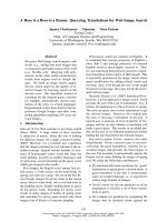
in the portal areas. MVI was identified in 83 (18.1%)
patients. MVI was observed at a magnification of 200×
with cluster tumor cells gathering in a vascular space
with/without smooth muscle wall, and mixed with blood
cells (Figure 1).
Impact factors of long-term survival
Among 458 patients, 102 (22.3%) patients experienced
HCC recurrence or de novo tumors. Eighty-four (18.3%)
patients died within 5 years follow-up, among which 50
Du et al. BMC Cancer 2014, 14:38
/>
Page 3 of 7
Table 1 Clinicopathological characteristics of 458 patients
with single SHCC
Clinicopathological features
Valuea
Age(yr), mean (median)
52.65 (52)
Table 1 Clinicopathological characteristics of 458 patients
with single SHCC (Continued)
Peritumor liver fatty change
Sex
Y
90 (19.7%)
N
368 (80.3%)
Male
384 (84.0%)
PFS(mo),median(range)
53.00 (4-75)
Female
74 (16.0%)
OS(mo),median(range)
54.00 (21-75)
Pre-operation AFP value ≥200 ug/mla
147 (33.0%)
Post-operation AFP value ≥20 ug/mlb
178 (63.6%)
Virus infectionc
No virus
30 (6.6%)
HBV
421 (92.0%)
HCV
4 (1.0%)
HBV&HCV
1 (0.2%)
HBV&HEV
1 (0.2%)
Cirrhosis
Y
411 (89.7%)
N
47 (10.3%)
Capsulary formationd
Y
249 (55.5%)
N
200 (44.5%)
e
Tumor border
Clearly
434 (97.3%)
Vaguely
12 (2.7%)
Tumor histological differentiation
Well differentiated
14 (3.1%)
Moderately differentiated
345 (75.3%)
Poorly differentiated
99 (21.6%)
Tumor microscopic manifestation
MVI
Y
83 (18.1%)
N
375 (81.9%)
Tumor infiltrative lymphocytes
≥50/HPF
84 (18.3%)
<50/HPF
374 (81.7%)
Clear cell subtype
Y
23 (5.0%)
N
435 (95.0%)
Tumor fatty change
Y
15 (3.3%)
N
443 (96.7%)
Large cell dyplastic change
Y
36 (7.9%)
N
422 (92.1%)
a
Missing data in 12 cases.
Missing data in 178 cases.
Missing data in 1 cases.
d
Missing data in 9 cases.
e
Missing data in 12 cases.
AFP a-fetoprotein, MVI microvascular invasion.
a
Values are expressed as n(%) or median(range).
b
c
(59.5%) patients had HCC recurrence. The median PFS and
OS durations were 53 and 54 months, respectively. The
5- year PFS and OS rate was 77.7% and 81.7%, respectively.
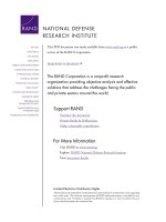
Univariate analysis suggested that SHCC patients with
MVI had shortened PFS and OS (p = 0.012, 0.028, respectively) (Figure 2). Kaplan-Meier survival analyses revealed
that MVI was associated with poorer PFS and OS for
females (p < 0.001, PFS&OS, both), patients with G2 or
G3 histological differentiation (p = 0.017, 0.008, PFS&OS,
respectively), pre-operation serum AFP level ≥ 200ug/ml
(p = 0.022, 0.014, PFS& OS, respectively), or post-operation
serum AFP level ≥ 20 ug/ml (p = 0.014, 0.003, PFS&OS,
respectively), as well as patients with TIL ≥ 50/HPF in
tumor areas (p = 0.001, p = 0.006, PFS&OS, respectively)
(Table 2).
Impact factors of microvascular invasion
Pearson X2 analysis indicated that histological differentiation (p = 0.009) was closely related with MVI. In well or
moderately differentiated tumors, 15.6% with MVI, compared to 27.0% in poorly differentiated tumors. Increasing
tumor size was also associated with higher rates of MVI.
Discussion
Evidence had suggested that tumor size is one of the
most important prognostic factors of patients with hepatocellular carcinoma (HCC) [6]. The latest Consensus on
Diagnosis and Treatment of Primary Liver Cancer in
China adopted 3 cm as the definition of small HCC [2].
Increasing research showed that when HCC is about
3 cm in size may be important as changes occur in DNA
stemline and biological characteristics, and HCC > 3 cm
exhibited a tendency towards more aggressive behavior
and may reach an important turning point for critical
transformation with a resultant change to a more infiltrative behavior [7,8].
It is generally accepted that the incidence of HCC is
higher in men than in women. Gender was not an independent prognostic factor for SHCC patients in our study,
however, MVI displayed survival differences in female
Du et al. BMC Cancer 2014, 14:38
/>
Page 4 of 7
A
B
C
D
Figure 1 Mrophological features of MVI. Tumor thrombus were found in vessels of surrounding liver in A SHCC without capsule (HE ×100); B SHCC
with infiltrative lymphocytes ≥ 50/HPF (HE ×200); C SHCC with invasive border and incomplete capsule (HE ×100); D poorly differentiated SHCC (HE ×50).
p=0.012
A
p=0.028
B
Figure 2 Cox Regression analysis of SHCC patients with MVI. 5 year cumulative survival of patients with MVI after hepatectomy, A PFS
(p = 0.012); B OS (p = 0.028).
Du et al. BMC Cancer 2014, 14:38
/>
Page 5 of 7
Table 2 Impact of MVI and following factors for SHCC
patients’ survival
Variables
With MVI Without MVI
p value1 p value 2
(log-rank) (log-rank)
Sex
Male
69
315
0.257
0.223
Female
14
60
0.002
0.000
≥50
44
218
0.311
0.081
<50
39
157
0.028
0.095
Age(yr)
Pre-operation AFP
value (ug/ml)
≥200
25
122
0.022
0.014
<200
55
244
0.370
0.330
≥20
34
144
0.021
0.003
<20
19
82
0.339
0.633
Y
75
336
0.023
0.056
N
8
39
0.747
0.174
Y
39
211
0.798
0.810
N
41
159
0.008
0.006
Y
78
356
0.012
0.035
N
4
8
0.051
0.091
Post-operation AFP
value (ug/ml)
Cirrhosis
Capsule
Tumor border
Tumor infiltrative
lymphocytes(/HPF)
≥50
10
74
0.001
0.006
<50
73
301
0.065
0.114
Tumor
differentiation
G1
2
12
0.752
0.752
G2&G3
81
363
0.008
0.017
p value1: p value of factors affecting patients’ PFS.
p value2: p value of factors affecting patients’ OS.
AFP a-fetoprotein, MVI microvascular invasion.
patients other than in males. The comparison of variables
showed that AFP had a positive correlation with gender.
Differences in several aspects of medical management
may contribute to the gender disparity in survival rates.
Underlying mechanism is still obscure and optimal
therapeutic regimens or hormonal mechanisms regarding HCC development should be elucidated to improve
clinical outcomes [9].
AFP has served as a representative tumor marker of
HCC for more than 40 years. Liu et al. confirmed that
AFP levels were remarkably higher in patients with vascular invasion (P < 0.001) [10]. High pre-operation and
post-operation AFP level has certain relationship with
long-term survival and MVI. While AFP level is positively
associated with tumor size, the larger tumor the higher
AFP, it is reasonable that AFP level failed to show a prognostic value for survival of SHCC patients.
Microvascular invasion is a histological feature of hepatocellular carcinoma related to aggressive biological behavior.
HCC is characterized by a tendency for vascular invasion.
During the past decades, many studies have addressed
the prognostic significance of MVI in HCC, either as a
primary or secondary object. Nevertheless, the prognostic
significance of MVI remains controversial, and there is
a significant interobserver and intraobserver variability
in the assessment of MVI. Junichi et al claimed that
microvascular invasion does not affect survival of SHCC
(up to 2 cm) [11], others with opposite opinion that
microscopic vascular invasion was an independent factor
for SHCC (up to 3 cm) [12]. Our findings indicate that
MVI has an adverse impact on long-term survival in
SHCC patients. Presence of MVI led to a significant
decrease in PFS and OS at 5 years. Combined our results
with other four studies included in Manuel RodrıguezPeralvarez et al meta-analysis (n = 1959), the RR of
MVI to PFS survival was 1.34 (95% CI =1.24-1.51) [5].
An international consensus delineating what is meant by
MVI in HCC could provide a more consistent evaluation
and, therefore, a more reliable prediction of prognosis
and a better understanding of the pathophysiology of
HCC angioinvasion.
Because microvascular invasion is a histopathologic
diagnosis, it cannot be made prior to the resection of
the tumor. Given the fact that MVI has a significant
impact on recurrence and survival after hepatectomy,
preoperative means of assessing the probability of MVI
are needed. Increasing tumor size and AFP level were
also recorded to be associated with higher rates of MVI
[13,14] in others research. In current study, we found
that tumor grade is a strong predictor of MVI. It’s wellproved that histological grade was an important prognostic factor for survival of HCC patients [15]. It has a
strong relationship with MVI but has no statistically
significance on survival, since most of the patients were
moderately differentiated in our study. It indicated that
SHCC may be not an early-stage of HCC, it certainly
represents an earlier lesion. A significant correlation
between infiltrative tumor margin in preoperative CT
and MVI was claimed, indicating tumor margin may serve
as a radiological sign in prediction of MVI in HCC
patients [16].
Tumor gross features including cirrhosis, capsule and
border failed to show prognostic impact on patients’ survival. Liver resection in the presence of compensated
liver cirrhosis is feasible but associated with a significantly unfavored prognosis for overall and progression-
Du et al. BMC Cancer 2014, 14:38
/>
free survival. Therefore preventing the progression of
cirrhosis are important methods to improve the survival
of HCC patients [17,18].
None of morphological features of surrounding liver
correlated with MVI and patients’ survival, including
peritumor large cell dysplastic foci (LCD), fatty change
of hepatocytes. LCD was originally considered a precursor
lesion of HCC and, accordingly, was referred to as dysplastic. Based on genetic and animal tests, Ferrell thought
that LCD were pathogenetically linked to and associated
with HCC but do not represent a direct precursor of HCC
[19]. Tumor fatty change was more easily observed in
patients with well differentiation (Grade I-II), a significant proportion of small (<2 cm) or “early” HCCs that
are only vaguely nodular have been observed to have
diffuse fatty change [20].
HCC is an example of inflammation-related cancer and
represents a paradigm of the relation occurring between
tumor microenvironment and tumor development. Stromal
cells in tumor microenvironment secrete cytokines and
proteins that promote angiogenesis, metastasis may
contribute to MVI.
The prognosis of HCC patients remains unsatisfactory
although it has been improved much in the past decades.
For SHCC patients, resection is considered the most
effective treatment. However, recurrence is the leading
cause of death during the initial 5-year period after intensive radical resection. High possibility of intrahepatic
recurrence remains one major obstacle for further
improving the survival and prognosis of SHCC patients
after curative resection. As MVI was a pivotal impact
factor for SHCC, the detection and prevention for MVI
will be a target for the therapy of SHCC patients. We will
dedicated in developing the model for prediction of
HCC patients’ survival and exploring measures to prevent
recurrence or metastasis. For those patients with high
risk factors of recurrence, intensive follow-up with
serum AFP and radiology is one of the best methods to
be recommended.
Conclusions
In conclusion, the present study confirmed that microvascular invasion has adverse effect on single small hepatocellular carcinoma patients’ survival and more easily
discovered in worse differentiated and large tumor patients,
we wish to establish a model which can predict and improve the survival of HCC patients.
Consent
Written informed consent was obtained from the patient
for the publication of this report and any accompanying
images.
Page 6 of 7
Competing interests
All the authors declare that they have no competing interests.
Authors’ contributions
Conception and design: YJ. Acquisition of data: MD, HCS. Analysis and
interpretation of data: MD, JZ. Draft the manuscript: MD. Statistical analysis:
MD. Critical revision of the manuscript for important intellectual content: YJ.
Technical or material support: LLC, HYZ, JZ, FT. Study supervision: YJ. All
authors read and approved the final manuscript.
Acknowledgements
We thank the participants to the workshop for encouraging comments and
insights. Weiping Li, Zhixin Qiu and Weishan Zhao for reviewing the manuscript
and important comments helped us in shaping the manuscript in its
present form.
Author details
1
Department of Pathology, Zhongshan Hospital, Fudan University, Shanghai
200032, China. 2Liver Cancer Institution, Zhongshan Hospital, Fudan
University, Shanghai 200032, China.
Received: 4 August 2013 Accepted: 21 January 2014
Published: 24 January 2014
References
1. Jemal A, Bray F, Center MM: Global cancer statistics. CA Cancer J Clin 2011,
61(2):69–90.
2. Guan Z, Yang B, Cong W, Zhou X, Chen X: Concenous on diagnosis and
treatment of primary liver cancer. J Clin Tumor 2009, 14(03):259–269.
3. Choi KK, Kim SH, Choi SB, et al: Portal venous invasion: the single most
independent risk factor for immediate postoperative recurrence of
hepatocellular carcinoma. J Gastroenterol Hepatol 2011, 26(11):1646–1651.
4. Zhou L, Rui JA, Ye DX, et al: Edmondson-Steiner grading increases the
predictive efficiency of TNM staging for long-term survival of patients
with hepatocellular carcinoma after curative resection. World J Surg 2008,
32(8):1748–1756.
5. Rodriguez-Peralvarez M, Luong TV, Andreana L, et al: A systematic review
of microvascular invasion in hepatocellular carcinoma: diagnostic and
prognostic variability. Ann Surg Oncol 2013, 20(1):325–339.
6. Huo TI, Hsu CY, Huang YH, et al: Prognostic prediction across a gradient
of total tumor volume in patients with hepatocellular carcinoma
undergoing locoregional therapy. BMC Gastroenterol 2010, 10:146.
7. Lu XY, Xi T, Lau WY, et al: Pathobiological features of small hepatocellular
carcinoma: correlation between tumor size and biological behavior.
J Cancer Res Clin Oncol 2011, 137(4):567–575.
8. Groeschl RT, Gamblin TC, Turaga KK: Ablation for hepatocellular
carcinoma: validating the 3-cm breakpoint. Ann Surg Oncol 2013. Epub
ahead of print.
9. Tangkijvanich P, Mahachai V, Suwangool P, et al: Gender difference in
clinicopathologic features and survival of patients with hepatocellular
carcinoma. World J Gastroenterol 2004, 10(11):1547–1550.
10. Liu C, Xiao GQ, Yan LN, et al: Value of alpha-fetoprotein in association
with clinicopathological features of hepatocellular carcinoma. World J
Gastroenterol 2013, 19(11):1811–1819.
11. Shindoh J, Andreou A, Aloia TA, et al: Microvascular invasion does not
predict long-term survival in hepatocellular carcinoma up to 2 cm:
reappraisal of the staging system for solitary tumors. Ann Surg Oncol
2013, 20(4):1223–1229.
12. Zhou YM, Yang JM, Li B, et al: Risk factors for early recurrence of small
hepatocellular carcinoma after curative resection. Hepatobiliary Pancreat
Dis Int 2010, 9(1):33–37.
13. Esnaola NF, Lauwers GY, Mirza NQ, et al: Predictors of microvascular
invasion in patients with hepatocellular carcinoma who are candidates
for orthotopic liver transplantation. J Gastrointest Surg 2002, 6(2):224–232.
14. McHugh PP, Gilbert J, Vera S, et al: Alpha-fetoprotein and tumour size are
associated with microvascular invasion in explanted livers of patients
undergoing transplantation with hepatocellular carcinoma. HPB (Oxford)
2010, 12(1):56–61.
15. Han DH, Choi GH, Kim KS, et al: The prognostic significance of the worst
grade in hepatocellular carcinoma with heterogeneous histologic grades
of differentiation. J Gastroenterol Hepatol 2013. Epub ahead of print.
Du et al. BMC Cancer 2014, 14:38
/>
Page 7 of 7
16. Chou CT, Chen RC, Lee CW, et al: Prediction of microvascular invasion of
hepatocellular carcinoma by pre-operative CT imaging. Br J Radiol 2012,
85(1014):778–783.
17. Gassmann P, Spieker T, Haier J, et al: Prognostic impact of underlying liver
fibrosis and cirrhosis after curative resection of hepatocellular
carcinoma. World J Surg 2010, 34(10):2442–2451.
18. Yan T, Zhao JJ, Bi XY, et al: Prognosis of hepatocellular carcinoma: a study
of 832 cases. Zhonghua Zhong Liu Za Zhi 2013, 35(1):54–58.
19. Esquivel CO, Gutierrez C, Cox KL, et al: Hepatocellular carcinoma and liver
cell dysplasia in children with chronic liver disease. J Pediatr Surg 1994,
29(11):1465–1469.
20. Farinati F, Sergio A, Baldan A, et al: Early and very early hepatocellular
carcinoma: when and how much do staging and choice of treatment
really matter? A multi-center study. BMC Cancer 2009, 9:33.
doi:10.1186/1471-2407-14-38
Cite this article as: Du et al.: Microvascular invasion (MVI) is a poorer
prognostic predictor for small hepatocellular carcinoma. BMC Cancer
2014 14:38.
Submit your next manuscript to BioMed Central
and take full advantage of:
• Convenient online submission
• Thorough peer review
• No space constraints or color figure charges
• Immediate publication on acceptance
• Inclusion in PubMed, CAS, Scopus and Google Scholar
• Research which is freely available for redistribution
Submit your manuscript at
www.biomedcentral.com/submit