Cinicomycological study of otomycosis with antifungal drug susceptibility testing of candida isolates using disk diffusion method in Kota region, Rajasthan, India
Bạn đang xem bản rút gọn của tài liệu. Xem và tải ngay bản đầy đủ của tài liệu tại đây (364.93 KB, 11 trang )
Int.J.Curr.Microbiol.App.Sci (2017) 6(6): 3356-3366
International Journal of Current Microbiology and Applied Sciences
ISSN: 2319-7706 Volume 6 Number 6 (2017) pp. 3356-3366
Journal homepage:
Original Research Article
/>
Cinicomycological study of otomycosis with antifungal drug susceptibility
testing of Candida isolates using disk diffusion method in
Kota region, Rajasthan, India
Rawat Sarita*, Saxena Naveen, Chand–E-Anita, Garg Namita,
Verma Vikas and Sharma Khushboo
Department of Microbiology, GMC Kota, Rajasthan, India
*Corresponding author
ABSTRACT
Keywords
Otomycosis,
Aspergillus niger,
Antifungal drug
susceptibility
Article Info
Accepted:
30 May 2017
Available Online:
10 June 2017
Otomycosis or fungal otitis externa is a superficial, sub-acute or chronic infection of the
external auditory canal. Its prevalence has been quoted to range from 9% to 27%. The aim
of the study is to find the prevalence of otomycosis along with its clinicomycological
profile and to detect antifungal drug susceptibility of Candida isolates by disk diffusion
method. A total of 100 samples were collected using swabs from ENT OPD based on
symptoms and otoscopic finding suggestive of otomycosis and were processed in
mycology lab. Otomycosis was diagnosed in 94 % of the cases with highest prevalence in
11-30 years of age group. Male to female ratio was 1.12:1. Aspergillus niger (58%) was
the predominant isolate followed by Aspergillus flavus (23%), Candida spp (12%),
Aspergillus fumigatus (4%), Penicillium (1%), Geotrichum (1%) and scopulariopsis (1%).
Among Candida species, Candida albicans (50%), C. tropicalis (25%), Candida glabrata
(16.66%) and Candida kefyr (8.33%) were isolated. Antifungal drug susceptibility testing
results shows 100% sensitivity to Amphotericin B for all candida isolates. Resistance
against fluconazole was present in 16 % of C.albicans isolates, 25% of C. tropicalis
isolates. Nystatin was resistance among 16% of C.albicans & 25% of C. tropicalis,
whereas clotrimazole resistance was present in 23% of C.albicans, 25% of C. tropicalis &
50% of C.glabrata isolates. Ketoconazole was resistant among 16% of C. albicans. Our
study showed a high prevalence of otomycosis in the Kota region, thus proper diagnosis
and treatment by aseptic techniques for this disease is required.
Introduction
Otomycosis or fungal otitis externa is a
superficial, sub-acute or chronic infection of
the external auditory canal, usually unilateral,
which is characterized by inflammation,
pruritis, scaling and otalgia1.The fungal
agents responsible for this clinical entity are
found as saprotrophic in the environment and
true fungal pathogens are rarely recovered
from these patients. The fungi are usually
secondary invaders of tissue already rendered
susceptible by bacterial infections, physical
injury or excessive accumulation or lack of
cerumen in the external auditory canal. As
such no age group is immune to this disease
but it is commonly seen between 2nd and 3rd
decades of life (2). Otomycosis is one of the
common conditions encountered in a general
otolaryngology clinic setting and its
prevalence has been quoted to range from 9%
3to 27.2% (4, 5) among patients who present
with signs and symptoms of otitis externa and
3356
Int.J.Curr.Microbiol.App.Sci (2017) 6(6): 3356-3366
up to 30% (6, 7) in patients with discharging
ears. The fungal agents responsible for this
clinical entity are A. niger, A. flavus, A.
fumigatus, A. terreus, A. sydowii, C. albicans,
Penicillium,
scopulariopsis
and
dermatophytes
like
Epidermophyton
floccosum, Trichophyton mentagrophytes and
Trichophyton violaceum (8). Other organisms
like Malassezia sympodialis (9) and
Pseudallescheria boydii (10) have also been
reported.
Most patients suffering from early otomycosis
complains of severe itching which often
progresses to pain, hearing loss, and may lead
to tympanic membrane perforations (11-13).
Manifestations are usually unilateral2.
Various factors have been proposed as
predisposing factors for otomycosis, including
a humid climate, excessive presence or
absence of cerumen, swimming, evidence of
fungal infection somewhere else in the body,
instrumentation of the ear, immune
compromised host and more recently
increased use of topical antibiotic/steroid
preparations14. It is more common in
individuals with lower socioeconomical status
with poor hygienic conditions. The infection
has also been observed in people who do not
clean their ears after taking bath or
swimming. The wetness predisposes to fungal
infection.
The present study was carried out to ascertain
the etiologic agents, epidemiologic patterns of
otomycosis in Kota region and to detect
antifungal drug susceptibility of the Candida
isolates using disk diffusion method.
Materials and Methods
The study involved 100 patients who
presented with symptoms of otomycosis at
ENT OPD of MBS hospital, Kota from 2015
to 2016. Clinical details such as chief
complaint, name, age, gender, suspected risk
factors, occupation, history of infection,
address, and other relevant information were
recorded. Informed written consent was
obtained from all subjects. After establishing
a clinical diagnosis, specimen and clinical
materials from the external auditory canal
were collected from all patients by means of
sterile cotton swabs. Materials were divided
into two samples for mycological processing.
With one sample slide KOH(10%) was perfo
rmed. The morphology(yeast and mold), and
other
relevant characteristics (spores,
arthroconidia, septate and non-septate hyphae,
etc.) were identified. The second sample was
inoculated on SDA and SCCA(Himedia).The
media were incubated at room temperature
(25°C) and observed for 3 weeks. Cultures
were examined every day to determine the
probable growth of fungi colonies and their
identification.
The identification process of the isolated
fungi was done based on macroscopic and
microscopic morphology. Lactophenol cotton
blue preparations were made from the
cultures and then examined microscopically.
The slide culture technique was also used
where morphological details of various fungi
was necessary for exact identification. The
isolated yeast species were identified using
various test- germ tube production, Corn meal
agar
morphology,
assimilation
and
fermentation of carbohydrates and Chrome
agar morphology. Antifungal sensitivity of
various fungal isolates was performed by the
disc diffusion method on Mueller Hinton
Agar supplemented with 2% glucose and
0.5mg/ L Methylene Blue (HiMedia make)
and tested for Fluconazole 10μg,Nystatin
100U, Amphotericin 100U, Ketoconazole
10μg, Clotrimazole 10μg disk (HiMedia).
After the measurement of zone of inhibition,
the results of antifungal sensitivity were
interpreted according to criteria given with
HiMedia antifungal discs. All mycological
investigation was carried out in the mycology
section of the Microbiology department.
3357
Int.J.Curr.Microbiol.App.Sci (2017) 6(6): 3356-3366
Results and Discussion
Otomycosis was diagnosed in 94 % of the
cases with highest prevalence in 11-30 years
of age group. Male to female ratio was
1.12:1(Male 53%, Female47%). 98 % cases
were unilateral (Right ear57%, Left ear 41%)
and rest 2 % were bilateral in presentation.
Out of 94 culture positive cases 90 has single
type of growth whereas rest 4 has mixed
growth. Total fungal isolates were 100 as 2
cases were of bilateral otomycosis (both were
culture positive) and 4 cases has mixed
growth. Among various isolates, Aspergillus
niger (58%) was the predominant isolate
followed by Aspergillus flavus (23%),
Candida spp (12%), Aspergillus fumigatus
(4%), Penicillium (1%), Geotrichum (1%) and
scopulariopsis (1%) [Table 1]. Among
Candida spp. isolated most common species
was Candida abicans (50%), followed by
Candida tropicalis (25%), Candida glabrata
(16.66%) and Candida kefyr (8.33%).
Maximum incidence of cases were recorded
in the rainy season from July to September
with peak number of cases in the August
month. As per the occupation field workers
(40%) were the most commonly affected
group, followed by Housewives (24%), Office
workers (15%) and rest 23 % in students and
retired
personnel.
Among
various
predisposing factor, use of oils like mustard
oil, coconut oil and instillation of other form
of ear drops like antibiotic drops, wax
dissolving drops was present in 56 % of the
cases. Following it, use of wooden, metallic
and paper roll was present in 35% of the cases
as predisposing factor. Cerumen was absent in
70 % of the patients [Table2]. History of
swimming was present in 4% cases, 7% of the
cases were diabetic and 5 % cases had history
of covering their heads.
Whereas in 10 % no such predisposing factors
and any chronic illness was present. In our
study itching (86%) was the most common
symptom followed by ear pain (40%),
sensation of ear blockage (42%), tinnitus
(22%), decreased hearing (15%) and
discharge (12%) [Table3]. Antifungal drug
susceptibility testing results shows sensitivity
to Amphotericin B by all candida isolates.
Resistance against fluconazole was present in
16 % of C. albicans isolates, 25% of C.
tropicalis isolates. Nystatin was resistance
among 16% of C. albicans & 25% of C.
tropicalis, whereas clotrimazole resistance
was present in 23% of C. albicans, 25% of C.
tropicalis & 50% of C. glabrata isolates.
Ketoconazole was resistant among 16% of C.
albicans
Otomycosis, a fungal infection of the ear, is
found throughout the world. It is worldwide
in distribution with a higher prevalence in the
hot, humid, and dusty areas of the tropics and
subtropics (4) Itching and pain in the ear are
the most common presenting symptoms of
otomycosis 14, 15. This usually progresses to
discomfort, irritation, sensation of sound in
the ear, sense of blocked ear, hearing loss and
aural discharge. Tympanic membrane
perforation can occur, but is rare. In our
study, the prevalence of otomycosis was 94%,
which is higher than the results found in other
studies, including work by Kumar16 who
found otomycosis in 75.9% of patients;
Pardhan et al., (7), who found otomycosis in
79.4% of patients, Kaur et al., 8, who found
the disease in 74.7% of patients. An analysis
of the age group suggested that otomycosis
can occur at any age. In our study highest
incidence of cases were found in 11-30yrs
(48%) of age group, and lowest among
extreme of ages <10 years(4%) and > than 60
years(4%). The same observation was made
by Paulose et al., (17), HS Satish et al., 18
and RP Rao et al., 19 study.
The people in age group from 11-30 years
usually spend more time in the outdoors and
are more exposed to the fungal spores due to
occupational exposure, travelling etc. making
them more vulnerable to otomycosis. In our
3358
Int.J.Curr.Microbiol.App.Sci (2017) 6(6): 3356-3366
study, otomycosis cases were found to be
more common in males (53%) than
females(47%).These findings were relatively
close to Kaur et al.,8, HS Satish et al.,18, SC
Prasad et al., 14 and A. Kazemi et al., 15
study. As males usually spend more time
outside, so are more exposed to dust, fungal
spores. These result could also be attributed to
the difference in surface lipids between males
and females, as surface lipids are under the
control of sex hormones20. So males are
supposed to have more lipids contents in the
skin of the external auditory canal, thus
making it more favorable for the growth of
fungus. The percentage of females was 47%
in our study, which may be due to the
household work like dusting, cleaning or
gardening thus exposing them to the fungal
spores. Although in RP Rao et al., 19 study
females percentage was higher than males.
Whereas, in Chander J et al., 21 study male to
female ratio was 1:1.This may be due to more
number of females attending the OPD as
compared to males.
Most of the studies revealed otomycosis to be
unilateral disease. In our study 98% of the
cases were unilateral. Out of which right ear
was involved in 57% and left ear in 42 %.
These results correlates with Paulose et
al.,(17), Kaur et al., 8 and H S Satish et
al.,(18) study. The unilateral nature of the
disease may be attributed to the habits like
self-manipulation of ear canal with wicks or
inserting fingers and as majority of the
population in our study were coincidentally
right handed, so more chances of
manipulating right ear may be present. In our
study bilateral otomycosis was present in
patients who had history of swimming. Our
study revealed higher rate of occurrence of
otomycosis cases from july to September.
Rainy season in our study area also
commences at july with peak rainfall during
July and August. In August 2016, Kota city
witnessed a whopping 193 mm of rainfall,
against the previous all-time highest rainfall
of 122.1 mm for the month. Also 100%
humidity was reported in the month of August
2016. The air borne fungal spores are carried
by water vapors, a fact which correlates the
higher rates of infection in monsoon when
relative humidity rises. SC Prasad et al., 14
noticed similar results. Whereas, in Than KM
et al., 27, Barati22 and Ahmad et al., 23 study
they had more occurrences of cases in the
dusty dry season or in autumn. This
difference may be due to the fact that the
symptoms may have started in the rainy
season; but patients did not present on time to
the clinic until the dry season.
In our study field workers (40%) were found
to be the highest affected occupation followed
by housewives (24%), Office workers (13%).
Whereas, the rest 23% of cases were students
and retired personnels. This finding correlates
with the Jaiswal et al., 24study. As the field
worker are more exposed to the
environmental fungal spores, so are the
highest affected group. In our study, history
of instillation of oil (mustard & coconut) and
ear drops like antibiotic drops, steroid drops,
wax removal drops was present in 56% of
total cases. Similar finding was found in HS
Satish et al., 18, M. abdelazeem et al., 20 and
RP Rao et al., 19 study. History of using
wicks was present in 35%, followed by
association with diabetes in 7% and
swimming history in 4%. This findings
correlates with HS Satish et al., 18 study.
Whereas no predisposing factor was present
in 10 % of the cases which is similar to
Lakshmipathi et al., 25, HS Satish et al., 18
study. This may be due to improper history
given by the patient. Oils have fatty acids that
provides a suitable medium for the growth of
fungus, which explains the higher incidence
of otomycosis in people who instill oils
regularly. Recurrent use of antibiotic drops,
steroids, antiseptics or wax solvent ear drops
applications alters the local environment of
3359
Int.J.Curr.Microbiol.App.Sci (2017) 6(6): 3356-3366
the external ear canal and allows super
infection by fungus. Use of metallic
/wooden/paper roll commonly used for
cleaning ear canal, often leads to trauma of
the canal skin into which the fungal spores
may seed in.
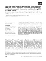
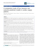
Fig.1 A.niger a) macroscopy (SDA growth), b) microscopy (40x, LCB)
Fig.2 A.flavus a) macroscopy (SDA growth), b) microscopy (40x, LCB)
Fig.3 A.fumigatus a) macroscopy (SDA growth), b) microscopy (40x, LCB)
3360
Int.J.Curr.Microbiol.App.Sci (2017) 6(6): 3356-3366
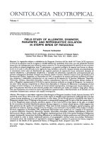
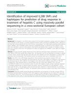
Fig.4 Penicillium a) macroscopy (SDA growth), b) microscopy (40x, LCB)
Fig.5 Scopulariopsis a) macroscopy (SDA growth), b) microscopy (40x, LCB)
Fig.6 Geotrichum a) macroscopy (SDA growth)
Fig.7 Candida spp a) macroscopy (SDA growth), b) microscopy (100x, gram)
3361
Int.J.Curr.Microbiol.App.Sci (2017) 6(6): 3356-3366
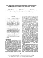
Table.1 Showing various fungal isolates
Fungal Isolated (N=100)
A.niger
A.flavus
A.fumigatus
Candida spp
Penicillium
Scopulariopsis
Geotrichum
No. of cases
58
23
4
12
1
1
1
Table.2 Showing the distribution of cases as per the Predisposing
Factors present Among the Study Population
Predisposing Factors
Use of Oils/ antibiotic drops
Use of wooden/metallic wick/paper roll
Covering of head as per customs
Diabetes
Swimming
No Cerumen
No predisposing factor
Percentage of cases (N=100)
56%
35%
5%
7%
4%
70%
10%
Table.3 Showing distribution of case as per the presenting Symptoms
Among the Study Population
Symptoms
Itching
Ear pain
Tinnitus
Discharge
Sensation of Blockage
Decreased hearing
Percentage of the cases (N=100)
86%
40%
22%
12%
42%
15%
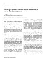
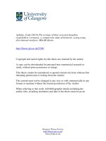
Fig.8 Antifungal drug susceptibility for candida isolates using disk diffusion method
3362
Int.J.Curr.Microbiol.App.Sci (2017) 6(6): 3356-3366
In our study 7% cases had diabetes. Similar
results were in seen in HS Satish et al., 18
where 16% cases had diabetes. History of
swimming in local ponds and swimming
pools was present in 4% of cases. This
findings was similar to HS Satish et al., 18
study where 8% of the total cases had history
of swimming. The lipid mantle layer formed
by the cerumen in the external canal is
considered as the key factor for the protection
of the canal wall, and its removal by frequent
irrigation of the external canal while
swimming, frequent bathing is incriminated
as a cause of recurrent otomycosis. In our
study, history of covering of head was present
in 5 % of cases. Mostly these patients were
Muslim females who used to wear burka.
Head covers increases moisture, heat and
humidity around the ears thus predisposing to
fungal infection. In Aneja KR study 26 the
major predisposing factors responsible for the
otomycosis have been found as the wearing of
traditional customary clothes. In 70 % of
patients, cerumen was lacking. This is in
correlation with M. abdelazeem et al., 20,
Pontes et al., 4 and SC Prasad et al., 14 study.
Absence of cerumen may lead to infection, as
cerumen serves an antimicrobial role by
physically protecting the external auditory
canal skin, establishing a low pH, making
inhospitable environment for pathogens by
producing antimicrobial compounds such as
lysozyme.
In our study itching (86%) was the most
common
presenting
symptom
which
correlates with Than KM, Naing KS and Min
M27, SC Prasad et al., 14, Abdolhassan
Kazemi et al., 15 and M. abdelazeem et al.,
20 study. Sense of ear blockage was present
in 42% of cases, similar to Than KM, Naing
KS and Min M27, SC Prasad et al., 14,
Abdolhassan Kazemi et al., 15studies. Otalgia
was present in 40% of cases and tinnitus in
22% of the cases. The aforementioned
symptoms in similar percentage were found in
HS Satish et al., 18 study. Discharge was
present in 12 % of the cases. In Abdolhassan
Kazemi et al., 15 study similar results were
found. Decreased in hearing was present in 15
% of the cases which correlates with M.
abdelazeem et al., 20 study. Discharge was
present in 12 % of the cases. In Abdolhassan
Kazemi et al., 15 study similar results were
found. Decreased in hearing was present in 15
% of the cases which correlates with M.
abdelazeem et al., 20 study. The mycosis of
external ear canal results in superficial
epithelial exfoliation, inflammation of the ear
canal skin, formation of masses of debris
containing
hyphae
and
suppuration.
Inflammation of the ear canal skin results in
itching and pain..In addition, symptoms like
tinnitus, aural fullness and decreased hearing
are as a result of accumulation of fungal
debris in the ear canal thus obstructing the ear
canal. Discharge is usually a more common
symptom in bacterial origin otitis externa. In
our study discharge was present maximally in
candida origin otomycosis.
In our study out of 100 samples, 94 were
culture positive. The negative cultures might
have been the result of previous treatment
before these patients entered our study. Single
fungal isolate was present in 90 cases whereas
mixed growth was present in 4 cases and two
cases had bilateral otomycosis, making a total
isolates to 100. In our study Aspergillus niger
was the most common isolate accounting for
58% which simulates to results of Yassin et
al., 28, Chander J et al., 21, Kaur et al., 8, HS
Satish et al., 18, Abdolhassan Kazemi et al.,
15 and RP Rao et al.,19 studies. Next to
Aspergillus niger, the most common isolates
were Aspergillus flavus (23%) and
Aspergillus fumigatus (4%). These findings
correlates with that of Abdolhassan Kazemi et
al., 15 and RP Rao et al., 19 study. In our
study Candida was isolated in 12 % of the
cases which is similar to Kaur et al., 8 and RP
Rao et al., 19 study. Penicillium isolated was
3363
Int.J.Curr.Microbiol.App.Sci (2017) 6(6): 3356-3366
1%. In Kaur et al., 8, RP Rao et al., 19 and
HS Satish et al., 18 study also similar results
were obtained. Geotrichum (1%) 29 and
Scopulariopsis
30(1%)
were
also
isolated.Scopulariopsis is a rare isolate found
to be associated with otomycosis. Among 12
% of the Candida spp isolated Candida
albicans (50%) was the major isolate followed
by Candida tropicalis (25%), Candida
glabrata (16.66%) and Candida kefyr (8.3%).
Aspergillus is abundant in soil or sand which
contains decomposing vegetable matter.
Whereas, Aspergillus niger is a common food
contaminant, a black mold which often grows
on a variety of fruits and vegetables. There
conidia being aerodynamic in nature are
dessicated rapidly in tropical sun and blown
in wind as small dust particles and are carried
by water vapors, a fact which correlates the
higher rates of infection, in monsoon when
relative humidity rises to 80%. And also the
human external auditory canal is an ideal
environment for this fungus to grow and
abundance of proteins and carbohydrates and
favorable humidity and temperature explains
this finding. Also Aspergillus are found to be
more common in hot and humid countries
whereas
Candida
spp.
has
more
preponderance of infections in temperate
regions13.Our study area comes under
subtropical zone, so Candidal isolates were
less in our study. The secretion of aspartic
proteinases (Sap1p to Sap10p) is an important
virulence determinant of C. albicans. Saps
facilitate invasion and colonization of host
tissue by disrupting host mucosal membranes
and degrading important immunological and
structural defense proteins.
Antifungal drug susceptibility testing results
shows sensitivity to Amphotericin B by all
candida
isolates.
Resistance
against
fluconazole was present in 16 % of C.
albicans isolates, 25% of C. tropicalis
isolates. Nystatin was resistance among 16%
of C.albicans & 25% of C. tropicalis, whereas
clotrimazole resistance was present in 23% of
C.albicans, 25% of C. tropicalis & 50% of
C.glabrata isolates. Ketoconazole was
resistant among 16% of C. albicans.
Clotrimazole which is the most commonly
prescribed drug for the treatment of
otomycosis was found to be resistant in 23 %
of the candida isolates. This clearly reflects
that all cases of otomycosis should not be
treated on just OPD basis, but rather should
be sent for fungal culture and antifungal drug
susceptibility testing and then should be
treated accordingly.
Acknowledgement
To all the patients who are the part of this
study.
References
1. Jadhav VJ, Pal M, Mishra GS. Etiological
significance of Candida albicans in
otitis externa. Mycopathologia 2003;
156(4):313-5.
2. Chander Jagdish, Textbook of Medical
Mycology, 3rd Edition. Chandigarh:
Mehta publishers; January 2009. p. 4189,343-345,279-280.
3. T. Mugliston and G. O'Donoghue,
―Otomycosis—a continuing problem,‖
Journal of Laryngology and Otology,
vol. 99, no. 4, pp. 327–333, 1985.
4 Pontes ZB, Silva AD, Lima Ede O, Guerra
Mde H, Oliveira NM, Carvalho Mde F,
et al., Otomycosis: a retrospective
study. Braz J Otorhinolaryngol. 2009;
75(3):367–70.
5 J. Fasunla, T. Ibekwe, and P. Onakoya,
―Otomycosis in western Nigeria,‖
Mycoses, vol. 51, no. 1, pp. 67–70,
2008.
6. P. Kurnatowski and A. Filipiak,
―Otomycosis:
prevalence,
clinical
symptoms, therapeutic procedure,‖
3364
Int.J.Curr.Microbiol.App.Sci (2017) 6(6): 3356-3366
Mycoses, vol. 44, no. 11-12, pp. 472–
479, 2001.
7 B. Pradhan, N. Ratna Tuladhar, and R. Man
Amatya, ―Prevalence of otomycosis in
outpatient department of otolaryngology
in Tribhuvan University Teaching
Hospital, Kathmandu, Nepal,‖.Annals
of
Otology,
Rhinology
and
Laryngology, vol. 112, no. 4, pp. 384–
387, 2003.
8. Kaur R, Mittal N. Kakkar M, Aggarwal
AK.Mathur MD. Otomycosis: A
clinicomycological study. Ear, Nose and
Throat Journal 2000; 79: 606-9.
9. Chai FC, Auret K, et al.,Malignant otitis
externa by Malassezia sympodialis. I J
Head Nec, 2000:22:87-9.
10. Bhally HS et al.,Otitis caused by
Scedosporium apiospermum in an
immunocompetent child. Int J Pediatric
Otorhinolaryngology. 2004; 68:975-8.
11. B. Viswanatha, D. Sumatha, and M. S.
Vijayashree,
―Otomycosis
in
immunocompetent
and
immunocompromised
patients:
comparative study and literature
review,‖ Ear, Nose & Throat Journal,
vol. 91, pp. 114–121, 2012.
12. W. B. Hurst, ―Outcome of 22 cases of
perforated tympanic membrane caused
by otomycosis,‖ Journal of Laryngology
and Otology, vol. 115, no. 11, pp. 879–
880, 2001.
13. J. C. Stern and F. E. Lucente,
―Otomycosis,‖ Ear, Nose and Throat
Journal, vol. 67, no. 11, pp. 804–810,
1988.
14. SC Prasad et al., Primary Otomycosis in
the Indian subcontinent: Predisposing
Factors,
Microbiology
and
classification.Int J Microbiol 2014;
2014:636493.
15. A Kazemi et al., Etiologic Agents of
Otomycosis in the North -Western Area
of Iran. Jundishapur Journal of
Microbiol.2015 Sep; 8(9):e21776.
16. Kumar KR. Silent perforation of tympanic
membrane and otomycosis. Indian
Journal
of
otolaryngology.1984;
36(4):161-162.1997 1997-1998.
17. Paulose KO, AL Khalifa S, Shenoy P,
Sharma RK. Mycotic infection of the
ear (otomycosis): A prospective Study.
J Laryngol Otol 1989; 103:30-5.
18.
H.S.
Satish,
Viswanatha.B,
Manjuladevi.M.‖ A Clinical Study of
Otomycosis‖. IOSR Journal of Dental
and
Medical
Sciences.
22790861.Volume 5, Issue 2 (Mar. - Apr.
2013), PP 57-62
19. Rajeshwari Prabhakar Rao, Rishmitha
Rao.‖ A Mycological Study of
Otomycosis ―.IJCMR, Vol.3, Issue 7,
July 2016:2454-7379.
20. Metwally ABDELAZEEM, Ahmed
GAMEA, Hanan MUBARAK, Nessma
ELZAWAWY.
―Epidemiology,
causative agents, and risk factors
affecting human otomycosis infections‖.
Turk J Med Sci (2015) 45: 820-826.
21. Chander J et al.,‖Otomycosis-A
Clinicomycological study and efficacy
of
mercurochrome
in
its
treatment‖.Mycopathologia1996;
135(1):9-12.
22. Barati B, Okhovvat SAR, Omrani MR.
Otomycosis in Central Iran: A Clinical
and Mycological Study. Iran Red
Crescent J. 2011; 13(12):873–76.
23. Mogadam Ahmad Yegane, Asadi
Mohammad Ali, Dehghani Rohullah,
Hooshyar Hossein. The prevalence of
otomycosis in Kashan, Iran, during
2001–2003. Jundishapur Jo Microbiol.
2009; 2(1):18–21
24. Jaiswal SK. Fungal Infection of Ear and
its
Sensitivity
Pattern.Indian
J
Otolaryngol 1990; 42(1):19-2.
25. Laksmipati G, Murti RB. Otomycosis.J
Indian Med Assoc 1960; 34:439-1.
26. Aneja KR, Sharma C, Joshi R. Fungal
infection of the ear: a common problem
3365
Int.J.Curr.Microbiol.App.Sci (2017) 6(6): 3356-3366
in the north eastern part of Haryana. Int
J Pediatr Otorhinolaryngol. 2010;
74(6):604–7.
27. Than KM et al.,‖Otomycosis in Burma
and its treatment‖ American Journal of
Tropical Medicine & Hygiene1980;
29:620-3.
28. Yassin A, Maher A, Moawad MK.
―Otomycosis: A survey in the eastern
Province of Saudi Arabia.‖J Laryngol
Otol1978; 92:869-76.
29. Degerli K, Ecemis T, Gunhan K,
Baskesen T, Kal E. [Agents of
otomycosis in Manisa region, Turkey,
1995-2011]. Mikrobiyol Bul. 2012;
46(1):79-84.
30. Hennequin C, el-Bez M, Trotonx J,
Simonet M. Ann Otolaryngol Chir
Cervicofac. 1994; 111(6):335-4.
How to cite this article:
Rawat Sarita, Saxena Naveen, Chand–E-Anita, Garg Namita, Verma Vikas and Sharma
Khushboo. 2017. Cinicomycological study of otomycosis with antifungal drug susceptibility
testing of Candida isolates using disk diffusion method in Kota region, Rajasthan.
Int.J.Curr.Microbiol.App.Sci. 6(6): 3356-3366.
doi: />
3366