Isolation and characterization of microorganisms from agriculture soil of Magnifera indica Orchard
Bạn đang xem bản rút gọn của tài liệu. Xem và tải ngay bản đầy đủ của tài liệu tại đây (265.59 KB, 7 trang )
Int.J.Curr.Microbiol.App.Sci (2017) 6(6): 2707-2713
International Journal of Current Microbiology and Applied Sciences
ISSN: 2319-7706 Volume 6 Number 6 (2017) pp. 2707-2713
Journal homepage:
Original Research Article
/>
Isolation and Characterization of Microorganisms from Agriculture Soil of
Magnifera indica Orchard
Y. C. Rashmi1*, R. Reshmi1, R. Poornima2 and Sujeet Kumar2
1
2
Mount Carmel College, Palace Road Bangalore, India
Department of Plant Biotechnology, University of Agricultural Sciences, GKVK,
Bengaluru-560065, India
*Corresponding author
ABSTRACT
Keywords
Mango, Bacterial
pathogen, Fungal
pathogen,
Biochemical test.
Article Info
Accepted:
26 May 2017
Available Online:
10 June 2017
A broad range of microorganisms are present in soil of mango orchard which
involved in various mango plant diseases. In order to preliminary study for plant
pathogenesis the soil samples were collected from GKVK, University of
Agricultural Science, Bangalore, Karnataka, India. A number of bacterial and
fungal isolates were obtained from soil sample. The bacterial isolates were
characterized by Gram staining, Catalase test, MR test, VP test, IND test and
Citrate test and fungal isolates were characterized by staining. These analyses
revealed the presence of various bacterial pathogen including Klebsiella
pneumoniae, Enterobacter aerogenes, Shigella species, Bacillus anthracis,
2007). species,
Bacillus
subtilis,
Staphylococcus
Streptococcus
species,
Corynebacterium, Micrococcus, Azomonas species and Rhizobium species.
Identification of Aspergillus niger, Aspergillus flavus, Fusarium oxysporum,
Penicillium and Rhizopus species characterized as a fungal pathogen. The present
study provided baseline information regarding the phytopathogenic bacteria and
fungus which associated with soil of mango orchard.
Introduction
Mango (Mangifera indica L.) is an important
fruit crop of the tropical and subtropical
countries (Litz, 2009). The mango tree is
considered to have evolved in the rainforests
of South and South-east Asia (Knight, 1980;
Krishna and Singh, 2007). India is the largest
producer of mango in the world, contributing
to nearly 46% of the total world production.
The major constrain of mango production is
many devastating diseases (Lim and Khoo,
1985; Iqbal et al., 2006; Rajput and Rao,
A range of microorganisms are involved in
these diseases such as fungi, algae and
bacteria (Litz, 2009). These microbes cause
sets of symptoms including dieback, spots,
necrosis, mildew scab, blotch, anthracnose
and rots in mango trees (Ploetz, 2003;
Freeman et al., 1999; Haggag and Abd ElWahab, 2009). Pseudomonas syringae and
Xanthomonas sp. (causing apical necrosis and
bacterial black spot respectively) are among
the few known bacterial pathogens of mango
2707
Int.J.Curr.Microbiol.App.Sci (2017) 6(6): 2707-2713
trees (Cazorla et al., 1998; Pruvost et al.,
2005; Ah-You et al., 2007). Currently, mango
trees in India are suffering from a disease
with symptoms like Dieback, Powdery
Mildew, Anthracnose/Blossom Blight, Mango
Malformation, Alternaria Leaf Spot, Bacterial
Canker, Stem End Rot, Gummosis and Root
Rot (Kumar et al., 1993; Ploetz, 2001;
Khanzada et al., 2004; Youssef et al., 2007).
There exists a lot of diversity of regarding the
prevalence of microorganism in mango
orchard soil of various parts of the world. In
India, however, scant information is available
about the prevalence of microorganism strains
in various parts of the country. Understanding
local pathogen genetic diversity is the first
step in a successful integrated disease
management programme. One of the purposes
of the present investigation on isolation of
microorganism form mango orchard soils of
Karnataka is to characterize biochemically.
Materials and Methods
of PDA is dissolved in 1000 ml of distilled
water and sterilized in autoclave for 15 min at
1210C. Streaking plate method was used to
get single colonies of pure culture.
Sample inoculums
One ml of 10-5 dilution of soil suspension was
plated out as innocula onto freshly prepared
sterile nutrient agar medium in petridishes
(Bacterial growth).
The innocula were evenly spread on the
surface of the nutrient agar plates by using a
sterile bent glass rod. After incubation for 2448 hrs at 370C, mucous colonies were formed
over the plates. Similarly for fungal growth
1ml of 10-7 dilution of soil suspension were
plated out as innocula onto freshly prepared
sterile Potato Dextrose Agar (PDA) medium
in Petri dishes. The innocula were evenly
spread on the surface of the PDA plates by
using a sterile bent glass rod. After incubation
for 48-72 hrs at 280C, fungus colonies were
formed over the plates.
Soil Sample collection
Gram staining
Soil samples were collected from the six sites
as unmoist soil, moist soil, shaded soil,
unshaded soil, Aged soil and new sapling soil
of mango orchard of University of
Agricultural Sciences, Bangalore, Karnataka,
India. These different sites helpful for capture
the diversity of the microorganisms. The soil
samples (0-15cm depth) were collected from
each site into freshly unused polythene bags.
Pure culture
For reducing microbial population, 1 g of soil
was dissolved in 10 ml of sterile distilled
water to make soil suspension. Serial dilution
was carried out for getting isolated single
colony. In this research, nutrient agar medium
was used for bacterial growth and PDA for
fungal growth. 28 g of nutrient agar was
dissolved in 1000 ml distilled water and 39 g
A loop full of the bacterium was spread on a
glass slide and fixed by heating on a very low
flame. Aqueous crystal violet (Himedia)
solution (0.5%) was spread over the smear for
30 seconds and then gently washed with slow
running tap water for one minute. It was then
flooded with iodine for one minute, rinsed in
tap water and decolorized with 95% ethanol
until colorless runoff. After washing, the
specimen was counter-stained with safranin
(Himedia) for approximately 10 seconds,
washed with water, dried and observed under
microscope at 40X using immersion oil
(Schaad, 1980).
Biochemical tests
Biochemical tests such as Indole test, Catalase
test, MR test, VP test, IND test and citrate test
2708
Int.J.Curr.Microbiol.App.Sci (2017) 6(6): 2707-2713
were carried out to find the enzymatic activity
of isolated organism.
Indole test
One percent (1%) of tryptone broth was
inoculated with a bacteria colony. Incubate
inoculated tubes at 370C for 48 hours. After
48 hours of incubation, add 1ml of Kovac’s
reagent and then shake the tubes gently and
allow standing for 20 minutes. The formation
of the red coloration at the top layer indicated
positive and yellow coloration indicates
negative.
Catalase test
This was carried out by putting a drop of
Hydrogen peroxide on all 6 clean slides. With
the edge of another slide, a colony of
organism was picked and allowed to be in
contact with the hydrogen peroxide. Presence
of bubbles indicates positive reaction and
absence of bubbles indicates negative
reaction.
MR-VP test
Prepare a MR-VP broth of pH 6.9 and then
pour the 5ml of broth in each of 6 test tubes
and sterilize by autoclaving at 15 lb pressure
for 15 min. Inoculate the test tubes with test
organism and incubate all the tubes at 370C
for 48-72 hrs, after which add 5 drops of
methyl red indicator to all the tubes, a red
color formation signifies a positive methyl red
test and yellow color signifies a negative
methyl red test. To the rest of the broth tubes
add 5 drops of 4% potassium hydroxide
(KHO) were added followed by some 15
drops of 5% alpha naphtol in ethanol. Shake
the tubes gently for 1min and allow the
reaction to complete for about 30-45 min. The
red color formation indicates a VP positive
test while no color change indicates VP
negative test.
Citrate utilization test
Prepare the Simmon’s citrate agar pH 6.9.
This was carried out by inoculating the test
organism in all test tubes containing simmon
citrate medium and after inoculation, these
test tubes were incubated at 370C for 48-72
hrs. The development of deep blue color after
incubation indicates a positive result.
IND test
Inoculate the tryptophan broth with broth
culture or emulsify isolated colony of the test
organism in tryptophan broth. Incubate at
370C for 24-48 hrs in incubator. Add 0.5 ml
of Kovacs reagent to the broth culture. The
positive result will show a red color ring
formation after the addition of Kovacs
reagent. The negative result will show a
brown color ring formation after the addition
of Kovacs reagent.
Results and Discussion
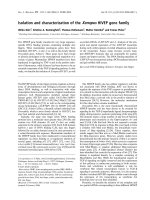
This study revealed that soil samples were
analysed with respect to different types of
bacteria and fungi. The bacteria found in all
six soil samples were biochemically
characterized
as
Staphylococcus
sp.,
Streptococcus sp., Klebsiella pneumoniae,
Enterobacter aerogenes, Shigella sp.,
Micrococcus sp., Bacillus anthracis, Bacillus
subtilis and Cocci sp., Azomonas sp.,
Corynebacterium sp., Rhizobium sp. are the
dominating species of the soil samples (Table
1 and Fig. 2). This result also supported by
previous researcher (Holding, 1971; Kumar et
al., 1993; Ploetz, 2001; Khanzada et al.,
2004; Youssef et al., 2007; Musliu
Abdulkadir and Salawudeen Waliyu, 2012;
Khan et al., 2014; Rupali, 2015). Gram
staining result reveals that Cocci, Klebsiella
pneumoniae,
Enterobacter
aerogenes,
Azomonas sp., Rhizobium sp. are Gramnegative (G-ve) and Micrococcus sp.,
2709
Int.J.Curr.Microbiol.App.Sci (2017) 6(6): 2707-2713
Staphylococcus sp., Streptococcus sp.,
Cornybacterium sp., Bacillus anthracis,
Bacillus subtilis are Gram-positive (G+ve).
Similarly, when the soil samples were tested
for different types of fungi, Penicillium,
Aspergillus
niger, Aspergillus
flavus,
Rhizopus, Fusarium oxysporoum.
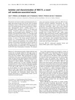
In this study the isolated fungi were identified
on the basis of cultural, microscopic and
morphological characteristics (Fig. 1). Nayak
(2015) also isolated similar type of fungus
from the rhizoshere of mango plant.
Fig.1 Identified fungi and their microscopic image. A and B; Aspergillus niger,
C; Aspergillus flavus D. Fusarium sp.
2710
Int.J.Curr.Microbiol.App.Sci (2017) 6(6): 2707-2713
Fig.2 Biochemical characterization of the soil isolate
Table.1 Morphology and biochemical characterization of bacterial isolates
Sl.No.
1
2
3
4
5
6
7
8
9
10
11
Identified
Bacteria
Klebsiella
Enterobacter
aerogenes
Micrococcus
Staphylococcus
Streptococcus
Corynebacterium
Azomonas
Rhizobium
Shigella sp.
Bacillus
anthracis
Bacillus
subtilis
Gram stain
MR test VP
test
-ve
+ve
-ve
+ve
IND test
-ve(bacilli)
-ve(bacilli)
Catalas
e test
+ve
+ve
-ve
-ve
Citrate
test
+ve
+ve
+ve(cocci)
+ve(cocci)
+ve(cocci)
+ve(bacilli)
-ve (cocci)
-ve (Rod)
-ve (Rod)
+ve (bacilli)
+ve
+ve
+ve
+ve
+ve
+ve
+ve
+ve
+ve
-ve
-ve
-ve
-ve
+ve
-ve
-ve
-ve
-ve
-ve
-ve
-ve
+ve
-ve
-ve
-ve
-ve
-ve
-ve
-ve
+ve
-ve
-ve
-ve
+ve
-ve
-ve
-ve
+ve (bacilli)
+ve
-ve
+ve
-ve
+ve
2711
Int.J.Curr.Microbiol.App.Sci (2017) 6(6): 2707-2713
The isolation of various fungal and bacteria
species of soil sample is quite rich in
microbial flora. In agriculture process soil
microorganisms such as bacteria and fungi
may play important roles in soil fertility and
pathogenesis in the form of loss and gain in
the production of grains, fruits, and
vegetables. Moreover, it also helps to
maintain or enhance the environment quality
and conserve natural resources. Identification
and characterization of isolated bacteria were
performed
by
morphological,
microscopically, biochemical tests such as
shape, arrangement, colonies, growth, indole
production test, methyl red and VogesProskauer test, citrate utilization test, catalase
test, growth at 37 °C. This study provides
knowledge on microorganisms present in
GKVK mango orchid soil habitat.
In conclusion the goal of this research was to
collect and characterize the soil sample from
mango orchard of Karnataka. In this study, we
collected soil sample from six sites of mango
orchard and characterized Bacillus anthracis,
Bacillus
subtilis,
Cocci,
Klebsiella
pneumoniae,
Enterobacter
aerogenes,
Micrococcus species, Staphylococcus sp.,
Streptococcus sp., Corynebacterium sp.,
Azomonas sp., Rhizobium sp. as a bacterial
pathogen and fungi pathogen as an
Aspergillus
niger, Aspergillus
flavus,
Fusarium oxysporium and Penicillium sp.
Acknowledgement
We thank Department of Biotechnology
University of Agricultural Science, Bengaluru
and Mount Carmel College, Bengaluru for
providing the labs and for the support to do
the work.
References
Ah-You, N., Gagnevin, L., Chiroleu, F.,
Jouen, E., Neto, J.R. and Pruvost, O.
2007. Pathological variations within
Xanthomonas
campestris
pv.
Mangiferae
indicae
support
its
separation into three distinct pathovars.
Phytopathol, 97(12): 1568-1577.
Cazorla, F.M., Tores, J.A., Olalla, L., PerezGarcia, A., Farre, J.M. and Vicente, A.
1998. Bacterial apical necrosis of
mango in southern Spain: a disease
caused by Pseudomonas syringae pv.
syringae. Phytopathol, 88: 614-620.
Freeman, S., Maimon, M. and Pinkas, Y.
1999. Use of GUS transformants of
Fusarium subglutinans for determining
etiology of mango malformation
disease. Phytopathol, 89: 456-461.
Haggag, W.M. and Abd El-Wahab, M.E.
2009. First report of Fusarium
sterilihyphosum and F. proliferatuminduced malformation disease of mango
in Egypt. J. Plant Pathol, 91(1): 231240.
Holding, A.J. and Collee J.G. 1971. Routine
biochemical tests, In Methods in
Microbiology, (Norris JR and Ribbons
DW, eds), Academic Press Inc. Ltd,
London, pp 1-32.
Iqbal, Z., Mehboob-ur-Rahman, A.A., Dasti,
Saleem, A. and Zafar, Y. 2006. RAPD
analysis of Fusarium isolates causing
‘‘Mango Malformation’’ disease in
Pakistan.
World
J.
Microbiol.
Biotechnol, 22: 1161-1167.
Khan, I. A., Khan, A., Asif, H., Jiskani, M.
M., Mühlbach, H. P. and Azim, M. K.
2014. Isolation and 16S rDNA sequence
analysis of bacteria from dieback
affected mango orchards in southern
Pakistan. Pak. J. Bot., 46(4): 14311435.
Khanzada, M.A., Lodhi, A. M. and Shahzad,
S., 2004. Pathogenicity of Lasiodiplodia
theobromae and fusarium solani on
mango. Pak. J. Bot., 36(1): 181-189.
Knight, R.J.R. 1980. Origin and world
importance of tropical and subtropical
2712
Int.J.Curr.Microbiol.App.Sci (2017) 6(6): 2707-2713
fruit crops. In: Tropical and Subtropical
Fruits. (Eds.): S. Nagy, P.E. Shaw. AVI,
Westport, CT, USA.
Krishna, H. and Singh, S.K. 2007.
Biotechnological advances in mango
(Mangifera indica L.) and their future
implication in crop improvement: a
review. Biotechnol. Adv., 25: 223-243.
Kumar, J., Singh, U.S., Beniwal, S.P.S. 1993.
Mango malformation: one hundred
years
of
research.
Ann.
Rev.
Phytopathol. 31: 217–32.
Lim, T.K. and Khoo, K.C. 1985. Diseases and
disorders of mango in Malaysia.
Tropical Press, Kuala Lumpur.
Litz, R.E. 2009. The mango botany,
production and uses (2nd edition) CBI
International Wallingford.
Musliu A. and Waliyu, S. 2012. Screening
and Isolation of the Soil Bacteria for
Ability
to
Produce
Antibiotics,
European J. App. Sci., 4: 211-215.
Nayak, B. K., 2015. Isolation and
identification of phylloplane and
endophytic fungi from one ornamental
plant, Mangifera indica. Int. J. Techno.
Chem. Res., 1(3): 188-192.
Ploetz, R.C, 2001. Malformation: a unique
and important disease of mango,
Mangifera indica L. In: Summerell,
B.A., Leslie, J.F., Backhouse, D.,
Bryden, W.L., Burbess, L.W. eds.
Fusarium: Paul E. Nelson Memorial
Symposium. St Paul, MN, USA: APS
Press, 233–47.
Ploetz, R.C. 2003. Diseases of mango. In:
Diseases of Tropical Fruit Crops. (Ed.):
R.C. Ploetz. CAB International,
Wallingford, pp 327-363.
Pruvost, O., Roumagnac, P., Gaube, C.,
Chiroleuand, F. and Gagnevin, L. 2005.
New media for the semiselective
isolation
and
enumeration
of
Xanthomonas
campestris
pv.
Mangiferae indicae, the causal agent of
mango bacterial black spot. J. Appl.
Microbiol., 99: 803-815.
Rajput, K.S. and Rao, K.S. 2007. Death and
decay in the trees of Mango (Mangifera
indica L.). Microbiol. Res., 162: 229237.
Rupali, D. 2015. Screening and Isolation of
Protease Producing Bacteria from Soil
Collected from Different Areas of
Burhanpur Region (MP) India. Int. J.
Curr. Microbiol. App. Sci., 4(8): 597606.
Schaad, N.W. 1980. Laboratory guide for the
identification of plant pathogenic
bacteria. Am. Phytopathol. Soc. St.
Paul. Minn., 28-45.
Youssefa, S.A., Maymonb, M., A. Zveibilb,
A., D. Klein-Guetab, D., Sztejnbergc,
A., Shalabya, A. A. and Freemanb, S.
2007. Plant Pathology, 56: 257–263.
How to cite this article:
Rashmi, Y.C., R. Reshmi, R. Poornima and Sujeet Kumar. 2017. Isolation and Characterization
of Microorganisms from Agriculture Soil of Magnifera indica Orchard.
Int.J.Curr.Microbiol.App.Sci. 6(6): 2707-2713. doi: />
2713