Optical imaging of tumor vascularity associated with proliferation and glucose metabolism in early breast cancer: Clinical application of total hemoglobin measurements in the
Bạn đang xem bản rút gọn của tài liệu. Xem và tải ngay bản đầy đủ của tài liệu tại đây (2.41 MB, 9 trang )
Ueda et al. BMC Cancer 2013, 13:514
/>
RESEARCH ARTICLE
Open Access
Optical imaging of tumor vascularity associated
with proliferation and glucose metabolism in
early breast cancer: clinical application of total
hemoglobin measurements in the breast
Shigeto Ueda1, Noriko Nakamiya1, Kazuo Matsuura1, Takashi Shigekawa1, Hiroshi Sano1, Eiko Hirokawa1,
Hiroko Shimada1, Hiroaki Suzuki4, Motoki Oda4, Yutaka Yamashita4, Osamu Kishino3, Ichiei Kuji2, Akihiko Osaki1
and Toshiaki Saeki1*
Abstract
Background: Near-infrared optical imaging targeting the intrinsic contrast of tissue hemoglobin has emerged as a
promising approach for visualization of vascularity in cancer research. We evaluated the usefulness of diffuse
optical spectroscopy using time-resolved spectroscopic (TRS) measurements for functional imaging of primary
breast cancer.
Methods: Fifty-five consecutive TNM stageI/II patients with histologically proven invasive ductal carcinoma and
operable breast tumors (<5 cm) who underwent TRS measurements were enrolled. Thirty (54.5%) patients
underwent 18F-fluoro-deoxy-glucose (FDG) positron emission tomography with measurement of maximum tumor
uptake. TRS was used to obtain oxyhemoglobin, deoxyhemoglobin, and total hemoglobin (tHb) levels from the
lesions, surrounding normal tissue, and contralateral normal tissue. Lesions with tHb levels 20% higher than those
present in normal tissue were defined as “hotspots,” while others were considered “uniform.” The findings in either
tumor type were compared with clinicopathological factors.
Results: “Hotspot” tumors were significantly larger (P = 0.002) and exhibited significantly more advanced TNM stage
(P = 0.01), higher mitotic counts (P = 0.01) and higher levels of FDG uptake (P = 0.0004) compared with “uniform”
tumors; however, other pathological variables were not significantly different between the two groups.
Conclusions: Optical imaging for determination of tHb levels allowed for measurement of tumor vascularity as a
function of proliferation and glucose metabolism, which may be useful for prediction of patient prognosis and
potential response to treatment.
Keywords: Breast cancer, Diffuse optical imaging, Total hemoglobin, Glucose metabolism
Background
Tumor angiogenesis is a vital process in the early phases
of cancer progression [1-3]. Of late, functional imaging
using near-infrared (NIR) diffuse optical spectroscopy
(DOS) has been used to develop noninvasive measurements for detection of primary breast cancer [4-6]. NIR
time-resolved DOS (NIR–TRS) systems are portable,
* Correspondence:
1
Department of Breast Oncology, International Medical Center, Saitama
Medical University, Hidaka City 350-1298, Saitama, Japan
Full list of author information is available at the end of the article
have high data acquisition rates, and can detect variations in photon transit times resulting from varying
levels of oxyhemoglobin (O2Hb) and deoxyhemoglobin
(HHb), which characterize optical properties of the tissue in terms of absorption coefficient (μa) and decreased
scattering coefficient (μs’) [7]. Quantification of O2Hb
and HHb levels in breast tissue allows for the measurement of total hemoglobin (tHb) levels (tHb = O2Hb +
HHb). Blood volume is directly related to tHb levels,
and abnormal tumor vascularization is believed to
contribute to local elevation in tHb levels [8]. Optical
© 2013 Ueda et al.; licensee BioMed Central Ltd. This is an open access article distributed under the terms of the Creative
Commons Attribution License ( which permits unrestricted use, distribution, and
reproduction in any medium, provided the original work is properly cited.
Ueda et al. BMC Cancer 2013, 13:514
/>
imaging provides excellent contrast of tHb levels in malignant tumor tissue and surrounding normal tissues,
and it has been considered useful for detecting tumor
vascularity and differentiating tumors from neighboring
tissues [9].
Zhu et al. first reported that an ultrasonography (US)guided optical imaging device could be used to distinguish early-stage breast cancer from benign lesions and
that the lesions showed two-fold higher tHb levels than
those observed in benign lesions [10,11]. On the other
hand, in a study of 276 patients in a readers-blinded
comparison study, Collettini et al., a radiologists’ study
group in Germany, reported no significant improvement
in diagnostic performance of initial NIR optical tomography for the detection of primary breast cancer compared
with the performance of a combination of mammography
and optical tomography. This was because of the low
spatial resolution of optical imaging [12].
Although the sensitivity for optical detection of tumoral lesions cannot be expected to be excellent, intrinsic optical contrast of malignant tumors, especially local
elevation of tHb levels, should correlate with biological
and physiological features. We hypothesized that optically visible tumors with locally elevated tHb levels
relative to those in the surrounding normal breast tissue
have increased angiogenesis and that optically low-contrast
tumors are less aggressive. In this study, we prospectively
enrolled consecutive, operable, TNM stageI/II patients with
relatively small tumors (<5 cm) that were initially diagnosed
as invasive ductal carcinoma (IDC) using biopsy. We investigated the potential clinical application of optical imaging
as a means of differentiating the unique features of breast
cancer.
Methods
Patients
We enrolled 88 patients from July 2012 to December 2012
at the Department of Breast Oncology, International
Medical Center, Saitama Medical School (Saitama,
Japan). Seventy women were diagnosed with IDC using
vacuum-assisted biopsy (Mammotome®, Johnson &
Johnson, USA) after identification of tumors by X-ray
mammography, ultrasonography (US), and/or dynamic
magnetic resonance imaging (MRI). Specialized breast
radiologists used US and/or MRI to determine the clinical size of the lesions. Histopathological analysis of
breast cancer, including determination of grade and intrinsic subtype, was performed by at least two experienced pathologists who used hematoxylin–eosin-stained
and immunohistochemical-stained slides of all core biopsy and surgical specimens. Two patients with bilateral
lesions, eight with large lesions (diameter ≥5 cm), 10
who had already received neoadjuvant therapy, and 13
who were diagnosed with non-IDC or special types of
Page 2 of 9
breast carcinoma were excluded. The final study group
comprised 55 consecutive TNM stageI/II breast cancer
women (62.5%) with IDC (diameter <5 cm) who ranged
in age from 22 to 81 years (mean, 58.6 years). The study
protocol was approved by the Institutional Review Board
of Saitama International Medical Center (Saitama, Japan).
Informed consent was obtained from all patients prior to
the study.
TRS breast imaging system
A dual-channel TRS system (TRS20, Hamamatsu
Photonics K.K., Japan) was used to measure the optical
properties of breast tissue at three wavelengths (760 nm,
800 nm, 834 nm). This system uses a time-correlated
single-photon counting (TCSPC) method for measuring
temporal response profiles of tissue against optical pulse
inputs and enables quantitative analysis of light absorption and scattering in tissue as per the Photon Diffusion
Theory [13]. The nonlinear least squares method was
used to fit the solution of the photon diffusion equation
in the reflectance mode to the observed temporal profiles. The coefficients μa and μs’ were obtained at three
wavelengths, and the O2Hb and HHb levels were calculated from the spectroscopic O2Hb and HHb data [14].
Then, the tHb levels were calculated by adding the
O2Hb and HHb levels.
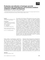
The TRS imaging system is presented in Figure 1. A
handheld probe with a 3-cm source–detector distance
was used to measure the breasts with the patients in
a supine position. On the basis of the information
obtained from the US system (HI VISION Preirus™,
Hitachi, Japan) in which the probe was combined with
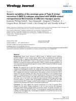
an optical probe as shown in Figure 1, a 10-mm square
grid map (Figure 2) was constructed on the lesion and
surrounding normal tissue. The points of maximum
tumor size were arrayed in the center of the map. The
grid map of a tumor-burdened breast basically comprised 7 × 7 points with a 10-mm interval between two
points in the x–y dimension. A minimum of 49 measurement points was obtained for each breast map. Because the spatial resolution of diffused light is poorer
than that of US, a lesion region of interest (ROI) used
for two-dimensional (2D) image reconstruction of tHb
distribution that was at least two-fold larger than that
observed by using US in the x–y dimension was chosen.
For the contralateral normal breast, a grid map comprising 5 × 5 points with a total of 25 points in the x–y
dimension was constructed in the quadrant region
corresponding to the lesion. For spline interpolation,
2D image processing, and analysis, custom software
(DataGridViewer, version 12; SincereTechnology Corp.,
Kanagawa, Japan) was used.
Average lesion tHb levels were calculated from tissue
O2Hb and HHb levels obtained using TRS measurement
Ueda et al. BMC Cancer 2013, 13:514
/>
Page 3 of 9
Figure 1 A dual-channel TRS system. The patient lies in the supine position on the bed. A US-guided optical probe from the TRS imaging
system (TRS20, Hamamatsu Photonics K.K., Japan) is used to acquire measurements of a patient’s breast and define an ROI in which the breast
lesion can be measured.
of breast tissue corresponding to the ROI. The measurement procedure and grid maps of tHb levels are shown
in Figure 3.
Nuclear grading system
The nuclear grade of IDC was determined by at least
two pathologists according to General Rules for Clinical
and Pathological Recording of Breast Cancer, 15th edition [15]. Nuclear atypia and mitotic count scores were
classified as low (1) and high (2 and 3).
Immunohistochemistry
The expression of estrogen receptor (ER), progesterone
receptor (PgR), and human epidermal growth factor
receptor-2 (HER2) were immunohistochemically examined as a routine for all specimens. Monoclonal
anti-ER antibody (clone ID5) (1:100), monoclonal antiPgR antibody (clone PgR636) (1:100), and the Herceptest
kit for HER2 were purchased from Dako (Grostrup,
Denmark) and used for immunohistochemical analysis.
The method used for immunohistochemistry was as described previously [16]. In brief, the 4 μm-thick sections
were deparaffinized in xylene, and dehydrated in a
graded ethanol series. Antigen retrieval was carried out
by incubation of the tissue sections in a microwave oven
in 10 mM sodium citrate (pH 6.0) with 0.1% Tween40 at
120°C for 45 min. After antigen retrieval, the tissue sections were incubated in 0.3% hydrogen peroxide in
methanol for 30 min, reacted with the primary antibody for
1–3 h, incubated with dextran polymer reagent conjugated
with peroxidase and secondary antibody (envision; Dako,
Glostrup, Denmark) for 1 h, and subsequently reacted
with 3,3-diaminobenzidine tetrahydrochloride-hydrogen
peroxide as the chromogen.
In the present study, a hormone receptor status score
of 3+ (≥10% nuclear staining) was regarded as positive
while a score of 2+/1+/0 (<10%) was regarded as negative [17]. With regard to HER2 expression, cases with a
score of 3+ were judged as showing overexpression. If a
score was 2+, fluorescent in situ hybridization (FISH)
was performed. When amplification of the HER2 gene
using FISH was observed, it was considered to be a positive result [18]. Others were considered to be negative.
18
F-fluoro-deoxy-glucose-PET/CT
Thirty enrolled patients (54.5% total) agreed to undergo 18Ffluoro-deoxy-glucose (FDG)-positron emission tomography
(PET)/computed tomography (CT) scans (Biograph-16,
Siemens–Asahi Medical Technologies, Tokyo, Japan) at
the Department of Nuclear Medicine of our institution.
Ueda et al. BMC Cancer 2013, 13:514
/>
Page 4 of 9
Binary classification of spatial distribution patterns of
lesion tHb
Unique features of tumoral lesions were determined
from an evaluation of tissue tHb distribution patterns in
the breast map. Spatial variations in the lesion tHb map
allowed us to easily locate the maximum optical contrast
corresponding to the tumor site. Figure 3(a) shows representative 2D images of tHb distribution patterns in
breasts with tumors. We found two qualitative features
that enabled differentiation of optically and visually detectable tumors from undetectable ones on the basis of
the distribution pattern of tHb. In this study, approximately half the tumors showed excellent tHb contrast
against the surrounding normal breast tissue. Others
showed equivocal results because of poor contrast
between the tumor and surrounding normal tissue.
Considering the results presented in Table 1 and from
visual assessment, we defined a visually detectable tumor
with at least ≥20% local elevation in tHb levels compared with those in both the contralateral breast tissue
and surrounding normal tissue as a “hotspot” tumor. The
others were described as “uniform” tumors, which did not
form a hotspot in the lesion and exhibited a more uniform
distribution pattern of tHb or a <20% increase in tissue tHb
levels compared with those in the surrounding normal tissue and/or contralateral breast tissue.
Figure 3(b) shows the result of TRS line measurement
of the breast through the tumor center. Ratios of the
tumor tHb and background normal tissue tHb (relative
tHb level) were compared between the two groups. The
line scan “hotspot” tumors showed a clear maximum on
the lesion.
Statistical analysis
Figure 2 TRS measurement procedure and 2D hemoglobin
map construction. Optical measurements comprising a grid map
over tumor and normal breast tissue are obtained using a handheld
probe. The tumor is always located in the center of a map.
Details of the measurement procedure are as previously
described [19,20]. Patients fasted for at least 6 h before
the 18F-FDG PET/CT study. One hour after intravenous
administration of 3.7 Mbq/kg 18F-FDG, a transmission
scan using CT for attenuation correction and anatomical
imaging was acquired for 90 s. PET data were reconstructed via a combination of Fourier rebinning and the
ordered subsets expectation maximization at iteration
number 3 and subset 8 with attenuation correction based
on CT data. An ROI was placed on the primary lesion, including the highest uptake area (circle ROI, diameter 1 cm),
and the maximum standardized uptake value (SUVmax)
in the ROI was calculated. SUV was calculated according
to the following formula: SUV = ROI activity (MBq/ml)/
injected dose (MBq/kg of body weight).
Student’s t test was used to calculate significance for comparison between continuous variables because the data
followed a normal distribution. The Fisher’s exact test and
Pearson’s chi-square test were used to test the statistical significance of the relationship between the independent
groups. The Pearson’s correlation coefficient was used to
analyze the degree of association between two continuous
variables. A level of P < 0.05 was considered to indicate
statistical significance. Logistic regression analysis was performed to find the best-fitting model to describe the relationship between dichotomous characteristics of tumor
tHb distribution (“hotspot” and “uniform” patterns) and a
set of the possible discriminators of clinicopathological factors. Statistical software (MedCalc Software, Broekstraat,
Belgium) was used for calculation.
Results
Baseline characteristics of the patients
Measurement data from a total of 55 tumors were evaluated in this study. There was a minimum 14-day interval
Ueda et al. BMC Cancer 2013, 13:514
/>
Page 5 of 9
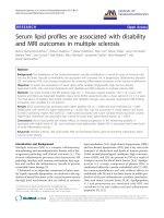
Figure 3 Optical imaging of tHb level in the breast. (a) A 2D image of the breast total hemoglobin level (tHb) is constructed by applying a
spline interpolation algorithm to the raw data. In this example, maps of “hotspot” and “uniform” patterns are shown. (b) A line scan shows that
compared with tumors with a “uniform” pattern, tumors with a “hotspot” pattern exhibit a significantly higher ratio of tHb levels to that in the
background normal breast tissue.
(average, 29.5 days; range, 15–55 days) between the
diagnostic core needle biopsy and baseline TRS measurements before surgery. Clinicopathological data
were obtained from medical records and pathological
reports of the surgical specimens. For nine patients
(16.4%) who received neoadjuvant endocrine treatment
after undergoing TRS scans, pathological data regarding the diagnostic core needle biopsy specimens were
obtained for this study. Table 2 presents the patient
and tumor characteristics.
Comparison of Hb levels in tumors and normal tissue
Absolute values of tHb, O2Hb, and HHb levels were
compared between the lesions and the surrounding normal tissue and between the lesions and the normal
contralateral breast tissue (Table 1). The mean tHb
levels of lesions were 27.6% higher than those in the surrounding normal tissue and 24.6% higher than those in
the contralateral tissue.
According to our tHb distribution pattern criteria, 31 tumors (56.4%) were “hotspot” tumors and 24 (43.6%) were
Ueda et al. BMC Cancer 2013, 13:514
/>
Page 6 of 9
Table 1 Comparison of hemoglobin parameters of lesion, surrounding tissue, and contralateral tissue
Optical parameters mean
(μM) ± SD
Lesion (n = 55)
Surrounding tissue
(n = 55)
Contralateral tissue
(n = 55)
p value*
O2Hb
20.4 ± 10.9
14.8 ± 9.2
15.3 ± 12.8
Lesion vs. surrounding tissue:
p = 0.001
Lesion vs. contralateral tissue:
p = 0.03
HHb
8.9 ± 4
6.7 ± 2.8
6.8 ± 4.6
Lesion vs. surrounding tissue:
p = 0.001
Lesion vs. contralateral tissue:
p = 0.01
tHb
29.3 ± 14.5
21.2 ± 11.7
22.1 ± 17.2
Lesion vs. surrounding tissue:
p = 0.001
Lesion vs. contralateral tissue:
p = 0.02
SD, standard deviation; *Student's t test.
“uniform” tumors. There were no significant differences between the two groups in absolute values of O2Hb (P = 0.9),
HHb (P = 0.7), or tHb (P = 0.8).
Comparison of clinicopathological factors between
“hotspot” and “uniform” tumors
Table 2 shows the patient age, TNM stage, tumor size,
nuclear atypia score, mitotic counts, nodal status, ER
staining, PgR staining, and HER2 status for the “hotspot”
and “uniform” tumors. The “hotspot” tumors showed
significantly more advanced stage than “uniform” tumors
(P = 0.01). The diameters of the “hotspot” tumors were
significantly higher than those of the “uniform” tumors
(P = 0.002). There were no significant differences between the two groups in any other clinicopathological
factors: age (P = 0.3), nuclear atypia score (P = 0.8),
nodal stage (P = 0.4), ER staining (P = 0.3), PgR staining
(P = 0.2), or HER2 status (P = 0.9). The number of “hotspot” tumors showing high mitotic count scores (52.2%)
was significantly higher than that of “uniform” tumors
Table 2 Clinicopathological factor and biomarker results in hotspot versus uniform-patterned breast cancers assigned
by tHb optical imaging
Variables
Values
Total
Hotspot
Uniform
p value
Age
Mean(year) ± SD
58.6 ± 12.3
59.9 ± 11.7
56.5 ± 13
NS*
TNM Stage
I
25
9
16
p = 0.01†
II
30
22
8
Tumor size
Mean(mm) ± SD
21.5 ± 9
24.8 ± 9.9
17.4 ± 5.6
p = 0.002*
Nuclear atypia
High
37
20
17
NS**
Low
5
3
2
Mitosis
High
14
12
2
Low
28
11
17
Nodal status
Positive
10
4
6
Negative
33
20
13
ER
Positive
45
24
21
Negative
6
5
1
PgR
Positive
41
21
20
Negative
10
8
2
HER2
Positive
6
4
2
Negative
44
24
20
FDG SUVmax
Mean ± SD
5 ± 3.3
6.6 ± 3.2
2.6 ± 1.7
†
SD, standard deviation; NS, not statistically significant; Student’s t test; Fisher’s exact test, Pearson’s chi square.
*
**
p = 0.01**
NS**
NS**
NS**
NS**
p = 0.0004*
Ueda et al. BMC Cancer 2013, 13:514
/>
showing high scores (10.5%; P = 0.01). Tumor SUV
measured by FDG PET/CT was significantly higher in
“hotspot” tumors than in “uniform” tumors (P = 0.0004).
The relationship between FDG SUVmax and tHb
When tumor size, TNM stage, mitotic count, and FDG
SUVmax were loaded in logistic regression analysis,
none of these variables contributed significantly to the
prediction of “hotspot” tumor. Figure 4 shows FDG
SUVmax was significantly correlated with relative tHb
level of tumor (coefficient r = 0.49; 95% CI, 0.15-0.75,
P = 0.007).
Discussion
In this study, we investigated the clinical application of
functional NIR–DOS imaging for the measurement of
intrinsic contrast of early-stage breast cancer. Significantly higher tHb levels were observed in early-stage
breast cancer tumors than in the surrounding normal
breast tissue and contralateral normal breast tissue.
However, there were a wide range of tHb levels between
the individual tumors and the normal tissues, and >40%
tumors did not show a clear elevation in tumor tHb
levels because of the presence of equal tHb levels in the
normal tissue. This finding is understandable because
mammary glandular tissue has much denser vascularity
compared with fatty tissue, and the absolute values of
tissue tHb levels vary among individuals. Tumors progress with the growth of new vessels from pre-existing
vessels so that lesion tHb levels continue to correlate
with the extent of vascularity in the background normal
tissue. In addition, tumor Hb levels are reportedly more
Page 7 of 9
sensitive to hormonal fluctuations induced by the menstrual cycle compared with those in the normal breast
tissue, with 10%–14% deviation [21]. Therefore, we focused on increased ratios of tHb levels in lesions to tHb
levels in the surrounding normal tissue and eventually
established a certain criteria for “hotspot” tumors, which
were detectable with a ≥20% local elevation in tHb levels
relative to that in the normal tissue. The other tumors,
which were visually equivocal or not clearly detected by
optical imaging, were classified as “uniform” tumors.
The “hotspot” pattern of tHb level was detected in
56% patients with early-stage breast cancer. Tumor size
was significantly greater in “hotspot” tumors than in
“uniform” ones, but this finding did not act as a predictor of excellent optical contrast because of a remarkable overlap between the two groups. This indicated that
the size of a tumor did not dictate its clarity on optical
imaging.
Mitotic count score, evaluated as a proliferative marker,
was significantly higher in “hotspot” tumors (52.2%) than in
“uniform” ones (10.5%; P = 0.01). In addition, tumor
SUVmax measured by FDG PET/CT was a good index for
discriminating between “hotspot” and “uniform” tumors.
Therefore, high-metabolic tumors should be identifiable by
optical imaging because of progressive angiogenesis, but
some tumors with low metabolic activity may absorb the
NIR light for optical measurements to a lesser degree than
that absorbed by the surrounding normal tissue because of
less blood retention due to less aggressive neoangiogenesis.
A biomarker study conducted by Groves et al. revealed that
tumor FDG uptake was significantly associated with angiogenesis as measured by an immunohistochemical bioassay
Figure 4 Correlation between FDG uptake and tumor tHb patterns. A scatter diagram of two patient groups (“hotspot” and “uniform”)
showing a significant relationship between FDG SUVmax and relative tHb level (coefficient r = 0.49; 95%CI, 0.15–0.72, P = 0.007).
Ueda et al. BMC Cancer 2013, 13:514
/>
of CD105 for new vessel formation in patients with
early-stage breast cancer [22]. This finding suggests
that tumor vascularity is closely associated with tumor
glycolytic activity.
Cancer cells respond autonomously to hypoxia, switch
oxidative phosphorylation in mitochondria to glycolysis,
and positively amplify neoangiogenesis [3,23]. Paradoxically, the phenomenon by which these tumors acquire an
increased glycolytic rate despite normal tissue oxygen
tension is called the Warburg effect [24,25]. Recent research revealed that autonomous upregulation of several
oncogenic signaling mechanisms independent of hypoxia, including a PI3K–AKT pathway, transcriptional
activity of HIF1, and aberrant function of p53, affects
overexpression of glucose transporters and related enzymes. The activation of these mechanisms contributes
to hypermetabolism and neoangiogenesis of the tumor
[22,26]. Therefore, it is evident that increased glucose
metabolism and angiogenesis may be, to some extent,
different phenotypical expressions of common underlying genetic and/or physiological processes [27].
Currently, FDG PET/CT attracts the attention of oncologists because the biological basis of FDG uptake in
cancer metabolism could be the Warburg effect [28].
The result that elevation of tumor tHb levels relative to
those in background normal breast tissue was correlated
with high FDG uptake is consistent with the observation
of recent research that showed the coupling of increased
glucose metabolism of cancer cells to neoangiogenesis
and hypoxia. Therefore, these features of “hotspot” and
“uniform” patterns can add functional information regarding the physiology of the tumor. For example, earlystage breast cancer patients with “hotspot” tumors could
initially be considered chemotherapy candidates in terms
of cancer cell activity.
Furthermore, breast cancer is known to have heterogeneous characteristics of gene expression patterns that
are strongly associated with prognosis and response to
therapy [29]. In the future, we believe that breast cancer
may be further classified into types on the basis of spectral differences.
The strength of this study was that we enrolled a
homogeneous group of consecutive TNM stageI/II patients with small-size (mean, 21.5 mm) IDC tumors,
whereas previous studies on optical breast imaging have
included advanced-stage or various histological types of
breast cancers [9,11,30].
Functional imaging using DOS has limitations with regard to the identification of tumor location because intense light scattering in tissues leads to low spatial
resolution and in-depth information of tissue absorption
cannot be assessed [31]. The current study used data
from a small patient population. A large prospective
study is required to further validate the results.
Page 8 of 9
Conclusions
Optical imaging of breast cancer tHb levels can potentially contribute to the identification of unique functional features of tumor vascularity that add diagnostic
value to cancer management and may assist in the development of a monitoring tool for treatment.
Abbreviations
DOS: Diffuse optical spectroscopy; TRS: Time-resolved spectroscopy; FDG:
18
F-fluoro-deoxy-glucose; PET: Positron emission tomography;
O2Hb: oxyhemoglobin; HHb: Deoxyhemoglobin; tHb: Total hemoglobin;
NIR: Near-infrared; μa: absorption coefficient; μs: Reduced scattering
coefficient; US: Ultrasonography; IDC: Invasive ductal carcinoma;
TCSPC: Time-correlated single-photon counting; ROI: Region of interest;
2D: Two-dimensional; FISH: Fluorescent in situ hybridization;
SUV: Standardized uptake value; ER: Estrogen receptor; PgR: Progesterone
receptor; HER2: Human epidermal growth factor receptor-2.
Competing interests
SU, NN, KM, TS, HS, EH, HS, OS, IK, AO, and TS had no competing interests.
HS, MO, and YY are employees of Hamamatsu Photonics K.K. They have not
applied for any patents related to this study.
Authors’ contributions
SU conceived and designed the study, conducted measurements, analyzed
the data, and performed the statistical and graphical analysis. NN conducted
measurements and analyzed the data with SU. SU and NN acquired funding
in the form of a Hidaka research grant from Saitama Medical University
(SMU). KM, TS, HS, EH, HS, and AO registered patients eligible for the study.
HS and MO advised us on technical issues and maintained the TRS imaging
system. IK participated in FDG PET image acquisition. TS was a significant
contributor to the study design, manuscript content, and organization. All
authors read and approved the final manuscript.
Authors’ information
Shigeto Ueda, MD is a breast surgeon and completed his PhD at SMU.
Research interests include functional PET imaging and diffuse optical
spectroscopy. He currently works as an assistant professor at SMU. Noriko
Nakamiya, MD is a breast surgeon in the Department of Breast Oncology at
SMU. Her research interests are in early breast cancer detection using
mammography and optical spectroscopy. Kazuo Matsuura, MD, PhD is an
associate professor at SMU. He is a breast surgeon. His research interests
include molecular biology and cancer immunology.
Takashi Shigekawa, MD, PhD is an assistant professor in SMU. He is a breast
surgeon. Hiroshi Sano, MD, PhD is an assistant professor at SMU. He is a
breast surgeon. Eiko Hirokawa, MD is an assistant professor at SMU. She is a
breast plastic surgeon. Hiroko Shimada, MD is an assistant professor at SMU.
She is a breast surgeon. Hiroaki Suzuki, PhD is a researcher at Hamamatsu
Photonics K.K. He maintained the TRS imaging system. Motoki Oda, PhD is a
researcher at Hamamatsu Photonics K.K. He also developed and improved
the TRS imaging system. Yutaka Yamashita, PhD is the chief researcher in the
Central Research Laboratory of Hamamatsu Photonics K.K. He developed the
TRS imaging system. Ichiei Kuji, MD, PhD is a radiologist and a professor in
the Department of Nuclear Medicine at SMU. His research interests include
cancer imaging using functional PET. He aids in the detection and diagnosis
of breast cancer using FDG PET scans. Akihiko Osaki, MD, PhD is a breast
surgeon and a professor in the Department of Breast Oncology at SMU. His
research interests include early detection of breast cancer using
mammography and optical spectroscopy. Toshiaki Saeki is a vice president at
SMU and the chief professor in the Department of Breast Oncology. His
research interests include design of clinical trials, molecular biology of
cancer, cancer imaging, and development of molecular targeting agents. His
interests in the field of biophotonics are centered on research and
technology development of diffuse optical imaging for applications in breast
cancer research.
Acknowledgments
The authors would like to thank all staff members at the Central US unit of
the Saitama International Medical Center for their kind cooperation. This
Ueda et al. BMC Cancer 2013, 13:514
/>
work was supported by JSPS KAKENHI Grant Number 25830105 and Hidaka
Research Grant.
Author details
1
Department of Breast Oncology, International Medical Center, Saitama
Medical University, Hidaka City 350-1298, Saitama, Japan. 2Department of
Nuclear Medicine, International Medical Center, Saitama Medical University,
Hidaka City 350-1298, Saitama, Japan. 3Central US Service, International
Medical Center, Saitama Medical University, Hidaka City 350-1298, Saitama,
Japan. 4Central Research Laboratory, Hamamatsu Photonics K.K, Hamamatsu
City 434-8601, Japan.
Received: 9 March 2013 Accepted: 28 October 2013
Published: 31 October 2013
References
1. Folkman J: Tumor angiogenesis: therapeutic implications. N Engl J Med
1971, 285:1182–1186.
2. Relf M, LeJeune S, Scott PA, Fox S, Smith K, Leek R, Moghaddam A,
Whitehouse R, Bicknell R, Harris AL: Expression of the angiogenic factors
vascular endothelial cell growth factor, acidic and basic fibroblast
growth factor, tumor growth factor beta-1, platelet-derived endothelial
cell growth factor, placenta growth factor, and pleiotrophin in human
primary breast cancer and its relation to angiogenesis. Cancer Res 1997,
57:963–969.
3. Jain RK: Normalization of tumor vasculature: an emerging concept in
antiangiogenic therapy. Science 2005, 307:58–62.
4. Tromberg BJ, Pogue BW, Paulsen KD, Yodh AG, Boas DA, Cerussi AE:
Assessing the future of diffuse optical imaging technologies for breast
cancer management. Med Phys 2008, 35:2443–2451.
5. Ueda Y, Yoshimoto K, Ohmae E, Suzuki T, Yamanaka T, Yamashita D, Ogura
H, Teruya C, Nasu H, Ima E, Sakahara H, Oda M, Yamashita Y: Time-resolved
optical mammography and its preliminary clinical results. Technol Cancer
Res Treat 2011, 10:393–401.
6. Fantini S, Sassaroli A: Near-infrared optical mammography for breast cancer
detection with intrinsic contrast. Ann Biomed Eng 2012, 40:398–407.
7. Ohmae E, Oda M, Suzuki T, Yamashita Y, Kakihana Y, Matsunaga A, Kanmura
Y, Tamuraet M: Clinical evaluation of time-resolved spectroscopy by
measuring cerebral hemodynamics during cardiopulmonary bypass
surgery. J Biomed Opt 2007, 12:062112.
8. Tromberg BJ, Cerussi A, Shah N, Compton M, Durkin A, Hsiang D, Butler J,
Mehta R: Imaging in breast cancer: diffuse optics in breast cancer:
detecting tumors in pre-menopausal women and monitoring neoadjuvant chemotherapy. Breast Cancer Res 2005, 7:279–285.
9. Cerussi A, Shah N, Hsiang D, Durkin A, Butler J, Tromberg BJ: In vivo
absorption, scattering, and physiologic properties of 58 malignant breast
tumors determined by broadband diffuse optical spectroscopy. J Biomed
Opt 2006, 11:044005.
10. Zhu Q, Xu C, Guo P, Aguirre A, Yuan B, Huang F, Castilo D, Gamelin J:
Optimal probing of optical contrast of breast lesions of different size
located at different depths by US localization. Technol Cancer Res Treat
2006, 5:365–380.
11. Zhu Q, Hegde PU, Ricci A Jr, Kane M, Cronin EB, Ardeshirpour Y, Xu C,
Aguirre A, Kurtzman SH, Deckers PJ, Tannenbaum SH: Early-stage invasive
breast cancers: potential role of optical tomography with US localization
in assisting diagnosis. Radiology 2010, 256:367–378.
12. Collettini F, Martin JC, Diekmann F, Fallenberg E, Engelken F, Ponder S, Kroencke
TJ, Hamm B, Poellinger A: Diagnostic performance of a Near-Infrared Breast
Imaging system as adjunct to mammography versus X-ray mammography
alone. Eur Radiol 2012, 22:350–357.
13. Patterson MS, Chance B, Wilson BC: Time resolved reflectance and
transmittance for the non-invasive measurement of tissue optical
properties. Appl Opt 1989, 28:2331–2336.
14. Ijichi S, Kusaka T, Isobe K, Okubo K, Kawada K, Namba M, Okada H, Nishida
T, Imai T, Itoh S: Developmental changes of optical properties in
neonates determined by near-infrared time-resolved spectroscopy.
Pediatr Res 2005, 58:568–573.
15. Elston CW, Ellis IO: Pathological prognostic factors in breast cancer. I. The
value of histological grade in breast cancer: experience from a large
study with long-term follow-up. Histopathology 2002, 41:154–161.
Page 9 of 9
16. Ueda S, Tsuda H, Sato K, Takeuchi H, Shigekawa T, Matsubara O, Hiraide H,
Mochizuki H: Alternative tyrosine phosphorylation of signaling kinases
according to hormone receptor status in breast cancer overexpressing the
insulin-like growth factor receptor type 1. Cancer Sci 2006, 97:597–604.
17. Umemura S, Kurosumi M, Moriya T, Oyama T, Arihiro K, Yamashita H,
Umekita Y, Komoike Y, Shimizu C, Fukushima H, Kajiwara H, Akiyama H:
Immunohistochemical evaluation for hormone receptors in breast
cancer: a practically useful evaluation system and handling protocol.
Breast Cancer 2006, 13:232–235.
18. Cuadros M, Villegas R: Systematic review of HER2 breast cancer testing.
Appl Immunohistochem Mol Morphol 2009, 17:1–7.
19. Kuji I, Imabayashi E, Minagawa A, Matsuda H, Miyauchi T: Brown adipose
tissue demonstrating intense FDG uptake in a patient with mediastinal
pheochromocytoma. Ann Nucl Med 2008, 22:231–235.
20. Ueda S, Saeki T, Shigekawa T, Omata J, Moriya T, Yamamoto J, Osaki A,
Fujiuchi N, Misumi M, Takeuchi H, Sakurai T, Tsuda H, Tamura K, Ishida J,
Abe Y, Imabayashi E, Kuji I, Matsuda H: 18F-fluorodeoxyglucose positron
emission tomography optimizes neoadjuvant chemotherapy for primary
breast cancer to achieve pathological complete response. Int J Clin Oncol
2012, 17:276–282.
21. Tanamai W, Chen C, Siavoshi S, Cerussi A, Hsiang D, Butler J, Tromberget B:
Diffuse optical spectroscopy measurements of healing in breast tissue
after core biopsy: case study. J Biomed Opt 2009, 14:014024.
22. Groves AM, Shastry M, Rodriguez-Justo M, Malhotra A, Endozo R, Davidson
T, Kelleher T, Miles KA, Ell PJ, Keshtgar MR: (1)(8)F-FDG PET and biomarkers
for tumour angiogenesis in early breast cancer. Eur J Nucl Med Mol
Imaging 2011, 38:46–52.
23. Goel S, Fukumura D, Jain RK: Normalization of the tumor vasculature
through oncogenic inhibition: an emerging paradigm in tumor biology.
Proc Natl Acad Sci U S A 2012, 109:E1214.
24. Shaw RJ: Glucose metabolism and cancer. Curr Opin Cell Biol 2006, 18:598–608.
25. Hsu PP, Sabatini DM: Cancer cell metabolism: Warburg and beyond.
Cell 2008, 134:703–707.
26. Semenza GL: HIF-1: upstream and downstream of cancer metabolism.
Curr Opin Genet Dev 2010, 20:51–56.
27. Dang CV, Semenza GL: Oncogenic alterations of metabolism. Trends Biochem
Sci 1999, 24:68–72.
28. Larson SM: Positron emission tomography-based molecular imaging in
human cancer: exploring the link between hypoxia and accelerated
glucose metabolism. Clin Cancer Res 2004, 10:2203–2204.
29. Sorlie T, Perou CM, Tibshirani R, Turid A, Geislerg S, Johnsenb H, Hastiee T,
Eisenh MB, Rijni MVD, Jeffreyj SS, Thorsenk T, Quistl H, Matesec JC, Brownm
PO, Botsteinc D, Lønningg PE, Børresen-Daleet AL: Gene expression
patterns of breast carcinomas distinguish tumor subclasses with clinical
implications. Proc Natl Acad Sci U S A 2001, 98:10869–10874.
30. Brown JQ, Wilke LG, Geradts J, Kennedy SA, Palmer GM, Ramanujam N:
Quantitative optical spectroscopy: a robust tool for direct measurement
of breast cancer vascular oxygenation and total hemoglobin content
in vivo. Cancer Res 2009, 69:2919–2926.
31. Yang WT: Emerging techniques and molecular imaging in breast cancer.
Semin Ultrasound CT MR 2011, 32:288–299.
doi:10.1186/1471-2407-13-514
Cite this article as: Ueda et al.: Optical imaging of tumor vascularity
associated with proliferation and glucose metabolism in early breast
cancer: clinical application of total hemoglobin measurements in the
breast. BMC Cancer 2013 13:514.