Early pneumothorax as a feature of response to crizotinib therapy in a patient with ALK rearranged lung adenocarcinoma
Bạn đang xem bản rút gọn của tài liệu. Xem và tải ngay bản đầy đủ của tài liệu tại đây (1.28 MB, 4 trang )
Gennatas et al. BMC Cancer 2013, 13:207
/>
CASE REPORT
Open Access
Early pneumothorax as a feature of response to
crizotinib therapy in a patient with ALK
rearranged lung adenocarcinoma
Spyridon Gennatas1,2*, Susana J Stanway1,2, Robert Thomas1,2, Toon Min1,2, Riyaz Shah3, Mary ER O’Brien1,2
and Sanjay Popat1,2
Abstract
Background: Single arm phase 1 and 2 studies on Crizotinib in ALK-positive patients so far have shown rapid and
durable responses. Spontaneous pneumothoraces as a result of response to anti-cancer therapy are rare in
oncology but have been documented in a number of tumour types including lung cancer. This includes cytotoxic
chemotherapy as well as molecular targeted agents such as gefitinib and Bevacizumab. These often require chest
drain insertion or surgical intervention with associated morbidity and mortality. They have also been associated
with response to treatment. This is the first report we are aware of documenting pneumothorax as response to
crizotinib therapy.
Case presentation: A 48-year-old Caucasian male presented with a Stage IV, TTF1 positive, EGFR wild-type
adenocarcinoma of the lung. He received first line chemotherapy with three cycles of cisplatin-pemetrexed
chemotherapy with a differential response, and then second-line erlotinib for two months before further
radiological evidence of disease progression. Further analysis of his diagnostic specimen identified an ALK
rearrangement by fluorescence in situ hybridization (FISH). He was commenced on crizotinib therapy 250 mg orally
twice daily. At his 4-week assessment he had a chest radiograph that identified a large left-sided pneumothorax
with disease response evident on the right. Chest CT confirmed a 50% left-sided pneumothorax on a background
of overall disease response. A chest tube was inserted with complete resolution of the pneumothorax that did not
recur following its removal.
Conclusion: Our case demonstrates this potential complication of crizotinib therapy and we therefore recommend
that pneumothorax be considered in patients on crizotinib presenting with high lung metastatic burden and with
worsening dyspnoea.
Keywords: Lung cancer, Lung adenocarcinoma, ALK rearrangement, Pneumothorax, Early pneumothorax, Crizotinib,
ALK rearranged lung adenocarcinoma
Background
Crizotinib is approved for ALK rearranged relapsed nonsmall cell lung cancer with most patients treated in the
initial expanded cohort phase 1b study acquiring durable
responses. The overall response rate was 57% (CI 46-68)
and the disease-control rate was 87% [1]. A retrospective
study on the same population by Shaw et al. suggested
* Correspondence:
1
Royal Marsden Hospital, London, UK
2
Royal Marsden Hospital, Surrey, UK
Full list of author information is available at the end of the article
that overall survival for patients with advanced ALKpositive NSCLC was significantly longer in patients given
Crizotinib second- or third-line compared to Crizotinib-na
ïve patients, whose prognosis was very similar to the general NSCLC population. This evidence indicates Crizotinib
might prolong overall survival in ALK-positive NSCLC
patients [2]. Regarding the timing of responses, in both the
phase I and phase II trials, the majority were achieved
within 8 weeks of treatment initiation. The duration of
response was 48.1 and 41.9 weeks respectively indicating a
very rapid and prolonged response. Treatment was
© 2013 Gennatas et al.; licensee BioMed Central Ltd. This is an Open Access article distributed under the terms of the Creative
Commons Attribution License ( which permits unrestricted use, distribution, and
reproduction in any medium, provided the original work is properly cited.
Gennatas et al. BMC Cancer 2013, 13:207
/>
Page 2 of 4
generally well tolerated with gastrointestinal (Grade 1/2)
and visual disorders (Grade 1) being the commonest [3,4].
Case presentation
A 48-year-old Caucasian male ex smoker (10 pack-year
tobacco exposure) presented in 2011 with cough and back
pain. Computerized tomography (CT) imaging showed a
lung primary and metastases to both lungs, spine, and left
adrenal. A computerized tomography (CT)-guided biopsy
confirmed TTF1 positive, EGFR wild-type adenocarcinoma of the lung. He received three cycles of cisplatinpemetrexed chemotherapy with a differential response,
and then second-line erlotinib for two months before further radiological evidence of disease progression. Erlotinib
was administered based on the findings of the BR.21 phase
III trial, which demonstrated that erlotinib prolonged
survival in relapsed NSCLC unselected by EGFR genotype
following progression on first- or second-line chemotherapy compared to placebo (6.7 months vs 4.7 months) [5].
He subsequently received radiotherapy to the spine for
palliation (8Gy, single fraction) followed by radiotherapy
to the mediastinum due to lymphadenopathy (20Gy, 5
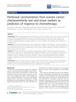
fractions). Thereafter, he was referred for further evaluation, where analysis of his diagnostic specimen identified
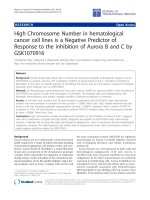
an ALK rearrangement by fluorescence in situ hybridization (FISH) using the Vysis (Abbott) LSI ALK dual
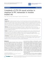
colour, break apart rearrangement probe (Abbott molecular, Illinois, Figure 1). Following a baseline staging CT
(Figure 2), which confirmed multiple metastatic sites,
including the lungs and adrenal gland, as well as a chronically collapsed left lung (first seen on CT prior to commencing erlotinib), he was commenced on crizotinib
therapy 250 mg orally twice-daily. At his 2-week assessment, he reported marginal clinical improvement. At his
4-week assessment he complained of worsening dyspnoea,
Figure 1 ALK rearrangement as visualised by fluorescence in
situ hybridization (FISH). FISH image of ALK re-arrangement using
the Vysis (Abbott) LSI ALK dual color, break apart rearrangement
probe (Abbott molecular, Illinois).
Figure 2 Baseline staging CT scan. CT scan of the chest prior to
commencing treatment with crizotinib exhibiting extensive
metastatic lung disease, mediastinal lymphadenopathy and a
partially collapsed left lung.
and a chest radiograph identified a large left-sided pneumothorax with disease response evident on the right. Chest
CT confirmed a 50% left-sided pneumothorax on a background of overall disease response (Figures 3 and 4) with
improvement in mediastinal nodal disease, lung metastases,
and re-canalization of the left main bronchus. The patient
was admitted and chest tube was placed with an underwater seal. Crizotinib was continued throughout. The left
lung re-expanded fully within 24 hours, with correspondent
symptomatic improvement and remained fully expanded
after removal of the chest tube (Figure 5 and 6) at which
point the patient was discharged. A few weeks later he deteriorated clinically due to likely disease progression, active
treatment was discontinued and his care was transferred to
the community palliative care team.
Figure 3 Chest CT showing disease response. Staging CT scan
after 4 weeks of crizotinib treatment showing disease response with
improvement in mediastinal nodal disease, lung metastases, and recanalization of the left main bronchus.
Gennatas et al. BMC Cancer 2013, 13:207
/>
Figure 4 Chest CT showing disease response and a 50%
left-sided pneumothorax. Staging CT scan following 4 weeks of
treatment showing disease response and a 50% left-sided
pneumothorax on ‘lung windows’.
Discussion
Case reports of remarkable responses to Crizotinib are
emerging. Kim et al., reported a case of a 14-year old girl
with extensive Stage IV (multiple lymph nodes and
lymphangitic lung metastasis), ALK-positive lung adenocarcinoma, who was treated with first-line Crizotinib.
An FDG-PET at Day 21 revealed an impressive partial
response across all disease sites. A 9-week CT revealed a
76.3% decrease in tumour bulk and at 64 weeks the patient remained at confirmed partial response [6].
Spontaneous pneumothoraces as a result of response
to anti-cancer therapy are rare in oncology and typically
Figure 5 Chest radiograph showing a 50% left-sided
pneumothorax. 50% left-sided pneumothorax as visualised on a
simple chest radiograph following 4 weeks of crizotinib treatment.
Page 3 of 4
Figure 6 Chest radiograph following removal of chest drain.
Complete re-expansion of the left lung as seen following removal of
left-sided chest drain for spontaneous pneumothorax secondary to
response to treatment with crizotinib.
occur in cases of metastatic osteosarcoma or germ cell
tumours, with case reports in other histologies eg. breast
cancer [7]. Although, spontaneous pneumothoraces have
also been described in primary lung cancer (including
small cell and NSCLC) as initial presentation or as complication, more commonly, of chest radiotherapy, they have
been very rarely associated with cytotoxic chemotherapy
[8,9]. A retrospective evaluation at a single centre from
Maniwa et al, demonstrated that patients with pneumothorax as a result of treatment for pulmonary malignancy
(primary or metastatic) often require prolonged chest tube
drainage and sometimes surgical drainage, which is associated with increased peri-operative morbidity and mortality,
highlighting the significance of early diagnosis and prompt
expert management of such cases [10].
Molecular targeted therapies have also been linked with
the development of spontaneous pneumothoraces. Mori
et al., reported a case of bilateral spontaneous pneumothoraces within four weeks of initiation of gefitinib treatment in a patient with multiple bilateral pulmonary
metastases. These were only small and resolved spontaneously. Interestingly there was associated treatment response in both lungs within the same time period as seen
on CT [11]. Yang et al., reported a case of Bevacizumabinduced pneumothorax in a patient treated for metastatic
colorectal cancer, including multiple lung metastases. This
required chest tube drainage for 5 days with complete
resolution of the pneumothorax, highlighting the importance of considering this diagnosis in patients with acute
chest discomfort while on Bevacizumab [12]. This is the
first report we are aware of documenting pneumothorax
as response to crizotinib therapy. Proposed mechanisms
include rapid tumour lysis and tissue necrosis in response
Gennatas et al. BMC Cancer 2013, 13:207
/>
to cytotoxics leading to rupture into the pleural cavity or
bronchopleural fistula development [8]. In our case, imaging did not suggest obvious macroscopic pleural disease
with no pleural nodularity evident, although several of his
lung metastases did abut the pleura.
Conclusion
Our case demonstrates that spontaneous pneumothoraces
are a potential complication of crizotinib therapy and we
therefore recommend that pneumothorax is considered in
patients on crizotinib presenting with high lung metastatic
burden and with worsening dyspnoea.
Consent
Page 4 of 4
8.
Lai RS, Perng RP, Chang SC: Primary lung cancer complicated with
pneumothorax. Jpn J Clin Oncol 1992, 22(3):194–197. Jun.
9. O’Connor BM, Ziegler P, Spaulding MB: Spontaneous pneumothorax in
small cell lung cancer. Chest 1992, 102(2):628–629. Aug.
10. Maniwa T, Nakagawa K, Isaka M, Ohde Y, Okumura T, Kondo H:
Pneumothorax associated with treatment for pulmonary malignancy.
Interact Cardiovasc Thorac Surg 2011, 13(3):257–261. Sep.
11. Mori M, Nakagawa M, Fujikawa T, Iwasaki T, Kawamura T, Namba Y, et al:
Simultaneous bilateral spontaneous pneumothorax observed during the
administration of gefitinib for lung adenocarcinoma with multiple lung
metastases. Intern Med 2005, 44(8):862–864. Aug.
12. Yang S-H, Lin J-K, Chen W-S, Lin T-C, Yang S-H, Jiang J-K, et al:
Pneumothorax after bevacizumab-containing chemotherapy: a case
report. Jpn J Clin Oncol 2011, 41(2):269–271. Feb.
doi:10.1186/1471-2407-13-207
Cite this article as: Gennatas et al.: Early pneumothorax as a feature of
response to crizotinib therapy in a patient with ALK rearranged lung
adenocarcinoma. BMC Cancer 2013 13:207.
Verbal informed consent was obtained from the patient
on the 24th of April 2012 for publication of this case report
and any accompanying images. His family were present at
the time. The patient is unfortunately now deceased.
Competing interests
The authors declare that they have no competing interests.
Authors’ contributions
TM carried out the fluorescence in situ hybridization (FISH) studies. RS
provided Figure 1 and RT provided the radiographic images. SG, SS and SP
examined, treated and observed the patient, including follow-up. SG, SS, MO
and SP participated in writing the manuscript. All authors read and approved
the final manuscript.
Acknowledgements
SP is in receipt of a Clinical Senior Lectureship Award from the Higher
Education Funding Council for England. All authors also acknowledge NHS
funding to the Royal Marsden Hospital/Institute of Cancer Research NIHR
Biomedical Research Centre.
Author details
1
Royal Marsden Hospital, London, UK. 2Royal Marsden Hospital, Surrey, UK.
3
Kent Oncology Centre, Maidstone and Tunbridge Wells NHS Trust, Kent, UK.
Received: 19 September 2012 Accepted: 16 April 2013
Published: 26 April 2013
References
1. Kwak EL, Bang YJ, Camidge DR, Shaw AT, Solomon B, Maki RG, et al:
Anaplastic lymphoma kinase inhibition in non–small-cell lung cancer.
N Engl J Med Mass Medical Soc 2010, 363(18):1693–1703.
2. Shaw AT, Yeap BY, Solomon BJ, Riely GJ, Gainor J, Engelman JA, et al: Effect
of crizotinib on overall survival in patients with advanced non-small-cell
lung cancer harbouring ALK gene rearrangement: a retrospective
analysis. Lancet Oncol 2011, 12(11):1004–1012. Oct.
3. Scagliotti G, Stahel RA, Rosell R, Thatcher N, Soria J-C: ALK translocation
and crizotinib in non-small cell lung cancer: an evolving paradigm in
oncology drug development. Eur J Cancer 2012, 48(7):961–973. May.
4. Gandhi L, Janne PA: Crizotinib for ALK-rearranged Non-Small Cell Lung
Cancer: a new targeted therapy for a new target. Clin Cancer Res 2012,
18(14):3737–42.
5. Shepherd FA, Rodrigues Pereira J, Ciuleanu T, Tan EH, Hirsh V, Thongprasert
S, et al: Erlotinib in previously treated non-small-cell lung cancer. N Engl J
Med 2005, 353(2):123–132. Jul 14.
6. Kim S-J, Kim D-W, Kim TM, Lee S-H, Heo DS, Bang Y-J: Remarkable tumor
response to Crizotinib in a 14-year-old girl with ALK-positive non-small
-cell lung cancer. J Clin Oncol 2012, 30(16):e147–e150. Jun 1.
7. Arora R, Singh P, Aggarwal RK, Virdi HK, Singh KK, Kang HS: Bilateral
spontaneous pneumothoraces in osteogenic sarcoma and extragonadal
germ cell tumour. Indian J Chest Dis Allied Sci 1994, 36(1):41–44. Jan.
Submit your next manuscript to BioMed Central
and take full advantage of:
• Convenient online submission
• Thorough peer review
• No space constraints or color figure charges
• Immediate publication on acceptance
• Inclusion in PubMed, CAS, Scopus and Google Scholar
• Research which is freely available for redistribution
Submit your manuscript at
www.biomedcentral.com/submit