Imatinib mesylate inhibits cell growth of malignant peripheral nerve sheath tumors in vitro and in vivo through suppression of PDGFR-β
Bạn đang xem bản rút gọn của tài liệu. Xem và tải ngay bản đầy đủ của tài liệu tại đây (974.18 KB, 11 trang )
Ohishi et al. BMC Cancer 2013, 13:224
/>
RESEARCH ARTICLE
Open Access
Imatinib mesylate inhibits cell growth of
malignant peripheral nerve sheath tumors in vitro
and in vivo through suppression of PDGFR-β
Jun Ohishi1,2, Mikiko Aoki1, Kazuki Nabeshima1*, Junji Suzumiya3, Tamotsu Takeuchi4, Akira Ogose5,
Michiyuki Hakozaki6, Yuichi Yamashita2 and Hiroshi Iwasaki1
Abstract
Background: Malignant peripheral nerve sheath tumors (MPNSTs) are highly aggressive and associated with poor
prognosis. Basic research to develop new treatment regimens is critically needed.
Methods: The effects of imatinib mesylate on MPNSTs were examined in six human MPNST cell lines and in a
xenograft mouse model.
Results: The results showed expression of platelet-derived growth factor receptor-β and suppression of its
phosphorylation by imatinib mesylate in all six cell lines. Imatinib mesylate effectively suppressed MPNST cell
growth in vitro at concentrations similar to those used clinically (1.46 − 4.6 μM) in three of six cell lines. Knockdown
of PDGFR-β by transfection with a specific siRNA also caused significant reduction in cell proliferation in the
sensitive cell lines, but not in the resistant cell lines. Furthermore, imatinib mesylate also significantly suppressed
colony formation within soft agar and tumor growth in xenograft models using two of the three sensitive MPNST
cell lines. There was excellent agreement between in vitro and in vivo sensitivity to imatinib mesylate, suggesting
possible selection of imatinib-sensitive tumors by in vitro analysis.
Conclusions: The results suggest that imatinib mesylate may be useful in the treatment of MPNST patients and
in vitro studies may help select cells that are sensitive to imatinib mesylate in vivo.
Background
Malignant peripheral nerve sheath tumor (MPNST) is
an uncommon malignancy defined as malignant tumor
arising from a peripheral nerve or showing nerve sheath
differentiation, and accounts for about 5% of all soft tissue sarcomas [1,2]. Approximately half of the MPNST
are found in patients with neurofibromatosis type 1
(NF1), while the rest develop de novo from peripheral
nerves. The incidence of MPNST is 0.001% in the general population, but is as high as 2% to 13% in NF1 patients [3,4]. MPNST cells appear to undergo several
genetic changes during their progression to the malignant phenotype, although the mechanisms involved in
this process remain unknown. MPNST is a very aggressive tumor, with a high rate of local recurrence and
* Correspondence:
1
Department of Pathology, Fukuoka University School of Medicine, 7-45-1
Nanakuma, Jonan-kuFukuoka 814-0180, Japan
Full list of author information is available at the end of the article
distant metastasis. Because chemotherapies and radiation therapies are unsuccessful, surgical removal is
presently the only effective treatment [5,6]. Patients with
unresectable primary tumors or those with clinically evident metastases have poor prognosis. The reported overall 5- and 10-year survival rates are 34% and 22%,
respectively [4]. Thus, there is a need to develop more
effective therapeutic modalities for MPNST.
We previously reported the identification of plateletderived growth factor-BB (PDGF-BB) as an MPNST cell
invasion-inducing factor by profiling eight motogenic
growth factors in two human MPNST cell lines [7]. We
also demonstrated higher mRNA and proteins expression levels of platelet-derived growth factor receptor-β
(PDGFR-β) in MPNST tissues than in benign peripheral
nerve sheath tumors, such as schwannomas and neurofibromas. The results also showed that PDGF-BB induced tyrosine phosphorylation of PDGFR-β, and that
imatinib mesylate inhibited MPNST cell invasion and
© 2013 Ohishi et al.; licensee BioMed Central Ltd. This is an Open Access article distributed under the terms of the Creative
Commons Attribution License ( which permits unrestricted use, distribution, and
reproduction in any medium, provided the original work is properly cited.
Ohishi et al. BMC Cancer 2013, 13:224
/>
proliferation by suppressing the phosphorylation of
PDGFR-β.
Overexpression of growth factors and/or their receptors is likely to play an important role in cellular transformation, and our previous results suggested that
PDGFR-β is a potentially promising target in the design
of novel treatments against MPNST. The objective of
this study was to analyze the cytotoxic effects of imatinib
mesylate on MPNST in vitro using six human MPNST
cell lines (FU-SFT8611, FU-SFT9817, HS-Sch-2, NMS-2,
NMS-2PC, and FMS-1) and its therapeutic effects in a
xenograft mouse model.
Methods
MPNST cell lines
The six human MPNST cell lines used in this study included FU-SFT8611, FU-SFT9817, HS-Sch-2, NMS-2,
NMS-2PC, and FMS-1. The FU-SFT8611 and FUSFT9817 cell lines were established in our department,
as described previously [8]. The HS-Sch-2 cell line was
established at Gifu University School of Medicine from a
left thigh MPNST in a 54-year-old woman with no
clinical evidence of NF1 [9]. The NMS-2 cell line was
established at Niigata University from an MPNST in the
right thigh of a 30-year-old man with NF1, and NMS2PC cells were derived from a retroperitoneal metastasis
diagnosed 9 months later in the same patient [10]. The
patient received pre- and post-operative chemotherapy.
The FMS-1 cell line was established at Fukushima
Medical University from a right axillary MPNST of a 69year-old woman with NF1 [11].
FU-SFT8611, FU-SFT9817, and HS-Sch-2 cells were
maintained in a 1:1 mixture of Dulbecco’s modified Eagle’s
medium (DMEM) (Gibco BRL, Rockville, MD, USA) and
Ham’s F12 (Nissui Seiyaku, Tokyo, Japan), while FMS-1,
NMS-2, and NMS-2PC cells were cultured in Roswell
Park Memorial Institute 1640 (RPMI1640) medium. Both
types of media, at pH 7.35, were supplemented with 10%
fetal calf serum (FCS), L-glutamine (746 μg/ml), sodium
bicarbonate (0.2%), streptomycin (90 μg/ml) and penicillin
G (90 μg/ml), used as the growth medium. All cell lines
were maintained under a humidified 5% CO2 atmosphere
at 37°C and the medium was replaced every 3 days.
Agents
Imatinib mesylate (STI571) was kindly provided by
Novartis Pharma AG (Basel, Switzerland). The drug was
diluted in sterile distilled water to a stock concentration
of 10 mM. The stock solution was stored at −20°C and
protected from light. Dilutions of this stock solution
were prepared immediately before use in cell culture
medium and added directly to the cells. Anti-PDGFR-β,
PDGFR-α, phospho-PDGFR-β, phospho-PDGFR-α, and
α-tubulin antibodies were purchased from Cell Signaling
Page 2 of 11
Technology (Danvers, MA, USA). Recombinant human
PDGF-BB was obtained from R&D Systems (Minneapolis,
MN, USA).
Detection of PDGFR and phosphorylated PDGFR by
western blotting
All six MPNST cell lines were cultured in growth
medium in 25 cm2 culture bottles. After stabilization of
the cells, the serum-containing medium was aspirated
and replaced with serum-free medium for 24 h. After
with or without pretreatment for 60 min with 10 μM
imatinib mesylate, the cells were stimulated for 30 min,
with 25 ng/ml of PDGF-BB in the presence or absence
of imatinib mesylate. After removal of the medium, the
cells were washed and scraped in phosphate-buffered saline (PBS) and collected by centrifugation at 1,000 rpm
for 5 min. The cells were then lysed with a cell lysis
buffer consisting of 50 mM Tris–HCl, pH 7.4, 150 mM
NaCl, 1 mM ethylenediaminetetraacetic acid (EDTA),
1% Triton X-100, 1 mM Na3VO4, and protease inhibitor
cocktail tablets (Complete Mini, Roche Applied Sciences,
Penzberg, Germany). Lysed cells were sonicated on ice for
15 min and centrifuged at 15,000 rpm for 20 min at 4°C.
The resultant supernatants were subjected to sodium
dodecyl sulfate-polyacrylamide gel electrophoresis (SDSPAGE) after measurement of their protein concentrations
using the Bio-Rad protein assay (Hercules, CA, USA). After
electrophoresis, the proteins were transferred electrophoretically to Immobilon membrane (Millipore, Bedford, MA,
USA). Non-specific sites were blocked with 2% bovine
serum albumin (BSA) in 0.05% Tween-20/Tris-buffered saline, pH 7.6 (TBS-T) at 37°C for 1 h and membranes were
incubated overnight at 4°C with antibodies against PDGFRα, PDGFR-β, phospho-PDGFR-α, phospho-PDGFR-β and
α-tubulin dissolved in TBS-T containing 1% BSA. After
washing with TBS-T, the membrane was incubated for 1 h
with peroxidase-conjugated goat anti-rabbit (for antiPDGFR-α, phospho-PDGFR-α, and phospho-PDGFR-β and
α-tubulin antibodies) or anti-mouse IgG (for anti-PDGFR-β
antibody). Color was developed with chemiluminescence
reagents according to the instructions supplied by the
manufacturer (DuPont NEN, Boston, MA, USA). The
bands on the film were subjected to image analysis (Image
J version 1.44 software, National Institute of Health,
Bethesda, MD, USA). Statistical analysis was performed
using the Student’s t-test.
RNA extraction and quantitative real-time reverse
transcription-polymerase chain reaction (RT-PCR) analysis
Total RNA was isolated from all six MPNST cell lines
using the High Pure RNA Tissue Kit, 2033674 (Roche
Applied Science, Indianapolis, IN, USA) according to
the instructions supplied by the manufacturer. Next,
One microgram of total RNA from each sample was
Ohishi et al. BMC Cancer 2013, 13:224
/>
reverse-transcribed using the PrimeScript II 1st standard cDNA Synthesis Kit (Takara Bio, Otsu, Japan). Realtime monitoring of PCR reactions was performed using
the Light-Cycler system (Roche Applied Science) and
SYBRVRPremix Ex Taq TMII (Takara Bio), according to
the instructions supplied by the manufacturer. Genespecific oligonucleotide primer pairs for PDGFR-β,
PDGFR-α and glyceraldehydes-3-phosphate dehydrogenase (GAPDH) were purchased from Takara Bio;
PDGFRB forward (F): GCCCTTATGTCGGAGCTGAA
GA, reverse (R): GTTGCGGTGCAGGTAGTCCA; PD
GFRA (F): CTGACATTGACCCTGTCCCTGA, (R): GA
TGAAGGTGGAACTGCTGGAAC; and GAPDH (F):
GCACCGTCAAGGCT GAGAAC, (R): TGGTGAAGA
CGCCAGTGGA.
Page 3 of 11
overnight, the cells were transfected with pooled siRNA
for PDGFR-βor negative control siRNA (B-Bridge International, Sunnyvale, CA, USA) using LipofectAMIN 2000
(Invitrogen, Carlsbad, CA) according to the instructions
supplied by the manufacturer. After 72 h transfection, the
cells were lysed and analyzed by western blotting with
anti-PDGFR-β, PDGFR–α, and α-tubulin antibodies to
confirm knock down at the protein level. To examine the
effect of siRNA for PDGFR-β on proliferation of MPNST
cells, cells were similarly transfected with siRNA in flatbottomed 96-well plate (3 × 10³ cells per well). Growth
medium containing 2% FCS was replaced every 72 h,
followed by assessment of cell proliferation at days 1, 3, 5,
and 7 using MTS assays as described above.
Soft agar colony formation assay
Cytotoxicity assay
MPNST cell proliferation was evaluated using the OneSolution Cell Proliferation Assay with the tetrazolium
compound 3-(4,5-dimethylthiazol-2-yl)-5-(3- carboxymethoxyphenyl)-2-(4-sulfophenyl)-2H-tetrazolium, inner salt
(MTS) (CellTiter 96 Aqueous, Promega, Madison, WI,
USA). The MTS compound is bioreduced to formazan by
reduced NADPH or reduced NADH produced by metabolically active dehydrogenases of cells, which can be
detected at 490 nm. After treatment of MPNST cells with
100 μl medium in each well of a flat-bottomed 96-well
plate, 20 μl of MTS solution was added to each well and
incubated at 37°C for 1 h. The 96-well plate was then
placed in a kinetic microplate reader (Benchmark, BioRad) and absorbance was measured at 490 nm.
The effects of imatinib mesylate on MPNST cell proliferation and survival were determined in 96-well plates
by the MTS assay. The six MPNST cell lines were
seeded onto the wells of flat-bottomed 96-well plate at
each appropriate number for proliferation (FU-SFT8611
and FU-SFT9817 cells: 1 × 10³ cells per well, HS-Sch-2,
NMS-2, NMS-2PC and FMS-1 cells: 3 × 10³ cells per
well) in growth medium. Cells were allowed to adhere to
the substratum overnight and then the medium was replaced by treatment medium containing 2% FCS in the
presence of different concentrations of imatinib mesylate
(1, 5, 10 and 20 μM) (day 0). The control groups were
incubated with fresh imatinib mesylate-free 2% FCS
containing medium. The test medium containing 2%
FCS without or with different concentrations of imatinib
mesylate was changed every 2 days. At days 0, 1, 3, and
5 of the experiment, the cell proliferation was determined by the MTS assay.
Small interfering RNA (siRNA)
MPNST cells were seeded in 6-well plates in antibioticfree medium containing 10% FCS at a density of 1.5 ×
105 cells per well. After stabilization of the cells
The effect of imatinib mesylate on the anchorageindependent growth of 3 MPNST cell line was examined
using a soft agar colony formation assay. After the
addition of 50 μl per well of 0.6% agar solution containing
10% FCS containing medium to the bottom of flatbottomed 96-well plate, as a basal agar layer, 75 μl per well
of 0.4% agar solution with 7% FCS containing 6.5 × 10³
MPNST cells were transferred onto the basal agar layer as
a cell agar layer. Imatinib mesylate was added to the cell
agar layer at final concentrations of 5 or 10 μM. After solidification of the cell agar layer, 2% FCS medium (100 μl)
with or without imatinib mesylate (5 or 10 μM) was overlaid onto the layers. After 7-days incubation at 37°C, the
numbers of anchorage-independent tumor cell colonies
growing in the soft agar were counted using a phase
contrast microscope.
Animal xenograft model and treatment with imatinib
mesylate
The experimental protocol was approved by the Ethics Review Committee for Animal Experimentation of Fukuoka
University School of Medicine. In these experiments, 5 ×
106 cells from each of three MPNST cell lines, HS-Sch-2,
FMS-1 and NMS-2PC, were transplanted into the subcutis of the right flank of 6–8 week-old female NOD/SCID
mice. When the tumor size reached 100 mm3 (about 28
days after cell inoculation), the mice were randomly divided into two groups: one group received water (control
group), and the other was treated orally with imatinib
mesylate (100 mg/kg/day) (treatment group). The latter
group was closely watched for unusual symptoms or behaviors in order to evaluate systemic toxicity of imatinib
mesylate, and the body weight was measured once a week.
Tumor size was measured once a week. The tumor
volume was calculated according to the formula: L ×
W2 × 0.5, where L is the longest diameter and W is the
width. At each time point, the mean tumor volume was
compared between the two groups with a two-tailed
Ohishi et al. BMC Cancer 2013, 13:224
/>
Page 4 of 11
Student’s t-test. The level of statistical significance was
set at P < 0.05. After euthanasia, the tumors and internal
organs were harvested and immediately fixed in 10% formalin. Non-necrotic portions of the tumor were excised
and frozen. Various organs were prepared for histopathological analysis.
DNA extraction, PCR and DNA sequencing
Genomic DNA was extracted from the cell lines by using
a GenElute Mammalian Genomic DNA Miniprep Kit
(Sigma-Aldrich, St. Louis, MO, USA) based on the procedure recommended by the manufacturer, and PDGFR-β
exons 12 and 18 were analyzed by PCR as described previously [7]. The primer sequences used were as follows;
PDGFR-β exon 12 primer forward (F): TGTCCTAGAC
GGACGAACCT, reverse (R): CCAACTTGAGTCCCCA
CACT; exon 18 (F): GAAGGGTCTTTCCCCACAAT,
(R): CACACTGGTCAGGAGGGAAT. The PCR products
were purified using a QiAquick PCR purification kit
(Qiagen Inc., Hilden, Germany). Direct sequencing of
PCR products was performed using Applied Biosystems
3730 DNA Analyzer (Applied Biosystems, Foster City,
CA, USA).
Fluorescence in situ hybridization (FISH)
FISH of 6 MPNST cell lines was performed by labeling
bacterial chromosomes (BACs) centromeric (RP11-1149B8
and RP11-348I17) and telomeric (RP11-101B10 and RP11434E5) to the PDGFB with Spectrum Green and Spectrum
Red (Abbott Molecular Inc., Des Plaines, IL, USA). Detection of the labeled probes with Spectrum Red and
Spectrum Green was performed with streptavidin Alexa
594 (Molecular Probes, Eugene, OR, USA) and fluorescein
isothiocyanate anti-diogoxigenin (Roche).
Results
Expression of PDGFR-β and PDGFR–α in six MPSNT cell
lines
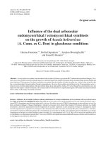
Western blotting detected the expression of PDGFR-β
protein in all six of the MPNST cell lines, and the expression of PDGFR-α protein in all but the HS-Sch-2
cells (Figure 1A). Quantitative real-time RT-PCR identified the expression of both PDGFR-α and PDGFR–β
mRNAs in all six of the MPNST cell lines. Interestingly,
the NMS-2 and NMS-2PC cell lines expressed both
mRNAs at higher levels than the other cell lines
(Figure 1B).
A
PDGFR-β
190 kDa
PDGFR-α
195 kDa
α-tubulin
52 kDa
B
PDGFR-α
5.0
4.0
3.0
2.0
1.0
0.0
*
Relative mRNA expression
Relative mRNA expression
PDGFR-β
6.0
300
*
250
*
200
150
100
≈
2.0
0.0
Figure 1 Expression of PDGFR proteins and mRNAs of in six MPNST cell lines analyzed by western blotting (A) and real-time RT-PCR
(B). The mRNA expression levels in each cell line were compared with the levels in FU-SFT8611 cells. PDGFR-β protein and mRNA were expressed
in all MPNST cell lines. The expression of PDGFR-α protein was observed in all but HS-Sch-2 cells. The PDGFR-α mRNA level in NMS-2 and NMS2PC cells was higher than in the other four cell lines. Data in (B) is the mean ± SEM (n = 3). Similar results were obtained in three independent
experiments. *P < 0.01, compared with the level in FU-SFT8611 cells (by Student’s t-test).
Ohishi et al. BMC Cancer 2013, 13:224
/>
Page 5 of 11
Effects of PDGF-BB and imatinib mesylate on PDGFR-β
phosphorylation in vitro
The effects of PDGF-BB and imatinib mesylate on the
phosphorylation of PDGFRs were investigated. In all six
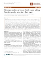
of the MPNST cell lines, PDGF-BB induced phosphorylation of PDGFR-β, but not of PDGFR-α (Figure 2). Furthermore, pretreatment with imatinib mesylate (10 μM)
almost completely suppressed PDGF-BB-induced phosphorylation of PDGFR-β in all six cell lines.
Effects of imatinib mesylate on MPNST cell proliferation
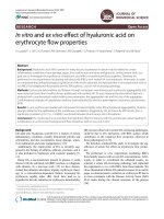
The effects of imatinib mesylate on MPNST cell proliferation were tested in all six MPNST cell lines at concentrations of 1, 5, 10, and 20 μM for 5 days, using the MTS
assay (Figure 3). One to five μM of imatinib mesylate,
which are equivalent to the concentrations achieved clinically in patients treated with imatinib mesylate, had a significant inhibitory effect in three cell lines (FU-SFT9817,
HS-Sch-2, and FMS-1 cells) at day 5, only a mild inhibitory effect in NMS-2 cells, but no effect in FU-SFT8611
or NMS-2PC cells. The 50% inhibitory concentration
(IC50) of imatinib mesylate at day 5 for FU-SFT8611, FUSFT9817, HS-Sch-2, NMS-2, NMS-2PC, and FMS-1 cells
were 16.6, 4.2, 3.2, 4.7, 14.7, and 4.2 μM, respectively.
Effects of siRNA-mediated inhibition of PDGFR-β
expression on MPNST cell growth
To clarify the role of PDGFR-β in MPNST cell proliferation, we examined the effects of siRNA for PDGFR-β on
cell proliferation in the two imatinib mesylate-sensitive
cell lines (HS-Sch-2 and FMS-1), and on one resistant
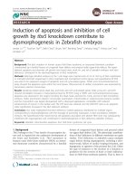
cell line (NMS-2PC). Transfection with PDGFR-β siRNA
specifically and effectively abrogated PDGFR-β protein
expression in all three cell lines (Figure 4). The specific
PDGFR-β knockdown significantly inhibited HS-Sch-2
and FMS-1 cell proliferation (Figure 4A, B), whereas
NMS-2PC cells showed no suppression of cell proliferation (Figure 4C).
Effects of imatinib mesylate on colony formation
In the anchorage-independent growth examined using the
soft agar colony formation assays, both imatinib mesylate-
FU-SFT8611
FU-SFT9817
HS-Sch-2
Phospho-PDGFR-β
190 kDa
PDGFR-β
190 kDa
Phospho-PDGFR-α
198 kDa
PDGFR-α
195 kDa
α-tubulin
52 kDa
PDGF-BB (25 ng/ml)
Imatinib mesylate (10 µM)
−
−
+
−
+
+
−
+
−
−
NMS-2
+
−
+
+
−
+
−
−
NMS-2PC
+
−
+
+
−
+
FMS-1
Phospho-PDGFR-β
190 kDa
PDGFR-β
190 kDa
Phospho-PDGFR-α
198 kDa
PDGFR-α
195 kDa
α-tubulin
52 kDa
PDGF-BB (25 ng/ml)
Imatinib mesylate (10 µM)
−
−
+
−
+
+
−
+
−
−
+
−
+
+
−
+
−
−
+
−
+
+
−
+
Figure 2 Effects of PDGF-BB and imatinib mesylate on phosphorylation of PDGFRs. PDGFR-β of all six MPNST cell lines was phosphorylated
in response to 25 ng/ml of PDGF-BB for 30 min. PDGF-BB stimulation did not induce any phosphorylation of PDGFR-α in all cell lines.
Pretreatment with 10 μM imatinib mesylate almost completely suppressed PDGF-BB-induced PDGFR-β phosphorylation in all cell lines.
Ohishi et al. BMC Cancer 2013, 13:224
/>
Page 6 of 11
FU-SFT8611
FU-SFT9817
0.50
1.00
2% FCS
2% FCS
1 µM Imatinib
1 µM Imatinib
0.75
Absorbance (490 nm)
Absorbance (490 nm)
5 µM Imatinib
10 µM Imatinib
20 µM Imatinib
0.50
*
0.25
5 µM Imatinib
0.40
10 µM Imatinib
20 µM Imatinib
0.30
*
0.20
*
*
0.10
*
*
0.00
0.00
0
1
3
5
0
1
5
NMS-2
HS-Sch-2
0.75
0.75
2% FCS
2% FCS
1 µM Imatinib
1 µM Imatinib
5 µM Imatinib
0.60
10 µM Imatinib
20 µM Imatinib
0.45
0.30
*
0.15
0
1
20 µM Imatinib
0.45
0.30
*
0.15
*
*
*
0.00
*
3
10 µM Imatinib
*
*
*
0.00
5 µM Imatinib
0.60
Absorbance (490 nm)
Absorbance (490 nm)
3
(Day)
(Day)
0
5
1
3
5
(Day)
(Day)
FMS-1
NMS-2PC
1.00
0.80
2% FCS
2% FCS
1 µM Imatinib
5 µM Imatinib
1 µM Imatinib
0.80
5 µM Imatinib
10 µM Imatinib
Absorbance (490 nm)
Absorbance (490 nm)
0.60
20 µM Imatinib
0.40
0.20
10 µM Imatinib
20 µM Imatinib
0.60
*
0.40
*
0.20
*
*
*
0.00
0.00
0
1
3
(Day)
5
*
0
1
3
5
(Day)
Figure 3 Effects of imatinib mesylate on the proliferation of six MPNST cell lines. MPNST cells were incubated for 5 days with imatinib
mesylate at the indicated concentrations in media containing 2% FCS. One to five μM of imatinib mesylate had significant inhibitory effects at
day 5 in three cell lines, FU-SFT9817, HS-Sch-2, and FMS-1 cells. Data are the means ± SEM (n = 4). *P < 0.01, compared with the control group
without imatinib mesylate (by Student’s t-test).
Ohishi et al. BMC Cancer 2013, 13:224
/>
Page 7 of 11
A
1.00
HS-Sch-2
Absorbance (490 nm)
Non treatment
PDGFR-β
(190 kDa)
PDGFR-α
(195 kDa)
α-tubulin
(52 kDa)
siRNA control
siRNA PDGFR-β
0
0
0.01
0
0.02
0
0.05
0
0
0
0.01 0.02
0
0.05
siRNA control
0.75
siRNA PDGFRB
0.50
0.25
*
0
1
(pmol/µl)
B
3
5
7
*
*
5
7
2.00
Absorbance (490 nm)
Non treatment
(190 kDa)
PDGFR-α
(195 kDa)
α-tubulin
(52 kDa)
0
0
0.01
0
0.02
0
0.05
0
0
0
0.01 0.02
0
0.05
siRNA control
1.50
siRNA PDGFRB
1.00
0.50
0.00
0
1
(pmol/µl)
C
3
(Day)
NMS-2PC
1.40
Absorbance (490 nm)
Non treatment
PDGFR-β
(190 kDa)
PDGFR-α
(195 kDa)
α-tubulin
(52 kDa)
siRNA control
siRNA PDGFR-β
*
(Day)
FMS-1
PDGFR-β
siRNA control
siRNA PDGFR-β
*
0.00
0
0
0.01
0
0.02
0
0.05
0
0
0
0.01 0.02
0
0.05
(pmol/µl)
siRNA control
1.05
siRNA PDGFRB
0.70
0.35
0.00
0
1
3
5
7
(Day)
Figure 4 Effects of siRNA-mediated inhibition of PDGFR-β expression on the growth of 3 MPNST cell lines. After 72 h transfection with
0.01, 0.02 and 0.05 pmol/μl siRNA, specific PDGFR-β knockdown was induced in all three cell lines. In the presence of 0.02 pmol/μl siRNA for
PDGFR-β, cell proliferation was significantly inhibited compared with the control, in two MPNST cell lines, HS-Sch-2 (A) and FMS-1 (B), but not
NMS-2PC (C). Data are the means ± SEM (n = 5). Similar results were obtained in at least two independent experiments. *P < 0.01 (by Student’s
t-test).
sensitive cell lines (HS-Sch-2 and FMS-1) were also responsive to imatinib mesylate (Figure 5A, B). Treatment
with both 5 and 10 μM imatinib mesylate significantly
inhibited anchorage-independent growth in these two cell
lines, whereas the NMS-2PC cell line was resistant to both
concentrations of imatinib mesylate.
Effects of imatinib mesylate on MPNST growth in the
xenograft model
To investigate the inhibitory effect of imatinib mesylate
on MPNST growth in vivo, three MPNST cell lines, two
imatinib mesylate-sensitive, HS-Sch-2 and FMS-1 cells,
and one -resistant, NMS-2PC cells, were each transplanted
subcutaneously into separate NOD/SCID mice (n = 5-7,
each). In HS-Sch-2 and FMS-1 cell-transplanted mice,
daily treatment with imatinib mesylate (100 mg/kg/day)
significantly suppressed tumor growth compared with the
control group (P < 0.01 for HS-Sch-2 group; P < 0.05 for
FMS-1 group. Figure 6A, B). In contrast, imatinib mesylate
did not suppress tumor growth in mice bearing NMS-2PC
cells (Figure 6C).
During imatinib mesylate treatment, the body weight
was only slightly affected, with less than 7% weight
reduction in all mice (Additional 1: Figure S1). Skin
dehydration, diarrhea, or abnormal behaviors were not
observed. Histopathologically, there were no apparent
differences between the treatment group and the control
group, such as inflammatory infiltrates, necrotic foci, the
Ohishi et al. BMC Cancer 2013, 13:224
/>
Page 8 of 11
A
B
HS-Sch-2
5 µM imatinib mesylate
No. of Colony-Forming Units
Medium only
50
*
40
10 µM imatinib mesylate
*
30
20
10
0
0
5
10
Imatinib mesylate (µM)
No. of Colony-Forming Units
50
FMS-1
*
40
30
20
10
*
No. of Colony-Forming Units
0
NMS-2PC
*
0
5
10
Imatinib mesylate (µM)
0
5
10
Imatinib mesylate (µM)
50
40
30
20
10
0
Figure 5 Soft agar colony formation assay. Anchorage-independent growth in soft agar was examined under different concentrations of
imatinib mesylate, and numbers of colonies were counted at day 7. (A) Numbers of anchorage-independent colonies of HS-Sch-2 cells growing
in the soft agar were scored using phase contrast microscopy. (B) Imatinib mesylate at 5 μM significantly decreased the number of colonies of
HS-Sch-2 and FMS-1 cells, but had no effect on NMS-2PC cells. Data are the means ± SEM (n = 6). *P < 0.01, compared with the control group
without imatinib mesylate (by Student’s t-test).
number of intratumoral blood vessels, or degenerated
cell changes (Additional 2: Figure S2). There were no
differences in histological findings of the intestine, spleen,
liver, and lungs between the treatment and control groups
(Additional 3: Figure S3).
Mutation of the PDGFRB gene and chromosomal
rearrangement including the PDGF-B gene
Because it is well known that the mutations of tyrosine kinase receptors affect the effectiveness of tyrosine kinase
inhibitors, we analyzed mutation in exons 12 and 18 of the
PDGFRB gene in all six MPNST cell lines. These exons correspond to the mutation-positive juxtamembrane and activation loop coding regions of the KIT and PDGFRA genes
[12]. Using FISH, we also examined whether MPNST cell
lines contained fusion genes involving the PDGF-B gene as
observed in dermatofibrosarcoma protuberans (DFSP) [13].
Neither mutation in exons 12 and 18 of the PDGFRB nor
split of the PDGF-B gene was detected in any of the 6
MPNST cell lines (Additional 4: Figure S4).
Ohishi et al. BMC Cancer 2013, 13:224
/>
Page 9 of 11
HS-Sch-2
A
400
Control group
Mean Tumor Volume (mm3)
Control group
Treatment group
300
*
200
Treatment group
100
0
0
7
14
(Day)
21
28
FMS-1
B
Mean Tumor Volume (mm3)
1600
Control group
Control group
Treatment group
1200
**
800
Treatment group
400
0
0
C
7
14
(Day)
21
28
NMS-2PC
600
Control group
Mean Tumor Volume (mm3)
Control group
Treatment group
450
300
Treatment group
150
0
0
7
14
(Day)
21
28
Figure 6 Effects of oral treatment with imatinib mesylate on tumor growth in mice of the xenograft model. One group of mice was
treated with 100 mg/kg/day imatinib mesylate (■; treatment group) while the other was treated with water (○; control group). (A, left) Xenografts
were established by injection of HS-Sch-2 cells in female NOD/SCID mice. The tumor volume was significantly smaller in the treatment group
than the control (■; n = 5, ○; n = 6, *P < 0.01, by Student’s t-test) on day 28. (B, left) Imatinib mesylate significantly reduced the growth of FMS-1
xenografts compared with the control groups (■; n = 7, ○; n = 6, **P < 0.05, by Student’s t-test). (C, left) Imatinib mesylate did not affect the
growth of NMS-2PC xenografts (■; n = 4, ○; n = 4). Representative picture of animals are shown at right for each experiment.
Ohishi et al. BMC Cancer 2013, 13:224
/>
Discussion
The present study is the first to show that imatinib
mesylate effectively suppressed cell growth in vitro at concentrations within the therapeutic range (1.46-4.6 μM), in
three of the six human MPNST cell lines. In two of these
three imatinib mesylate-sensitive cell lines (HS-Sch-2 and
FMS-1), we confirmed that imatinib mesylate also significantly suppressed tumor growth in the xenograft model.
There was excellent concordance between in vitro and
in vivo sensitivity to imatinib mesylate.
In the imatinib mesylate-sensitive cell lines, imatinib
mesylate suppressed MPNST cell growth via inhibition
of PDGFR-β phosphorylation. Knockdown of PDGFR-β
by transfection with a specific siRNA also caused significant reduction in cell proliferation in the sensitive cell
lines but not in resistant cell lines. Furthermore, although PDGF-BB-induced phosphorylation of PDGFR-β
was inhibited by imatinib mesylate in all cell lines, cell
growth was suppressed by imatinib mesylate only in the
imatinib mesylate-sensitive cell lines. These results support the conclusion that PDGFR-β is involved in the
main intracellular signal pathway for tumor cell growth
in imatinib mesylate-sensitive cell lines.
PDGF was first identified as a growth-promoting factor produced by human platelets for mesenchymal cells
such as dermal fibroblasts, smooth muscle cells, and
other connective tissue cells [14]. PDGF consists of four
chains (A, B, C, and D), which dimerize to form at least
five hetero- and homo-dimers: PDGF-AA, PDGF-AB,
PDGF-BB, PDGF-CC, and PDGF-DD [15]. PDGFR is a
transmembrane tyrosine kinase receptor and there are
two receptor types, termed PDGFR-α and PDGFR-β
[16]. The two different subunits of PDGFR have different
PDGF dimers binding specificities. Based on cell culture
experiments, the possible PDGF/PDGFR interactions are
multiple and complex: PDGFR-αα binds PDGF-AA,
PDGF-AB, PDGF-BB and PDGF-CC; PDGFR-ββ binds
PDGF-BB, and PDGF-DD; and PDGFR-αβ binds PDGFAB, PDGF-BB, PDGF-CC, and PDGF-DD. However,
in vivo, only a few interactions were demonstrated:
PDGFR-αα binds PDGF-AA and PDGF-CC, and PDGFRββ binds only PDGF-BB [15]. In the present study, like the
previous study [7], PDGF-BB induced tyrosine phosphorylation of PDGFR-β but not of PDGFR-α, although both
types of receptors were expressed in all but the HS-Sch-2
cell line. These results suggest that activation of PDGFR-β
may play a more important role in MPNST cell proliferation than PDGFR-α. The observation that PDGFR-β
mRNA and protein expression were higher in MPNST
than in benign peripheral nerve sheath tumors or
Schwann cells [7,17], also supports its important role.
It is well known that mutations of tyrosine kinase receptors affect the effectiveness of tyrosine kinase inhibitors, including imatinib mesylate [18]. Most patients
Page 10 of 11
with advanced gastrointestinal stromal tumors carry mutations in c-kit or PDGFR-α, and the response to imatinib
mesylate in these patients depends on the type of mutation. To our knowledge, PDGFRB mutations have never
been described in MPNST, although some somatic
PDGFRA mutations and several polymorphisms have been
reported [19]. We analyzed the sequences of exons 12 and
18 of the PDGFRB gene in all six MPNST cell lines, but
no PDGFRB mutations were detected. We also examined
the presence of chromosomal rearrangements including
in the PDGFB gene, because the fusion gene leads to autocrine PDGF receptor stimulation in dermatofibrosarcoma
protuberans (DFSP) [13]. However, no split of the PDGFB
gene was detected by FISH in all six MPNST cell lines
(Additional 4: Figure S4).
The PDGF/PDGFR autocrine loop has an important
role in other malignancies, such as glioma [20], osteosarcoma [21], breast cancer [22], and ovarian cancer [23],
but has yet to be completely described for MPNST cells.
The loop may be involved in the sensitivity of MPNST
cells to Imatinib, and we are currently exploring this
possibility.
Conclusions
These results suggest that imatinib mesylate may be useful in the treatment of MPNST patients. When primary
cultures of MPNST are available, analysis of sensitivity
to imatinib in vitro may help select patients with imatinibsensitive tumors.
Additional files
Additional file 1: Figure S1. During imatinib mesylate treatment, the
body weight was only slightly affected, with less than 7% reduction in all
mice.
Additional file 2: Figure S2. Transplanted tumors showed a
proliferation of atypical spindle or polygonal shaped cells with oval nuclei
and distinct nucleoli. These cells proliferated loosely as interconnected
cords or networks, or compactly in a sheet-like pattern. Mitotic figures are
frequently observed. There were no differences in histological findings
between the treatment and control groups.
Additional file 3: Figure S3. During imatinib mesylate treatment, there
were no differences in histological findings of the intestine, spleen, liver,
and lungs between the treatment group and the control group.
Additional file 4: Figure S4. Using fluorescence in situ hybridization
(FISH), we examined whether MPNST cell lines contained fusion genes
involving the PDGF-B. No slit of the PDGF-B gene was detected in any of
the six MPNST cell lines.
Competing interests
All authors have no conflicts of interest to declare.
Authors’ contributions
JO, MA carried out the experiments. KN, HI, JS, and YY participated in the
design of the study, its coordination, and helped to draft the manuscript. In
addition technical guidance from TT, AO, and MH lead to successful
experiments. All authors approved the final manuscript.
Ohishi et al. BMC Cancer 2013, 13:224
/>
Acknowledgments
We thank M Ishiguro, K Yano, H Fukagawa and M Onitsuka for their excellent
technical assistance.
Author details
1
Department of Pathology, Fukuoka University School of Medicine, 7-45-1
Nanakuma, Jonan-kuFukuoka 814-0180, Japan. 2Gastroenterological Surgery,
Fukuoka University School of Medicine, Fukuoka, Japan. 3Shimane University
Hospital Cancer Center, Shimane, Japan. 4Department of Immunopathology,
University School of Medicine, Gifu, Japan. 5Division of Orthopaedic Surgery,
Department of Regenerative Transplant Medicine, Niigata University
Graduate School of Medicine and Dental Science, Niigata, Japan.
6
Department of Orthopaedic Surgery, Fukushima Medical University School
of Medicine, Fukushima, Japan.
Received: 29 November 2012 Accepted: 25 April 2013
Published: 4 May 2013
References
1. Lewis JJ, Brennan MF: Soft tissue sarcomas. Curr Probl Surg 1996,
33(10):817–872.
2. Woodruff JM, Kourea HP, Louis DN, Scheithauer BW: Malignant peripheral
nerve sheath tumor (MPNST). In World health organization classification of
tumors pathology and genetics of tumor of the nervous system. Lyon, France:
IARC Press; 2000:172–174.
3. Evans DG, Baser ME, McGaughran J, Sharif S, Howard E, Moran A: Malignant
peripheral nerve sheath tumours in neurofibromatosis 1. J Med Genet
2002, 39(5):311–314.
4. Ducatman BS, Scheithauer BW, Piepgras DG, Reiman HM, Ilstrup DM:
Malignant peripheral nerve sheath tumors. A clinicopathologic study of
120 cases. Cancer 1986, 57(10):2006–2021.
5. Ferner RE, Gutmann DH: International consensus statement on malignant
peripheral nerve sheath tumors in neurofibromatosis. Cancer Res 2002,
62(5):1573–1577.
6. Wong WW, Hirose T, Scheithauer BW, Schild SE, Gunderson LL: Malignant
peripheral nerve sheath tumor: analysis of treatment outcome.
Int J Radiat Oncol Biol Phys 1998, 42(2):351–360.
7. Aoki M, Nabeshima K, Koga K, Hamasaki M, Suzumiya J, Tamura K, Iwasaki H:
Imatinib mesylate inhibits cell invasion of malignant peripheral nerve
sheath tumor induced by platelet-derived growth factor-BB. Lab Invest
2007, 87(8):767–779.
8. Aoki M, Nabeshima K, Nishio J, Ishiguro M, Fujita C, Koga K, Hamasaki M,
Kaneko Y, Iwasaki H: Establishment of three malignant peripheral nerve
sheath tumor cell lines, FU-SFT8611, 8710 and 9817: conventional and
molecular cytogenetic characterization. Int J Oncol 2006, 29(6):1421–1428.
9. Sonobe H, Takeuchi T, Furihata M, Taguchi T, Kawai A, Ohjimi Y, Iwasaki H,
Kaneko Y, Ohtsuki Y: A new human malignant peripheral nerve sheath
tumour-cell line, HS-sch-2, harbouring p53 point mutation. Int J Oncol
2000, 17(2):347–352.
10. Imaizumi S, Motoyama T, Ogose A, Hotta T, Takahashi HE: Characterization
and chemosensitivity of two human malignant peripheral nerve sheath
tumour cell lines derived from a patient with neurofibromatosis type 1.
Virchows Arch 1998, 433(5):435–441.
11. Hakozaki M, Hojo H, Sato M, Tajino T, Yamada H, Kikuchi S, Abe M:
Establishment and characterization of a novel human malignant
peripheral nerve sheath tumor cell line, FMS-1, that overexpresses
epidermal growth factor receptor and cyclooxygenase-2. Virchows Arch
2009, 455(6):517–526.
12. Sihto H, Franssila K, Tanner M, Vasama-Nolvi C, Sarlomo-Rikala M, Nupponen
NN, Joensuu H, Isola J: Platelet-derived growth factor receptor family
mutations in gastrointestinal stromal tumours. Scand J Gastroenterol 2006,
41(7):805–811.
13. Simon MP, Pedeutour F, Sirvent N, Grosgeorge J, Minoletti F, Coindre JM,
Terrier-Lacombe MJ, Mandahl N, Craver RD, Blin N, et al: Deregulation of
the platelet-derived growth factor B-chain gene via fusion with collagen
gene COL1A1 in dermatofibrosarcoma protuberans and giant-cell
fibroblastoma. Nat Genet 1997, 15(1):95–98.
14. Heldin CH, Westermark B: Growth factors: mechanism of action and
relation to oncogenes. Cell 1984, 37(1):9–20.
15. Andrae J, Gallini R, Betsholtz C: Role of platelet-derived growth factors in
physiology and medicine. Genes Dev 2008, 22(10):1276–1312.
Page 11 of 11
16. Hart CE, Forstrom JW, Kelly JD, Seifert RA, Smith RA, Ross R, Murray MJ,
Bowen-Pope DF: Two classes of PDGF receptor recognize different
isoforms of PDGF. Science 1988, 240(4858):1529–1531.
17. Badache A, De Vries GH: Neurofibrosarcoma-derived Schwann cells
overexpress platelet-derived growth factor (PDGF) receptors and are
induced to proliferate by PDGF BB. J Cell Physiol 1998, 177(2):334–342.
18. Demetri GD, Von Mehren M, Blanke CD, Van den Abbeele AD, Eisenberg B,
Roberts PJ, Heinrich MC, Tuveson DA, Singer S, Janicek M, et al: Efficacy and
safety of imatinib mesylate in advanced gastrointestinal stromal tumors.
N Engl J Med 2002, 347(7):472–480.
19. Holtkamp N, Okuducu AF, Mucha J, Afanasieva A, Hartmann C, Atallah I,
Estevez-Schwarz L, Mawrin C, Friedrich RE, Mautner VF, et al: Mutation and
expression of PDGFRA and KIT in malignant peripheral nerve sheath
tumors, and its implications for imatinib sensitivity. Carcinogenesis 2006,
27(3):664–671.
20. Strawn LM, Mann E, Elliger SS, Chu LM, Germain LL, Niederfellner G, Ullrich A,
Shawver LK: Inhibition of glioma cell growth by a truncated platelet-derived
growth factor-beta receptor. J Biol Chem 1994, 269(33):21215–21222.
21. Sulzbacher I, Traxler M, Mosberger I, Lang S, Chott A: Platelet-derived
growth factor-AA and -alpha receptor expression suggests an autocrine
and/or paracrine loop in osteosarcoma. Mod Pathol 2000, 13(6):632–637.
22. Jechlinger M, Sommer A, Moriggl R, Seither P, Kraut N, Capodiecci P,
Donovan M, Cordon-Cardo C, Beug H, Grunert S: Autocrine PDGFR
signaling promotes mammary cancer metastasis. J Clin Invest 2006,
116(6):1561–1570.
23. Matei D, Emerson RE, Lai YC, Baldridge LA, Rao J, Yiannoutsos C, Donner
DD: Autocrine activation of PDGFRalpha promotes the progression of
ovarian cancer. Oncogene 2006, 25(14):2060–2069.
doi:10.1186/1471-2407-13-224
Cite this article as: Ohishi et al.: Imatinib mesylate inhibits cell growth of
malignant peripheral nerve sheath tumors in vitro and in vivo through
suppression of PDGFR-β. BMC Cancer 2013 13:224.
Submit your next manuscript to BioMed Central
and take full advantage of:
• Convenient online submission
• Thorough peer review
• No space constraints or color figure charges
• Immediate publication on acceptance
• Inclusion in PubMed, CAS, Scopus and Google Scholar
• Research which is freely available for redistribution
Submit your manuscript at
www.biomedcentral.com/submit