Anti-cancer effects of newly developed chemotherapeutic agent, glycoconjugated palladium (II) complex, against cisplatin-resistant gastric cancer cells
Bạn đang xem bản rút gọn của tài liệu. Xem và tải ngay bản đầy đủ của tài liệu tại đây (674.18 KB, 9 trang )
Tanaka et al. BMC Cancer 2013, 13:237
/>
RESEARCH ARTICLE
Open Access
Anti-cancer effects of newly developed
chemotherapeutic agent, glycoconjugated
palladium (II) complex, against cisplatin-resistant
gastric cancer cells
Mamoru Tanaka1, Hiromi Kataoka1*, Shigenobu Yano2,3, Hiromi Ohi4, Keisuke Kawamoto5, Takashi Shibahara5,
Tsutomu Mizoshita1, Yoshinori Mori1, Satoshi Tanida1, Takeshi Kamiya1 and Takashi Joh1
Abstract
Background: Cisplatin (CDDP) is the most frequently used chemotherapeutic agent for various types of
advanced cancer, including gastric cancer. However, almost all cancer cells acquire resistance against CDDP,
and this phenomenon adversely affects prognosis. Thus, new chemotherapeutic agents that can overcome the
CDDP-resistant cancer cells will improve the survival of advanced cancer patients.
Methods: We synthesized new glycoconjugated platinum (II) and palladium (II) complexes, [PtCl2 (L)] and
[PdCl2 (L)]. CDDP-resistant gastric cancer cell lines were established by continuous exposure to CDDP, and gene
expression in the CDDP-resistant gastric cancer cells was analyzed. The cytotoxicity and apoptosis induced by
[PtCl2 (L)] and [PdCl2 (L)] in CDDP-sensitive and CDDP-resistant gastric cancer cells were evaluated. DNA doublestrand breaks by drugs were assessed by evaluating phosphorylated histone H2AX. Xenograft tumor mouse models
were established and antitumor effects were also examined in vivo.
Results: CDDP-resistant gastric cancer cells exhibit ABCB1 and CDKN2A gene up-regulation, as compared with
CDDP-sensitive gastric cancer cells. In the analyses of CDDP-resistant gastric cancer cells, [PdCl2 (L)] overcame
cross-resistance to CDDP in vitro and in vivo. [PdCl2 (L)] induced DNA double-strand breaks.
Conclusion: These results indicate that [PdCl2 (L)] is a potent chemotherapeutic agent for CDDP-resistant gastric
cancer and may have clinical applications.
Keywords: Glycoconjugated platinum (II) complex, Glycoconjugated palladium (II) complex, Cisplatin,
Drug resistance, Gastric cancer
Background
Cancer is a leading cause of death worldwide, and according to the WHO mortality database (as at November
2006), gastric cancer is the second leading cause of cancer
death after lung cancer.
Cisplatin (CDDP) is the most frequently used chemotherapeutic agent for various types of advanced cancer
and is used in combination regimens. Some CDDP* Correspondence:
1
Departments of Gastroenterology and Metabolism, Nagoya City University
Graduate School of Medical Sciences, Kawasumi, Mizuho-cho, Mizuho-ku
467-8601, Nagoya, Japan
Full list of author information is available at the end of the article
based combination chemotherapy regimens have also
shown high response rates [1]. Based on recent Japanese
phase III trials for metastatic gastric cancer, S1 plus cisplatin combination chemotherapy was established as the
standard first-line chemotherapy [2].
However, CDDP-based combination chemotherapy
regimens have several disadvantages, including side effects such as nephrotoxicity, neurotoxicity, ototoxicity
and vomiting. In addition, some tumors acquire resistance to CDDP, reducing its efficacy [3,4]. Several mechanisms are involved in CDDP resistance [5]. Such
mechanisms include decreased intracellular drug accumulation and/or increased drug efflux [6-9], drug inactivation
© 2013 Tanaka et al.; licensee BioMed Central Ltd. This is an Open Access article distributed under the terms of the Creative
Commons Attribution License ( which permits unrestricted use, distribution, and
reproduction in any medium, provided the original work is properly cited.
Tanaka et al. BMC Cancer 2013, 13:237
/>
by increased levels of cellular thiols [6,10], increased nucleotide excision-repair activity [9,11] and evasion of
apoptosis [6,12]. Thus, for continued progress in cancer
therapy, more effective drugs must be found.
Cancer cells take in higher levels of glucose than
normal cells, a phenomenon known as the Warburg effect [13]. To achieve lower undesired toxicity, enhanced
solubility and tumor selectivity, we have developed and
have reported several glycoconjugated drugs [14,15].
Another strategy to design new antitumor agents related
to CDDP is to change the nature of the central metal ion
[16,17]. As palladium (Pd) chemistry is similar to that of
platinum (Pt), Pd complexes (II) are expected to exhibit
antitumor activities similar to those of Pt. Attempts have
been made to synthesize Pd (II) complexes with such
activities, as Pd complexes are expected to have less
kidney toxicity than Pt complexes [18].
In this study, we synthesized a new glycoconjugated Pt
(II) complex and a new glycoconjugated Pd (II) complex,
and analyzed its cytotoxicity, ability to induce apoptosis,
and ability to induce DNA double-strand breaks in
CDDP-sensitive and CDDP-resistant gastric cancer cell
lines in vitro and in vivo.
Methods
Drugs
Reagents and solvents used in this study were commercial products of the highest available purity. The Pt (II)
and Pd (II) complexes were easily prepared using the
one-pot reaction of Pt (II) or Pd (II) salt, amino sugar
and pyridine aldehyde derivative without isolation of a
Schiff base ligand (L) as follows.
[PtCl2 (L)] (L = 2-deoxy-2-[(2-pyridinylmethylene)
amino]-α-D-glucopyranose):Dichloro (2-deoxy-2-[(2pyridinylmethylene)amino]-α-D-glucopyranose) Pt. An
aqueous (50 mL) solution of D (+)-glucosamine • hydrochloride (0.65 g, 3.0 mmol) was neutralized with NaHCO3
(0.26 g, 3.1 mmol). To this solution, a MeOH (50 mL) solution of 2-pyridinecarbaldehyde (0.32 g, 3.1 mmol) was
added, followed by stirring for 2 h and addition of K2
[PtCl4] (1.25 g, 3.0 mmol) in 30 mL of H2O. The reaction
was continued for another 41 h at room temperature. The
mixture was concentrated by evaporation and the resulting residue was purified by silica gel column chromatography (eluent: acetone) to give a pale yellow powder
(1.07 g, 67%). Single crystals were obtained by recrystallization from MeOH/Et2O. Anal. calcd for [PtCl2 (L)],
C12H18Cl2N2O5Pt, C; 26.98, H; 3.02, N; 5.24. found for C;
27.13, H; 2.97, N; 5.07. MS (FAB, pos): m/z = 498 [M–Cl]+.
[PdCl2 (L)] (L = 2-deoxy-2-[(2-pyridinylmethylene)
amino]-α-D-glucopyranose):Dichloro (2-deoxy-2-[(2pyridinylmethylene)amino]-α-D-glucopyranose) palladium. This complex was prepared by following a
Page 2 of 9
similar procedure as described above for [PtCl2 (L)]
using Na2 [PdCl4] instead of K2 [PtCl4]. The complex was
dissolved in MeOH and insoluble materials were removed
by filtration. The filtrate was concentrated by evaporation
to give a pale yellow powder (1.1 g, 83%). This complex
was purified by recrystallization from MeOH/Et2O. Anal.
calcd for [PtCl2 (L)], C12H18Cl2N2O5Pd, C; 32.35, H; 3.62,
N; 6.29. found for C; 32.02, H; 3.51, N; 6.01. MS (FAB,
pos): m/z = 431 [M–HCl + Na]+.
CDDP and CBDCA were purchased from BristolMyers Co. (Tokyo, Japan). L-OHP was purchased from
Yakult (Tokyo, Japan).
Measurements
Elemental analysis was carried out on a Perkin-Elmer
240C or a Fisons Instruments EA1108 Elemental Analyzer. 1H- and 13C-NMR spectra were recorded on a JEOL
JNM-GSX400 in N,N-dimethylformamide-d7 (DMF-d7)/
D2O. Mass spectra were obtained on a JEOL JMS-700 T
Tandem MS-station mass spectrometer.
Crystallography
Suitable crystals for X-ray crystallography were obtained
by slow recrystallization of [PtCl2 (L)] and [PdCl2 (L)]
from a minimal amount of methanol and ether mixtures.
Crystallographic data (excluding structure factors) for
the structure reported in this paper were deposited with
the Cambridge Crystallographic Data Center as supplementary publication no. CCDC-835397. Copies of the
data can be obtained free of charge on application to
CCDC, 12 Union Road, Cambridge CB21EZ, UK (Fax:
(+44) 1223-336-033; E-mail: ).
Cell culture
The human gastric cancer cell lines MKN28 (Japanese
Cancer Research Resources Bank, No. 0253) and MKN45
(Japanese Cancer Research Bank, No. 0254) were cultured
in RPMI1640 (Sigma-Aldrich, St. Louis, MO) supplemented with 10% fetal bovine serum (FBS) and 1% ampicillin
and streptomycin. Cells were cultured under an atmosphere of 5% CO2 at 37°C.
Establishment of CDDP-resistant sublines from MKN28
and MKN45
CDDP-resistant MKN28 (MKN28 (CDDP)) and CDDPresistant MKN45 (MKN45 (CDDP)) were established by
continuous exposure to CDDP starting at 0.5 μmol/L
and increasing in a stepwise manner to 10 μmol/L for
more than 5 months. Experiments with these sublines
were performed after maintenance in CDDP-free medium for 2–3 weeks.
Tanaka et al. BMC Cancer 2013, 13:237
/>
RT2 Profiler PCR arrays for human cancer drug resistance
& metabolism
Total RNA (1 μg) from MKN45 (0) or MKN45 (CDDP)
was converted to cDNA and used to screen inflammatory cytokines and receptors using quantitative real-time
PCR arrays according to the manufacturer’s instructions
(SuperArray Bioscience). Reactions were cycled in an ABI
Prism 7500 FAST sequence detector (Applied Biosystems)
and acquired data were analyzed using the DDCt method
to determine the expression levels of each transcript normalized against the expression level of housekeeping gene
controls. A gene-wise, two-sample t-test was performed
for each transcript to identify statistical differences in expression between MKN45 (0) or MKN45 (CDDP).
In vitro treatment
Cell viability was determined by WST-8 cell proliferation
assay. Gastric cancer cells were seeded into 96-well
culture plates at 5 × 10³ cells/100 μL/well and incubated overnight. Cells were treated for 48 h with graded
concentrations of CDDP (0–200 μmol/L), [PtCl2(L)]
(0–200 μmol/L), [PdCl2(L)] (0–200 μmol/L), L-OHP
(0–100 μmol/L) or CABDA (0–400 μmol/L). After treatment, cells were incubated with cell a counting kit-8
(Dojindo, Kumamoto, Japan) for 4 h and absorption at
450 nm was measured with a microscope reader (SPECTRA MAX340; Molecular Devices, Silicon Valley, CA).
Cell viability was expressed as a percentage vs. untreated
control cells and half maximal (50%) inhibitory concentration (IC50) was calculated. Resistance factor (RF) is
defined as the relative ratio of IC50 values in both cell
lines (MKN28 (CDDP)/MKN28 (0) or MKN45 (CDDP)/
MKN45 (0)).
Assessment of apoptosis
Apoptosis was assessed by analysis of activation of
caspase-3 and caspase-7 using the substrate DEVDaminoluciferin from the Caspase-Glo 3/7 Assay kit
(Promega) according to the manufacturer’s instructions.
Briefly, gastric cancer cells (104 per well) were plated on a
96-well culture plate with three replicates per treatment.
After 24 h of plating, cells were treated for 72 h with
graded concentrations of CDDP (0–200 μmol/L), [PtCl2
(L)] (0–200 μmol/L), [PdCl2(L)] (0–200 μmol/L), L-OHP
(0–100 μmol/L) or CABDA (0–400 μmol/L). Caspase-Glo
reagent was added to each well and incubated for 1 h,
and luminescence was measured using a LUMAT LB
9507 luminometer (Berthold Technologies). Results
were analyzed by Welch’s t-test between MKN45 (0)
and MKN45 (CDDP).
Assessment of DNA double-strand breaks
Cells were washed with PBS (−) and subsequently dissolved in 1 cell lysis buffer (Cell Signaling Technology)
Page 3 of 9
containing 20 mmol/L Tris–HCl (pH 7.5), 150 mmol/L
NaCl, 1 mmol/L Na2EDTA, 1 mmol/L EGTA, 1% Triton, 2.5 mmol/L sodium pyrophosphate, 1 mmol/L
h-glycerophosphate, 1 mmol/L Na3VO4, and 1 Ag/mL
leupeptin with the addition of 1 mmol/L phenylmethylsulfonyl fluoride. After disruption in an ice bath using a
Bio-ruptor sonicator (Cosmo Bio) for 15 s, lysates were
centrifuged at 15,000 rpm for 10 min at 4°C. Each sample was normalized as equal protein concentrations
using a protein assay kit (Bio-Rad Laboratories). An
equal quantity of 2 SDS-PAGE sample buffer [0.5 mol/L
Tris–HCl (pH 7.2), 1% SDS, 100 mmol/L β-mercaptoethanol, and 0.01% bromophenol blue] was added to
each sample, followed by boiling for 5 min at 100°C. Aliquots of sample were fractioned on 8% to 15% SDSPAGE and were then electroblotted onto nitrocellulose
membrane. The membrane was blocked with 5% skimmed milk in PBS (−) for 1 h at room temperature. The
membrane was incubated with primary antibodies, antiγH2AX (Bethyl Laboratories, Inc., 1:2000), overnight at
4°C and was then washed with 0.05% Tween 20 in PBS
(−) three times at 5-min intervals. The membrane was
incubated with secondary antibody for 1 h at room
temperature followed by three washes with 0.05% Tween
20 in PBS (−) three times at 5-min intervals. The
membrane was treated with enhanced chemiluminescence detection reagents (Amersham) for 1 min at room
temperature and exposed to scientific imaging films
(Eastman Kodak), and proteins were visualized as bands.
Filters were stripped and re-probed with monoclonal
β-actin antibody (Abcam plc) as an internal control.
Animals and tumor models
Pathogen-free female nude mice (BALB/c Slc-nu/nu)
aged 4 weeks and weighing 20–25 g were obtained from
Japan SLC (Kyoto, Japan). Animals were allowed to acclimatize for 2 weeks in the animal facility before any interventions were initiated. Xenograft tumor models were
established by subcutaneously implanting 3 × 10⁶ gastric
cancer cells (MKN45 (0), MKN45 (CDDP)) in 200 μL of
PBS. Experimental procedures were approved by the
Nagoya City University Center for Experimental Animal
Science, and mice were raised in accordance with the
guideline of the Nagoya City University Center for Animal Experiments.
In vivo treatment
At 7 days after tumor inoculation, mice were given an
intraperitoneal injection of CDDP, [PtCl2 (L)] or [PdCl2
(L)] at a dose of 40 μmol/kg. Tumor growth was monitored daily by measuring tumor volume with vernier
calipers. Tumor volume was calculated using the following formula: (length × width × depth)/2. Each group
Tanaka et al. BMC Cancer 2013, 13:237
/>
consisted of 5 mice. Results were analyzed by multiple
testing (Holm method) between groups.
Statistical analysis
Descriptive statistics and simple analyses were carried
out using the statistical package R version 2.4.1 (www.
r-project.org/). Apoptosis induction was analyzed by
Welch’s t-test. Antitumor effects were analyzed by the
Bonferroni-Holm method. P-values of <0.05 were considered to be statistically significant.
Results
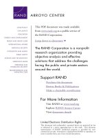
Crystal structure of [PtCl2 (L)] and [PdCl2 (L)]
The crystal structures of [PtCl2 (L)] and [PdCl2 (L)]
(Cambridge Crystallographic Data Center as supplementary publication no. CCDC-835397) show that each
metal atom is surrounded by four donor atoms, two
nitrogen atoms and two chloride ions, in a cis-configuration. As expected, the geometry around the metal
center is approximately square planar (Figure 1). The
pyranoid ring of the sugar unit adopts an unusual α-4C1
conformation. Thus, both complexes have similar
structures.
Conformational analysis of sugar units on [PtCl2 (L)] and
[PdCl2 (L)] by means of NMR measurements
1
H-NMR and 13C-NMR spectra of the two complexes
were obtained in DMF-d7/D2O and unambiguously assigned by 1H-1H and 13C-1H COSY two-dimensional
NMR spectroscopy. Conformation of the sugar ring in
both complexes was investigated by 1H-NMR spectroscopy in DMF-d7/D2O after OH proton exchange, which
Page 4 of 9
reveals signals originating from protons that are attached
to the carbon atoms of the sugar unit. The vicinal
proton-proton coupling constants for [PtCl2 (L)] (3 J1,2 =
3.2, 3 J2,3 = 11.2, 3 J3,4 = 8.4, 3 J4,5 = 10.0 Hz) and [PdCl2
(L)] (3 J1,2 = 3.4, 3 J2,3 = 11.2, 3 J3,4 = 8.6, 3 J4,5 = 10.0 Hz)
correspond to α-4C1 conformations as observed in the
X-ray crystallography, indicating the structural similarity
in the sugar unit in the solid and solution states.
Genes up-regulated in CDDP-resistant gastric cancer
sublines
The 20-fold changes in gene expression for MKN45 (0)
and MKN45 (CDDP) are presented in Table 1. Among
84 genes related to human cancer drug resistance and
metabolism, 8 genes were significantly altered with fold
changes larger than 20. Genes that were up-regulated by
greater than 20-fold were ABCB1, APC, ATM, BRCA2
and CDKN2A, whereas down-regulated genes were
CYP2B6, CYP2C19 and PPARγ.
[PdCl2 (L)] revealed minimum resistance to
CDDP-resistant gastric cancer cells
We investigated the cytotoxicity of CDDP, [PtCl2 (L)],
[PdCl2 (L)], L-OHP and CABDA in the gastric cancer
cell lines MKN28 (0), MKN28 (CDDP), MKN45 (0) and
MKN45 (CDDP), and summarize the results in Table 2.
In the parent cell lines (MKN28 (0) and MKN45 (0)),
[PtCl2 (L)] and [PdCl2 (L)] exhibited lower cytotoxicity
than CDDP and L-OHP, and higher cytotoxicity than
CABDA. Resistance factor (RF) was calculated as the
relative ratio of IC50 values in both cell lines (MKN28
(CDDP)/MKN28 (0) or MKN45 (CDDP)/MKN45 (0)).
(B)
(A)
[PtCl2(L)]
[PdCl2(L)]
Figure 1 Structures of complexes used in this study. (A) Perspective drawing of [PtCl2 (L)] with atomic numbering scheme in the crystal.
Selected bond length (Å) and angles (o), Pt(1)-Cl(1) 2.2985(8), Pt(1)-Cl(2) 2.2922(8), Pt(1)-N(1) 2.016(3), Pt(1)-N(2) 2.006(3); Cl(1)-Pt(1)-Cl(2) 89.77(3),
Cl(1)-Pt(1)-N(1) 94.84(7), Cl(2)-Pt(1)-N(2) 95.30(7), N(1)-Pt(1)-N(2) 80.14(10). (B) Perspective drawing of [PdCl2 (L)] with atomic numbering scheme in
the crystal. Selected bond length (Å) and angles (o), Pd(1)-Cl(1) 2.2940(8), Pd(1)-Cl(2) 2.2832(8), Pd(1)-N(1) 2.033(2), Pd(1)-N(2) 2.025(2); Cl(1)-Pd(1)-Cl
(2) 91.04(3), Cl(1)-Pd(1)-N(1) 94.02(7), Cl(2)-Pd(1)- N(2) 94.50(6), N(1)-Pd(1)-N(2) 80.49(8).
Tanaka et al. BMC Cancer 2013, 13:237
/>
Page 5 of 9
Table 1 Expression profiles of genes related to human cancer drug resistance and metabolism showing at least 20-fold
change in expression
Symbol
GenebankID
Incease
Gene name
ABCB1
NM_000927
122.73
ABC20,CD243,CLCS,GP170,MDR1,MGC163296,P-GP,PGY1
APC
NM_000038
27.25
BTPS2,DP2,DP2.5,DP3,GS
ATM
NM_000051
27.35
AT1,ATA,ATC,ATD,ATDC,ATE,DKFZp781A0353,MGC74674,TEL1,TELO1
BRCA2
NM_000059
34.61
BRCC2,BROVCA2,FACD,FAD,FAD1,FANCB,FANCD,FANCD1
CDKN2A
NM_000077
2689.53
ARF,CDK4I,CDKN2,CMM2,INK4,INK4a,MLM,MTS1,TP16,p14,Prop14ARF,
p16,p16INK4,p16INK4a,p19
CYP2B6
NM_000767
−39.27
CPB6,CYP2B,CYPIIB6,IIB1,P450
CYP2C19
NM_000769
−145.20
CPCJ,CYP2C,P450C2C,P450IIC19
PPARG
NM_015869
−29.31
CIMT1,NR1C3,PPARG1,PPARG2,PPARgamma
Similarly to CABDA, cells treated with [PtCl2 (L)]
showed cross-resistance to CDDP. On the other hand,
[PdCl2 (L)] overcame cross-resistance to CDDP, similarly
to L-OHP, although [PdCl2 (L)] showed a lower degree
of cross-resistance than L-OHP (Table 2).
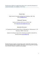
[PdCl2 (L)] induced apoptosis in CDDP-resistant gastric
cancer cell lines
We examined apoptosis induction by CDDP, [PtCl2 (L)],
[PdCl2 (L)], L-OHP and CABDA in the gastric cancer
cell lines MKN45 (0) and MKN45 (CDDP) (Figure 2A).
In the parental cell line (MKN45 (0)), all drugs tended
to induce apoptosis in a dose-dependent manner. In the
CDDP-resistant subline (MKN45 (CDDP)), induction of
apoptosis by CDDP, CABDA and [PtCl2 (L)] was lower
than in the parental cell line. On the other hand, [PdCl2
(L)] and L-OHP maintained apoptosis induction against
CDDP-resistant gastric cancer cells.
Table 2 In vitro cytotoxicity assay in CDDP-sensitive
and -resistant gastric cancer cell lines
MKN28
Resistance factor
IC50(μM)
MKN28(0)
MKN28(CDDP)
80.8 ± 6.6
[PdCl2(L)]
1.02
78.9 ± 4.0
L-OHP
1.19
46.4 ± 4.0
55.2 ± 3.8
[PtCl2(L)]
2.54
111.7 ± 27.1
283.9 ± 19.3
CDDP
3.37
19.4 ± 2.4
65.4 ± 4.6
CABDA
4.33
202.9 ± 17.2
878.3 ± 34.1
MKN45
Resistance factor
IC50(μM)
MKN45(0)
MKN45(CDDP)
[PdCl2(L)]
1.14
61.2 ± 6.8
69.7 ± 4.1
L-OHP
1.3
27.3 ± 1.1
35.6 ± 6.7
[PtCl2(L)]
2.18
129.5 ± 14.8
282.6 ± 34.5
CDDP
3.27
23.5 ± 2.2
77.0 ± 8.5
CABDA
3.42
152.8 ± 3.7
522.0 ± 27.4
[PdCl2 (L)] induced DNA double-strand breaks in
CDDP-resistant gastric cancer cells
Cells were labeled with an antibody against phosphorylated histone H2AX (γ-H2AX), which detects doublestrand breaks caused by drugs such as CDDP [19]. We
used Western blotting for evaluation of γ-H2AX protein
expression by CDDP and [PdCl2 (L)] in the gastric cancer cell lines MKN45 (0) and MKN45 (CDDP). In the
parental cell line (MKN45 (0)) treated with CDDP or
[PdCl2 (L)], γ-H2AX protein levels increased and were
the same by 24 and 48 h after treatment. In the CDDPresistant subline (MKN45 (CDDP)), γ-H2AX protein
levels increased with [PdCl2 (L)], but did not increase
with CDDP (Figure 2B). These results indicated that
[PdCl2 (L)], but not CDDP induced DNA double-strand
breaks in CDDP-resistant gastric cancer cells.
[PdCl2 (L)] significantly suppressed CDDP-resistant gastric
cancer cell proliferation
We examined the effects of CDDP, [PtCl2 (L)] and
[PdCl2 (L)] on xenograft tumor models established by
subcutaneously implanting the gastric cancer cell lines
MKN45 (0) and MKN45 (CDDP). At 7 days after tumor
inoculation, mice were given an intra-peritoneal injection of CDDP, [PtCl2 (L)] or [PdCl2 (L)] at a dose of
40 μmol/kg. In MKN45 (0) nude mice, CDDP, [PtCl2
(L)] and [PdCl2 (L)] suppressed tumor growth significantly as compared to controls (p < 0.01). In MKN45
(CDDP) nude mice, [PdCl2 (L)] suppressed tumor growth
significantly (p < 0.01) as compared to CDDP, but [PtCl2
(L)] did not (Figure 3). None of the therapies had any
obvious side effects, such as diarrhea or weight loss (data
not shown).
Discussion
[PtCl2 (L)] and [PdCl2 (L)] were developed as antitumor
drugs with sugar conjugated ligands, and were expected
to have a number of advantages, including significant reductions in side effects, improved water solubility, and
Tanaka et al. BMC Cancer 2013, 13:237
/>
Page 6 of 9
(A)
(B)
7
Relative apoptosis rate
CDDP
MKN45(0)
MKN45(CDDP)
6
Cont
MKN45(CDDP)
24h
48h
Cont
24h
48h
γ
**
**
5
**
4
**
3
MKN45(0)
β-actin
*
*
MKN45(0)
MKN45(CDDP)
**
2
1
[PdCl2(L)]
0
Cont
24h
48h
Cont
24h
48h
γ
50 100 200 50 100 200 50 100 200 25 50 100 100 200 400
(µmol/L)
[PtCl2(L)]
[PdCl2(L)]
L-OHP
CBDCA
control CDDP
β-actin
Figure 2 Investigation of cytotoxicity mechanism of [PtCl2 (L)] and [PdCl2 (L)]. (A) [PdCl2 (L)] induced apoptosis on CDDP-resistant gastric
cancer cell lines. Apoptosis was assessed by analyzing activation of caspase-3 and caspase-7. Mean of three independent experiments in triplicate;
bars, SE. Values for apoptosis of cells in FBS alone were used as controls. Significance was determined by Welch’s t-test. *, P < 0.05, **, P < 0.01
relative to parental cell line. (B) [PdCl2 (L)] induced DNA double-strand breaks in CDDP-resistant gastric cancer cells. Cells were labeled with
antibody against phosphorylated histone H2AX (γ-H2AX), which detects double-strand breaks caused by drugs such as CDDP. An evaluation of
γ-H2AX protein expression was investigated by Western blotting at 24 or 48 h after treatment.
In this report, we found that gastric cancer cell
lines adapted to growth in the presence of 10 μmol/L
CDDP (MKN45 (CDDP)) showed enhanced ABCB1 and
CDKN2A expression as compared with their CDDPsensitive parental cell lines (MKN45 (0)) (Table 1). Prolongation of the cell cycle at the G1-S transition allows
for DNA repair to occur. It is therefore unsurprising that
growth arrest mediated by CDKN2A is able to enhance
resistance to drugs whose mechanism of action is
dependent on DNA damage, such as CDDP [20]. ABCB1
is the most extensively studied ABC transporter [21].
greater cellular uptake. These complexes were very easily
prepared in good yields by one-pot reaction of Pt or Pd
salts, amino sugar and pyridine aldehyde derivative without isolation of Schiff base ligand, and were characterized by X-ray crystallography and 1H- and 13C-NMR
spectra. One-pot reaction is a strategy to improve the efficiency of a chemical reaction whereby a reactant is
subjected to successive chemical reactions. This saves
time and resources by avoiding lengthy separation processes and purification of the intermediate chemical
compounds while increasing chemical yield.
MKN45 CDDP
1500
2000
control n=5
control n=5
CDDP n=5
[PtCl2(L)] n=5
1000
**
[PdCl2(L)] n=5
500
0
**
**
Tumor volume mm3
Tumor volume mm3
2000
CDDP n=5
1500
[PtCl2(L)] n=5
**
[PdCl2(L)] n=5
1000
500
0
0
5
10
15
days
20
25
30
0
5
10
15
20
25
30
days
Figure 3 [PdCl2 (L)] significantly suppressed CDDP-resistant gastric cancer cell proliferation in xenograft model. Cells were inoculated in
dorsal skin at a concentration of 3 × 10⁶ gastric cancer cells (MKN45 (0), MKN45 (CDDP)) in 200 μL of PBS. At 7 days after tumor inoculation,
tumor-bearing mice were given intraperitoneal injection of CDDP, [PtCl2 (L)] or [PdCl2 (L)] at a dose of 40 μmol/kg (n = 5 for each). Tumor volumes
were monitored for 28 days in control mice (no treatment), and mice treated with CDDP, [PtCl2 (L)] or [PdCl2 (L)]. Data are means ± SE. Significance
was determined by the Bonferroni-Holm method. **, P < 0.01 relative to controls.
Tanaka et al. BMC Cancer 2013, 13:237
/>
The expression of P-glycoprotein ABCB1 is implicated
in multidrug resistance (MDR). MDR proteins confer
drug resistance by reducing intracellular drug accumulation due to active efflux of drugs [22,23]. The
CDDP-resistant cell line (MKN45 (CDDP)) was useful
for studying the resistance mechanisms of CDDP and
for studying the effects of other anticancer drugs for gastric cancer under CDDP resistance.
Many experiments have been performed in order to
develop new anti-cancer drugs that show preferential
accumulation within the target tumor tissue for various
active targeting approaches, such as liposomes [24],
polymer microspheres [25-27] and nanoparticles [28-31].
Our results indicate that the glucose-linked anticancer
drug is a useful drug delivery system for accumulation in
the target tumor.
In order to circumvent CDDP resistance, significant amounts of work have been devoted to preparing
anticancer complexes, including amine Pt complexes
[32,33], diamine Pt complexes [34,35], trans-Pt complexes [36-38], multinuclear Pt complexes [39-41] and
Pt (IV) coordination complexes [42-44]. Progress in the
field of anticancer chemistry of Pd-based transition
metal complexes has been reviewed [45]. [PdCl2 (L)] and
L-OHP overcame cross-resistance to CDDP, although
[PdCl2 (L)] showed a lower degree of cross-resistance
than L-OHP (Table 2). The cytotoxicity of L-OHP in
CDDP-resistant cell lines has been considered to be due
to the differences of DNA damage and/or recognition
processes between CDDP and L-OHP [46]. The DNA
damage caused by Pd (II) compounds is reportedly processed in a different manner from that induced by Pt (II)
complexes [47]. In the CDDP-resistant subline (MKN45
(CDDP)), [PdCl2 (L)] showed significantly higher
antitumor effects in vitro (Table 2) and in vivo (Figure 3)
as compared with CDDP and [PtCl2 (L)]. Apoptosis by
[PdCl2 (L)] did not decrease when compared with parental cells, although apoptosis induced by [PtCl2 (L)] decreased (Figure 2A). These results indicate that the
resistance mechanism of Pd (II) complexes might be different from those of Pt (II) complexes.
Phosphorylation of histone H2AX (γH2AX) has been
used as an indicator of exposure to a variety of DNAdamaging agents such as ionizing radiation [48], gemcitabine [49], topotecan [50], etoposide, bleomycin, and
doxorubicin [51]. The stimulus for γH2AX formation
after CDDP treatment is replication fork collapse and
subsequent double-strand break formation at sites of
inter-strand cross-links [52,53] immediately after formation of double-strand breaks [52,54]. The present results
revealed that [PdCl2 (L)] induced DNA double-strand
breaks in CDDP-resistant gastric cancer cells in which
CDDP could not induce DNA double-strand breaks
(Figure 2B).
Page 7 of 9
Conclusion
We demonstrated that a new glycoconjugated Pt (II)
complex, [PtCl2 (L)], and a new glycoconjugated Pd (II)
complex, [PdCl2 (L)], showed significant antitumor effects in CDDP-sensitive gastric cancer and executed
their biological effects by inducing apoptosis. In
addition, [PdCl2 (L)] overcame cross-resistance to CDDP
in CDDP-resistant gastric cancer, while [PtCl2 (L)] did
not. When compared with L-OHP, [PdCl2 (L)] showed a
lower degree of cross-resistance to CDDP and [PdCl2
(L)] is speculated to be less toxic to the kidney than Pt
complexes such as L-OHP and CDDP. Furthermore, glucose conjugation may increase drug solubility and tumor
selectivity. From these findings, we conclude that [PdCl2
(L)] is a potentially useful antitumor drug for CDDPresistant gastric cancer.
Competing interests
All the authors declare that there is no conflict of interest.
Authors’ contributions
Conception and design, MT and HK; Acquisition of data, MT; Analysis and
interpretation of data, MT and HK; Drafting of the manuscript, MT and HK;
Revising it critically for important intellectual content, SY, HO and KK; Final
approval of the version to be published, HK and TJ; General supervision of
research group, TJ. All authors read and approved the final manuscript.
Acknowledgements
We are grateful to Dr. Kimiko Hasegawa at RIGAKU for her helpful X-ray
crystallographic analysis and Yukimi Ito at Nagoya City University Graduate
School of Medical Sciences for technical assistance. This work was financially
supported in part by a Grant-in-Aid for Scientific Research from the Ministry
of Education, Culture, Sports, Science and Technology (MEXT) of Japan, the
Japan-German exchange program supported by the Japan Society for the
Promotion of Science (JSPS), a Grant-in-Aid from Kyoto University for Young
Scientists and the San-EiGen Foundation for Chemical Research. We would
also like to thank Yukimi Ito for technical assistance.
Financial support
This study was supported by a Grant-in-Aid for Scientific Research from the
Ministry of Education, Culture, Sports, Science and Technology (MEXT) of
Japan, the Japan-German exchange program supported by the Japan Society
for the Promotion of Science (JSPS), a Grant-in-Aid from Kyoto University for
Young Scientists, and the San-EiGen Foundation for Chemical Research.
Author details
1
Departments of Gastroenterology and Metabolism, Nagoya City University
Graduate School of Medical Sciences, Kawasumi, Mizuho-cho, Mizuho-ku
467-8601, Nagoya, Japan. 2Graduate School of Materials Science, Nara
Institute of Science and Technology, 8916-5 Takayama, Ikoma, Nara,
630-0192, Japan. 3Office of Society-Academia Collaboration for Innovation,
Kyoto University, Katsura, Nishikyo-ku, Kyoto, Japan. 4Department of Industrial
Chemistry, Kinki Polytechnic College, 1778 Inaba-cho, Kishiwada, Osaka
596-0103, Japan. 5Department of Chemistry, Okayama University of Science,
1-1Ridai-cho, Kita-ku 700-0005, Okayama, Japan.
Received: 23 July 2012 Accepted: 8 May 2013
Published: 14 May 2013
References
1. Hainsworth JD, Johnson DH, Greco FA: Cisplatin-based combination
chemotherapy in the treatment of poorly differentiated carcinoma and
poorly differentiated adenocarcinoma of unknown primary site: results
of a 12-year experience. J Clin Oncol 1992, 10(6):912–922.
2. Koizumi W, Narahara H, Hara T, Takagane A, Akiya T, Takagi M, Miyashita K,
Nishizaki T, Kobayashi O, Takiyama W, Toh Y, Nagaie T, Takagi S, Yamamura
Tanaka et al. BMC Cancer 2013, 13:237
/>
3.
4.
5.
6.
7.
8.
9.
10.
11.
12.
13.
14.
15.
16.
17.
18.
19.
20.
21.
22.
23.
Y, Yanaoka K, Orita H, Takeuchi M: S-1 plus cisplatin versus S-1 alone for
first-line treatment of advanced gastric cancer (SPIRITS trial): a phase III
trial. Lancet Oncol 2008, 9(3):215–221.
Rennicke A, Voigt W, Mueller T, Fruehauf A, Schmoll HJ, Beyer C,
Dempke W: Resistance mechanisms following cisplatin and oxaliplatin
treatment of the human teratocarcinoma cell line 2102EP. Anticancer Res
2005, 25(2A):1147–1155.
Timmer-Bosscha H, Mulder NH, De Vries EG: Modulation of cisdiamminedichloroplatinum(II) resistance: a review. Br J Cancer 1992, 66(2):227–238.
Kartalou M, Essigmann JM: Mechanisms of resistance to cisplatin. Mutat
Res 2001, 478(1–2):23–43.
Hall MD, Okabe M, Shen DW, Liang XJ, Gottesman MM: The role of cellular
accumulation in determining sensitivity to platinum-based
chemotherapy. Annu Rev Pharmacol Toxicol 2008, 48:495–535.
Brabec V, Kasparkova J: Modifications of DNA by platinum complexes.
Relation to resistance of tumors to platinum antitumor drugs. Drug Resist
Updat 2005, 8(3):131–146.
Florea AM, Busselberg D: Anti-cancer drugs interfere with intracellular
calcium signaling. NeuroToxicology 2009, 30(5):803–810.
Torigoe T, Izumi H, Ishiguchi H, Yoshida Y, Tanabe M, Yoshida T, Igarashi T,
Niina I, Wakasugi T, Imaizumi T, Momii Y, Kuwano M, Kohno K: Cisplatin
resistance and transcription factors. Curr Med Chem Anticancer Agents
2005, 5(1):15–27.
Xie XK, Yang DS, Ye ZM, Tao HM: Enhancement effect of adenovirusmediated antisense c-myc and caffeine on the cytotoxicity of cisplatin in
osteosarcoma cell lines. Chemotherapy 2009, 55(6):433–440.
Wang Z, Xu B, Lin D, Tan W, Leaw S, Hong X, Hu X: XRCC1
polymorphisms and severe toxicity in lung cancer patients treated
with cisplatin-based chemotherapy in Chinese population. Lung
Cancer 2008, 62(1):99–104.
Jordan P, Carmo-Fonseca M: Molecular mechanisms involved in cisplatin
cytotoxicity. Cell Mol Life Sci 2000, 57(8–9):1229–1235.
Warburg O: On the origin of cancer cells. Science 1956, 123(3191):309–314.
Hirohara S, Obata M, Alitomo H, Sharyo K, Ando T, Tanihara M, Yano S:
Synthesis, photophysical properties and sugar-dependent in vitro
photocytotoxicity of pyrrolidine-fused chlorins bearing S-glycosides.
J Photochem Photobiol B 2009, 97(1):22–33.
Tanaka M, Kataoka H, Mabuchi M, Sakuma S, Takahashi S, Tujii R, Akashi H,
Ohi H, Yano S, Morita A, Joh T: Anticancer effects of novel photodynamic
therapy with glycoconjugated chlorin for gastric and colon cancer.
Anticancer Res 2011, 31(3):763–769.
Brudzinska I, Mikata Y, Obata M, Ohtsuki C, Yano S: Synthesis, structural
characterization, and antitumor activity of palladium(II) complexes
containing a sugar unit. Bioorg Med Chem Lett 2004, 14(10):2533–2536.
Dallavalle F, Gaccioli F, Franchi-Gazzola R, Lanfranchi M, Marchio L,
Pellinghelli MA, Tegoni M: Synthesis, molecular structure, solution
equilibrium, and antiproliferative activity of thioxotriazoline and
thioxotriazole complexes of copper II and palladium II. J Inorg Biochem
2002, 92(2):95–104.
Divsalar A, Bagheri MJ, Saboury AA, Mansoori-Torshizi H, Amani M:
Investigation on the interaction of newly designed anticancer Pd(II)
complexes with different aliphatic tails and human serum albumin.
J Phys Chem B 2009, 113(42):14035–14042.
Foster ER, Downs JA: Histone H2A phosphorylation in DNA double-strand
break repair. FEBS J 2005, 272(13):3231–3240.
Grim J, D'Amico A, Frizelle S, Zhou J, Kratzke RA, Curiel DT: Adenovirusmediated delivery of p16 to p16-deficient human bladder cancer cells
confers chemoresistance to cisplatin and paclitaxel. Clin Cancer Res 1997,
3(12 Pt 1):2415–2423.
Li YT, Chua MJ, Kunnath AP, Chowdhury EH: Reversing multidrug
resistance in breast cancer cells by silencing ABC transporter genes with
nanoparticle-facilitated delivery of target siRNAs. Int J Nanomedicine 2012,
7:2473–2481.
Cole SP, Sparks KE, Fraser K, Loe DW, Grant CE, Wilson GM, Deeley RG:
Pharmacological characterization of multidrug resistant MRP-transfected
human tumor cells. Cancer Res 1994, 54(22):5902–5910.
Theou N, Gil S, Devocelle A, Julie C, Lavergne-Slove A, Beauchet A, Callard P,
Farinotti R, Le Cesne A, Lemoine A, Faivre-Bonhomme L, Emile JF:
Multidrug resistance proteins in gastrointestinal stromal tumors: sitedependent expression and initial response to imatinib. Clin Cancer Res
2005, 11(21):7593–7598.
Page 8 of 9
24. Vicent MJ, Duncan R: Polymer conjugates: nanosized medicines for
treating cancer. Trends Biotechnol 2006, 24(1):39–47.
25. Liu Z, Ballinger JR, Rauth AM, Bendayan R, Wu XY: Delivery of an
anticancer drug and a chemosensitizer to murine breast sarcoma by
intratumoral injection of sulfopropyl dextran microspheres. J Pharm
Pharmacol 2003, 55(8):1063–1073.
26. Lin R, Shi Ng L, Wang CH: In vitro study of anticancer drug doxorubicin in
PLGA-based microparticles. Biomaterials 2005, 26(21):4476–4485.
27. Foger F, Noonpakdee W, Loretz B, Joojuntr S, Salvenmoser W, Thaler M, BernkopSchnurch A: Inhibition of malarial topoisomerase II in Plasmodium falciparum
by antisense nanoparticles. Int J Pharm 2006, 319(1–2):139–146.
28. Ambruosi A, Khalansky AS, Yamamoto H, Gelperina SE, Begley DJ, Kreuter J:
Biodistribution of polysorbate 80-coated doxorubicin-loaded [14C]-poly
(butyl cyanoacrylate) nanoparticles after intravenous administration to
glioblastoma-bearing rats. J Drug Target 2006, 14(2):97–105.
29. Dong Y, Feng SS: Nanoparticles of poly(D, L-lactide)/methoxy poly
(ethylene glycol)-poly(D, L-lactide) blends for controlled release of
paclitaxel. J Biomed Mater Res A 2006, 78(1):12–19.
30. Farokhzad OC, Karp JM, Langer R: Nanoparticle-aptamer bioconjugates for
cancer targeting. Expert Opin Drug Deliv 2006, 3(3):311–324.
31. Cheng X, Kuhn L: Chemotherapy drug delivery from calcium phosphate
nanoparticles. Int J Nanomedicine 2007, 2(4):667–674.
32. Raynaud FI, Boxall FE, Goddard PM, Valenti M, Jones M, Murrer BA, Abrams
M: Kelland LR: cis-Amminedichloro(2-methylpyridine) platinum(II)
(AMD473), a novel sterically hindered platinum complex: in vivo
activity, toxicology, and pharmacokinetics in mice. Clin Cancer Res 1997,
3(11):2063–2074.
33. Yoshida M, Khokhar AR, Siddik ZH: Cytotoxicity and tolerance to DNA
adducts of alicyclic mixed amine platinum(II) homologs in tumor
models sensitive and resistant to cisplatin or tetraplatin. Oncol Rep 1998,
5(5):1281–1287.
34. Paul AK, Srivastava TS, Chavan SJ, Chitnis MP, Desai S, Rao KK: Synthesis,
characterization, cytotoxic, and DNA binding studies of some platinum
(II) complexes of 1,2-diamine and alpha-diimine with 2pyridinecarboxylate anion. J Inorg Biochem 1996, 61(3):179–196.
35. Monti E, Gariboldi M, Maiocchi A, Marengo E, Cassino C, Gabano E, Osella D:
Cytotoxicity of cis-platinum(II) conjugate models. The effect of chelating
arms and leaving groups on cytotoxicity: a quantitative structure-activity
relationship approach. J Med Chem 2005, 48(3):857–866.
36. Bierbach U, Qu Y, Hambley TW, Peroutka J, Nguyen HL, Doedee M, Farrell N:
Synthesis, Structure, Biological Activity, and DNA Binding of Platinum(II)
Complexes of the Type trans-[PtCl(2)(NH(3))L] (L = Planar Nitrogen Base). Effect
of L and Cis/Trans Isomerism on Sequence Specificity and Unwinding Properties
Observed in Globally Platinated DNA. Inorg Chem 1999, 38(15):3535–3542.
37. Ali MS, Khan SR, Ojima H, Guzman IY, Whitmire KH, Siddik ZH, Khokhar AR:
Model platinum nucleobase and nucleoside complexes and antitumor
activity: X-ray crystal structure of PtIV(trans-1R,2R-diaminocyclohexane)
trans-(acetate)2(9-ethylguanine)Cl]NO 3.H2O. J Inorg Biochem 2005,
99(3):795–804.
38. Coluccia M, Natile G: Trans-platinum complexes in cancer therapy.
Anticancer Agents Med Chem 2007, 7(1):111–123.
39. Manzotti C, Pratesi G, Menta E, Di Domenico R, Cavalletti E, Fiebig HH,
Kelland LR, Farrell N, Polizzi D, Supino R, Pezzoni G, Zunino F: BBR 3464: a
novel triplatinum complex, exhibiting a preclinical profile of antitumor
efficacy different from cisplatin. Clin Cancer Res 2000, 6(7):2626–2634.
40. Komeda S, Kalayda GV, Lutz M, Spek AL, Yamanaka Y, Sato T, Chikuma M,
Reedijk J: New isomeric azine-bridged dinuclear platinum(II) complexes
circumvent cross-resistance to cisplatin. J Med Chem 2003, 46(7):1210–1219.
41. Zhu J, Zhao Y, Zhu Y, Wu Z, Lin M, He W, Wang Y, Chen G, Dong L, Zhang
J, Lu Y, Guo Z: DNA cross-linking patterns induced by an antitumoractive trinuclear platinum complex and comparison with its dinuclear
analogue. Chemistry 2009, 15(21):5245–5253.
42. Kelland LR, Murrer BA, Abel G, Giandomenico CM, Mistry P, Harrap KR:
Ammine/amine platinum(IV) dicarboxylates: a novel class of platinum
complex exhibiting selective cytotoxicity to intrinsically cisplatin-resistant
human ovarian carcinoma cell lines. Cancer Res 1992, 52(4):822–828.
43. Yoshida M, Khokhar AR, Siddik ZH: Axial ligands and alicyclic ring size modulate
the activity and biochemical pharmacology of ammine/cycloalkylamineplatinum(IV) complexes in tumor cells resistant to cis-diamminedichloroplatinum
(II) or trans-1R,2R-1S,2S-diaminocyclohexanetetrachloroplatinum(IV). Cancer Res
1994, 54(17):4691–4697.
Tanaka et al. BMC Cancer 2013, 13:237
/>
Page 9 of 9
44. Kelland LR, Barnard CF, Mellish KJ, Jones M, Goddard PM, Valenti M,
Bryant A, Murrer BA, Harrap KR: A novel trans-platinum coordination
complex possessing in vitro and in vivo antitumor activity. Cancer Res
1994, 54(21):5618–5622.
45. Caires AC: Recent advances involving palladium (II) complexes for the
cancer therapy. Anticancer Agents Med Chem 2007, 7(5):484–491.
46. Chaney SG, Campbell SL, Bassett E, Wu Y: Recognition and processing
of cisplatin- and oxaliplatin-DNA adducts. Crit Rev Oncol Hematol 2005,
53(1):3–11.
47. Kruszewski M, Bouzyk E, Oldak T, Samochocka K, Fuks L, Lewandowski W,
Fokt I, Priebe W: Differential toxic effect of cis-platinum(II) and palladium
(II) chlorides complexed with methyl 3,4-diamine-2,3,4,6-tetradeoxyalpha-L-lyxo-hexopyranoside in mouse lymphoma cell lines differing in
DSB and NER repair ability. Teratog Carcinog Mutagen 2003, Suppl 1:1–11.
48. Tanaka T, Huang X, Halicka HD, Zhao H, Traganos F, Albino AP, Dai W,
Darzynkiewicz Z: Cytometry of ATM activation and histone H2AX
phosphorylation to estimate extent of DNA damage induced by
exogenous agents. Cytometry A 2007, 71(9):648–661.
49. Ewald B, Sampath D, Plunkett W: H2AX phosphorylation marks
gemcitabine-induced stalled replication forks and their collapse upon
S-phase checkpoint abrogation. Mol Cancer Ther 2007, 6(4):1239–1248.
50. Huang X, Okafuji M, Traganos F, Luther E, Holden E, Darzynkiewicz Z:
Assessment of histone H2AX phosphorylation induced by DNA
topoisomerase I and II inhibitors topotecan and mitoxantrone and by
the DNA cross-linking agent cisplatin. Cytometry A 2004, 58(2):99–110.
51. Banath JP, Olive PL: Expression of phosphorylated histone H2AX as a
surrogate of cell killing by drugs that create DNA double-strand breaks.
Cancer Res 2003, 63(15):4347–4350.
52. Olive PL, Banath JP: Kinetics of H2AX phosphorylation after exposure to
cisplatin. Cytometry B Clin Cytom 2009, 76(2):79–90.
53. Niedernhofer LJ, Odijk H, Budzowska M, Van Drunen E, Maas A, Theil AF,
De Wit J, Jaspers NG, Beverloo HB, Hoeijmakers JH, Kanaar R: The structurespecific endonuclease Ercc1-Xpf is required to resolve DNA
interstrand cross-link-induced double-strand breaks. Mol Cell Biol 2004,
24(13):5776–5787.
54. Banuelos CA, Banath JP, Kim JY, Aquino-Parsons C, Olive PL: gammaH2AX
expression in tumors exposed to cisplatin and fractionated irradiation.
Clin Cancer Res 2009, 15(10):3344–3353.
doi:10.1186/1471-2407-13-237
Cite this article as: Tanaka et al.: Anti-cancer effects of newly developed
chemotherapeutic agent, glycoconjugated palladium (II) complex,
against cisplatin-resistant gastric cancer cells. BMC Cancer 2013 13:237.
Submit your next manuscript to BioMed Central
and take full advantage of:
• Convenient online submission
• Thorough peer review
• No space constraints or color figure charges
• Immediate publication on acceptance
• Inclusion in PubMed, CAS, Scopus and Google Scholar
• Research which is freely available for redistribution
Submit your manuscript at
www.biomedcentral.com/submit