The effect of different dosing regimens of motesanib on the gallbladder: A randomized phase 1b study in patients with advanced solid tumors
Bạn đang xem bản rút gọn của tài liệu. Xem và tải ngay bản đầy đủ của tài liệu tại đây (419.6 KB, 11 trang )
Rosen et al. BMC Cancer 2013, 13:242
/>
RESEARCH ARTICLE
Open Access
The effect of different dosing regimens of
motesanib on the gallbladder: a randomized
phase 1b study in patients with advanced solid
tumors
Lee S Rosen1*, Lara Lipton2, Timothy J Price3, Neil D Belman4, Ralph V Boccia5, Herbert I Hurwitz6,
Joe J Stephenson Jr7, Lori J Wirth8, Sheryl McCoy9, Yong-jiang Hei10, Cheng-Pang Hsu11 and Niall C Tebbutt12
Abstract
Background: Gallbladder toxicity, including cholecystitis, has been reported with motesanib, an orally administered
small-molecule antagonist of VEGFRs 1, 2 and 3; PDGFR; and Kit. We assessed effects of motesanib on gallbladder
size and function.
Methods: Patients with advanced metastatic solid tumors ineligible for or progressing on standard-of-care
therapies with no history of cholecystitis or biliary disease were randomized 2:1:1 to receive motesanib 125 mg
once daily (Arm A); 75 mg twice daily (BID), 14-days-on/7-days-off (Arm B); or 75 mg BID, 5-days-on/2-days-off
(Arm C). Primary endpoints were mean change from baseline in gallbladder size (volume by ultrasound;
independent review) and function (ejection fraction by CCK-HIDA; investigator assessment).
Results: Forty-nine patients received ≥1 dose of motesanib (Arms A/B/C, n = 25/12/12). Across all patients,
gallbladder volume increased by a mean 22.2 cc (from 38.6 cc at baseline) and ejection fraction decreased by a
mean 19.2% (from 61.3% at baseline) during treatment. Changes were similar across arms and appeared reversible
after treatment discontinuation. Three patients had cholecystitis (grades 1, 2, 3, n = 1 each) that resolved after
treatment discontinuation, one patient developed grade 3 acute cholecystitis requiring cholecystectomy, and two
patients had other notable grade 1 gallbladder disorders (gallbladder wall thickening, gallbladder dysfunction) (all in
Arm A). Two patients developed de novo gallstones during treatment. Twelve patients had right upper quadrant
pain (Arms A/B/C, n = 8/1/3). The incidence of biliary “sludge” in Arms A/B/C was 39%/36%/27%.
Conclusions: Motesanib treatment was associated with increased gallbladder volume, decreased ejection fraction,
biliary sludge, gallstone formation, and infrequent cholecystitis.
Trial registration: ClinicalTrials.gov NCT00448786
Background
A key goal of early-phase studies of investigational cancer therapeutics is an assessment of the treatment’s toxicity [1]. However, such studies may be poorly powered
to assess the incidence of uncommon adverse events
(AEs) [2], which may be complicated further by inconsistent reporting practices [3,4]. Because infrequent AEs
* Correspondence:
1
Department of Medicine, University of California Los Angeles,
Santa Monica, CA, USA
Full list of author information is available at the end of the article
may be inadequately characterized or overlooked in
early-phase studies, their relationship to treatment dose
and/or schedule can remain undetermined.
Cholecystitis [5-10] and other gallbladder toxicities (including biliary colic, cholelithiasis, gallbladder enlargement, and gallbladder wall thickening/edema [7,8,11,12])
have been reported in clinical trials investigating
motesanib, an orally administered small-molecule antagonist of vascular endothelial growth factor receptors
(VEGFRs) 1, 2, and 3; platelet-derived growth factor
(PDGFR); and Kit for the treatment of advanced solid
© 2013 Rosen et al.; licensee BioMed Central Ltd. This is an Open Access article distributed under the terms of the Creative
Commons Attribution License ( which permits unrestricted use, distribution, and
reproduction in any medium, provided the original work is properly cited.
Rosen et al. BMC Cancer 2013, 13:242
/>
tumors. Conversely, cholecystitis was not reported as an
AE in other studies of motesanib as monotherapy [12,13]
or combined with cytotoxic chemotherapy [14] or other
agents [11,15,16]. However, it is unknown how many
patients who received motesanib in these studies had undetected or underreported gallbladder toxicity, particularly
given that abdominal pain was a frequently reported AE
[5-8]. Thus, the proportion of patients with changes in
gallbladder size and/or function is potentially greater than
the incidence of gallbladder AEs. The etiology of gallbladder toxicity associated with motesanib treatment is uncertain, but it is interesting to note that cholecystitis has been
reported among patients treated with other inhibitors of
tyrosine kinases [17-26].
The previous clinical studies of motesanib suggested that
a dosing regimen of 75 mg twice daily continuously may be
associated with an increased risk of gallbladder toxicities.
Therefore, to investigate more thoroughly the occurrence of
gallbladder toxicity associated with motesanib treatment, we
designed a randomized phase 1b study with three alternative
motesanib dosing regimens to directly assess the effects of
motesanib on both the size and function of the gallbladder
using ultrasound and hepatobiliary iminodiacetic acid scan
using cholecystokinin (CCK-HIDA), respectively.
Methods
Eligibility
Patients (≥18 years) had histologically confirmed advanced
metastatic solid tumors; measurable or nonmeasurable disease per Response Evaluation Criteria in Solid Tumors
(RECIST) [27] version 1.0; an Eastern Cooperative Oncology
Group performance status ≤2; an in situ gallbladder at
screening ultrasound; adequate cardiac, renal, hepatic, and
hematologic function; and were ineligible to receive or had
progressed on standard-of-care therapies. Key exclusion criteria were history of cholecystitis, prior biliary procedure, or
prior or ongoing biliary disease; uncontrolled central
nervous system metastases; uncontrolled hypertension
(>150/90 mmHg); peripheral neuropathy grade >1; arterial/
venous thrombosis within 1 year and bleeding diathesis or
bleeding within 14 days and major or minor surgery within
28 days or 7 days, respectively, of randomization; radiation
therapy within 14 days; active dosing with anticoagulation
therapy (except prophylactic low-dose warfarin; heparin or
heparin flushes); or prior treatment with small-molecule
VEGFR inhibitors. Prior treatment with bevacizumab was
permitted if the last dose was administered ≥42 days from
randomization. Patients provided written informed consent. Study procedures were approved by an institutional
review board at each site.
Study design and treatment
In this open-label phase 1b study (11 sites in the United
States and Australia), patients were randomized 2:1:1 to
Page 2 of 11
receive (in 21-day cycles) motesanib orally as follows:
125 mg once daily (QD; Arm A), 75 mg twice daily
(BID) for 2 weeks followed by a 1-week treatment-free
period (Arm B), or 75 mg BID for 5 days followed by a
2-day treatment-free period (Arm C). It was hypothesized that the treatment-free periods would prevent
chronic inhibition of the VEGF axis, thus limiting adverse events that may otherwise be associated with continuous dosing. In each arm, up to eight additional
patients (nonrandomly assigned) could be treated depending on the degree of variability in the primary endpoint measurements. Treatment continued until disease
progression or unacceptable toxicity. Motesanib doses
could be reduced (in 25-mg decrements) or withheld to
manage toxicity; treatment could be resumed at the
lower dose once toxicity had resolved (dose re-escalation
was not permitted). Treatment was discontinued in patients requiring >2 dose reductions. Hypertension,
thrombosis, gallbladder toxicity, and proteinuria were
managed using protocol-specific guidelines.
The primary endpoints were mean change from
baseline in gallbladder size (volume by ultrasound)
and function (ejection fraction by CCK-HIDA). Secondary endpoints included mean change from baseline in gallbladder size (volume) by computed
tomography (CT) scan, maximum change from baseline in gallbladder size (volume) and function (ejection fraction), changes in gallbladder dimensions
other than volume (by ultrasound), assessment of
gallbladder filling (by CCK-HIDA), change in gallbladder size and function between the last on-treatment
and the last available off-treatment measurement, objective response, pharmacokinetics of motesanib, and
incidence of treatment-emergent AEs.
Assessment of gallbladder size and function
Gallbladder volume was assessed by ultrasound after
a ≥8 hours fast at screening (within 21 days prior to
randomization) and before the motesanib morning dose
on days 8 and 15 of cycle 1, on day 1 of cycles 2 and 3,
every 6 weeks thereafter, and at the safety follow-up (30
to 33 days after the last dose). Ultrasound was
performed weekly when motesanib was withheld and
weekly for 4 weeks following treatment discontinuation.
Gallbladder ultrasound measurements were assessed by
independent central radiologic review (MedQIA, Los
Angeles, CA, USA). Gallbladder ejection fraction was
assessed by investigators or other study site personnel using
CCK-HIDA at screening (within 21 days of randomization),
on day 1 of cycle 2, day 1 of cycle 6 (±3 days), and at
the safety follow-up. Study-specific training and standard
operating procedures were supplied to all radiology
technicians.
Rosen et al. BMC Cancer 2013, 13:242
/>
Tumor assessments
Tumor response per RECIST [27] was assessed by the
investigators. Magnetic resonance imaging or CT scans
were performed at screening, every 6 weeks thereafter,
and at the safety follow-up. Complete or partial responses were confirmed >28 days after the initial response assessment throughout the study. Patients who
discontinued without a postbaseline tumor assessment
or confirmation were considered nonresponders.
Adverse events
AEs occurring during treatment and through the safety
follow-up were recorded and graded according to the
National Cancer Institute Common Terminology Criteria for Adverse Events (version 3.0).
Pharmacokinetic analysis
Blood samples were collected as follows: predose and at
30 minutes and 1, 2, 4, 6, 8, and 24 hours postdose on day
1 of weeks 1 and 4, and predose and 1 to 2 hours postdose
at weeks 2, 3, and 7 and every 3 weeks thereafter.
Noncompartmental analysis was performed on individual
plasma motesanib concentrations from week 1 (day 1 of
cycle 1) and week 4 (day 1 of cycle 2) using validated
WinNonlin Enterprise software (Version 5.1.1, Pharsight
Corporation, Mountain View, CA, USA) to estimate the
maximum observed plasma concentration (Cmax), the observed minimum (trough) plasma concentration at 24
hours postdose (Cmin), and the area under the plasma
concentration-time curve (AUC). Motesanib concentrations were assessed as described previously [14].
Statistical analysis
The sample size was 48 patients. Assuming a standard deviation of 110cc and a one-sided 95% confidence interval (CI),
a sample size of 24 patients for Arm A and 12 patients each
for Arms B and C would allow for an estimate of the overall
average change from baseline in gallbladder volume to
within ±37cc and ±52cc, respectively. Patients were randomized 2:1:1.
The ultrasound and CCK-HIDA gallbladder analysis
sets, which included all randomized patients who received
≥1 dose of motesanib and had baseline and ≥1 evaluable
follow-up ultrasound or CCK-HIDA, respectively, were used
for the principal analysis of endpoints related to gallbladder
size and characteristics. For each dosing scheme, estimates
for the mean and maximum change from baseline in gallbladder size (volume as measured by ultrasound) and function (ejection fraction as measured by CCK-HIDA scan)
were calculated. Mean change from baseline was calculated
by taking the difference between the baseline gallbladder
measurement and the average gallbladder measurement observed during study treatment. The mean (95% CI) difference was then calculated across all patients for each
Page 3 of 11
treatment arm, and for the whole study population. Maximum change from baseline in gallbladder size or volume
was calculated by taking the difference between the baseline
gallbladder measurement and the maximum gallbladder
measurement observed during study treatment. The mean
(95% CI) maximum change from baseline was then calculated across all patients for each treatment arm, and for the
whole study population. Reversibility of changes in gallbladder volume and ejection fraction were evaluated calculating
changes between the last on-treatment measurement and
the last available measurement following the discontinuation
of motesanib. Covariates (treatment, age, sex, body mass
index, and nonsteroidal anti-inflammatory drug [NSAID]
use) were explored in a linear regression model for potential
relationships with gallbladder volume. Objective response
was assessed for the safety analysis set, including only patients with measureable disease at baseline.
Results
Patients
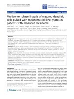
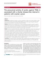
Between March 20, 2007, and December 12, 2008, 48 patients
were randomized to treatment with motesanib at
three different doses: Arm A (125 mg QD), n = 24;
Arm B (75 mg BID 2 weeks on/1 week off ), n = 12;
Arm C (75 mg BID 5 days on/2 days off), n = 12 (Figure 1).
As permitted per protocol, one additional patient was
nonrandomly assigned to Arm A for a total enrollment of
49 patients; all received ≥1 dose of motesanib. Thyroid
cancer was the most common tumor type (Table 1).
Demographics and baseline characteristics were generally
balanced among the treatment arms, although fewer patients received prior therapies in Arm A than in Arms B
and C (Table 1). The ultrasound gallbladder analysis set included 92% of patients; the CCK-HIDA gallbladder analysis set included 84% of patients. One patient (Arm A)
with mesothelioma had a cholecystectomy during the
study (see Adverse Events) but had baseline and evaluable
postbaseline assessments and was therefore included in
both gallbladder analysis sets. All patients discontinued
treatment (Figure 1). Twenty patients (80%) in Arm A,
8 (67%) in Arm B, and 8 (67%) in Arm C completed the
safety follow-up. Reasons for not completing the safety
follow-up were disease progression (Arms A and C, n = 1
each), death (Arm A, n = 2; both due to disease progression), AE (Arm C, n = 1), and withdrawn consent (Arm B,
n = 1). Median follow-up times in Arms A, B, and C were
17 (range, 6–57), 18 (1–58), and 22 (5–60) weeks,
respectively.
Effects of motesanib dose on gallbladder size and
function
Baseline gallbladder volume and ejection fraction were
similar across arms (Table 2). Across all patients, gallbladder volume increased by a mean 22.2 cc (median,
Rosen et al. BMC Cancer 2013, 13:242
/>
Page 4 of 11
Assessed for eligibility
(N=66)
Randomized (n=48)
Arm A: motesanib 125 mg QD (n=25*)
Received motesanib (n=25)
Arm B: motesanib 75 mg BID, 14 d on/7 d off
(n=12)
Received motesanib (n=12)
Arm C: motesanib 75 mg BID, 5 d on/2 d off (n=12)
Received motesanib (n=12)
Discontinued treatment (n=25)
Disease progression (n=18)
Adverse event (n=5)
Continuing treatment in rollover study† (n=2)
Consent withdrawn (n=0)
Discontinued treatment (n=12)
Disease progression (n=9)
Adverse event (n=2)
Continuing treatment in rollover study† (n=1)
Consent withdrawn (n=0)
Discontinued treatment (n=12)
Disease progression (n=5)
Adverse event (n=4)
Continuing treatment in rollover study† (n=2)
Consent withdrawn (n=1)
Gallbladder analysis set, ultrasound (n=23)
Excluded (n=2)
Gallbladder analysis set, CCK-HIDA (n=22)
Excluded (n=3)
Safety analysis set (n=25)
Gallbladder analysis set, ultrasound (n=11)
Excluded (n=1)
Gallbladder analysis set, CCK-HIDA (n=10)
Excluded (n=2)
Safety analysis set (n=12)
Gallbladder analysis set, ultrasound (n=11)
Excluded (n=1)
Gallbladder analysis set, CCK-HIDA (n=10)
Excluded (n=2)
Safety analysis set (n=12)
Figure 1 Disposition of patients in the study. *One patient was nonrandomly assigned to Arm A and received treatment with motesanib
125 mg QD. †Total shown does not reflect 2 additional patients who discontinued motesanib for other reasons but later were granted a waiver
to continue in a rollover study.
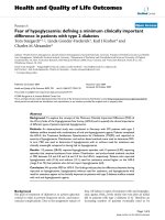
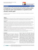
17.3 cc; range, −43.3 to 83.2 cc) from 38.6 cc at baseline
during motesanib treatment. Gallbladder volume increased from baseline in all dosing cohorts, starting before the end of the first 21-day motesanib treatment
cycle (Table 2; Figure 2A, B, C).
Motesanib treatment also affected gallbladder function. Across all patients, ejection fraction decreased by a
mean 19.2% (median, −18.0%; range, −81% to 67%) from
61.3% at baseline during the study. Gallbladder ejection
fraction during treatment was generally lower than baseline measurements (Table 2; Figure 2D, E, F).
Changes in gallbladder volume and function appeared
to be at least partially reversible. Among 45 patients in
the gallbladder volume analysis set, 33 had an evaluable
ultrasound after motesanib discontinuation. In each arm,
mean changes from last on-treatment to last available
off-treatment measurement indicated a decrease in gallbladder volume (Table 2). Similarly, among the 41 patients in the gallbladder ejection fraction analysis set
who had an evaluable CCK-HIDA after motesanib discontinuation (n = 10), gallbladder mean ejection fraction
increased between these two time points (Table 2).
To adjust for potential confounding factors, linear regression analyses were performed. The results were consistent with the data from the preplanned analysis,
showing a trend toward decreasing gallbladder volume
and increasing gallbladder ejection fraction over time
(data not shown).
Treatment, age, sex, body mass index, and NSAID use
were examined in a linear regression model as potential
covariates for gallbladder volume. Of those, only NSAID
use was positively associated with increased gallbladder
volume as assessed by ultrasound (P = .0133); the other
covariates were not significantly associated with gallbladder volume. Exploratory analyses did not show an association between pharmacokinetic exposure to motesanib
and gallbladder volume (data not shown). Covariate analyses and exploratory pharmacokinetic exposure analyses
for gallbladder ejection fraction could not be performed
because of insufficient ejection fraction data.
Changes in other gallbladder characteristics
Some patients in Arms A and B developed gallstones
and/or pericholecystic fluid while receiving motesanib
(Table 3), including two patients who developed de novo
gallstones; however, two patients with gallstones at baseline did not have gallstones at subsequent examinations.
Sludge occurred in all three treatment arms at relatively
high incidence rates (Arms A/B/C, 39%/36%/27%).
Adverse events
Adverse events considered related to treatment with
motesanib by investigators were generally consistent in
frequency and severity with what has been reported in
previous motesanib studies [5,7-9,12,14,15]. Incidence of
grade ≥3 treatment-related AEs in Arms A, B, and C
was 32%, 42%, and 33%, respectively. Two patients had
grade 4 AEs (one each in Arms B and C). Two deaths
occurred during the study; both were caused by disease
progression.
Rosen et al. BMC Cancer 2013, 13:242
/>
Page 5 of 11
Table 1 Patient demographics and baseline characteristics
Characteristics
Arm A
Arm B
Arm C
Motesanib
Motesanib
Motesanib
All
patients
125 mg QD
75 mg BID 2 wk on/1 wk off
75 mg BID 5 d on/2 d off
n = 25
n = 12
n = 12
N = 49
Women
10 (40)
6 (50)
5 (42)
21 (43)
Men
15 (60)
6 (50)
7 (58)
28 (57)
22 (88)
11 (92)
11 (92)
44 (90)
Sex, n (%)
Race, n (%)
White
Black
2 (8)
0 (0)
0 (0)
2 (4)
Hispanic
1 (4)
1 (8)
0 (0)
2 (4)
Native Hawaiian or other Pacific Islander
0 (0)
0 (0)
1 (8)
1 (2)
59 (28–70)
52 (30–70)
59 (22–81)
58 (22–81)
<65 y
18 (72)
10 (83)
9 (75)
37 (76)
≥65 y
7 (28)
2 (17)
3 (25)
12 (24)
≥75 y
0 (0)
0 (0)
1 (8)
1 (2)
Thyroid
1 (4)
7 (58)
4 (33)
12 (24)
Colon
3 (12)
0 (0)
1 (8)
4 (8)
4 (8)
Median age (range), y
Age group, n (%)
Tumor type, n (%)
Non–small-cell lung
3 (12)
0 (0)
1 (8)
Carcinoma of unknown origin
1 (4)
1 (8)
0 (0)
2 (4)
Cervix
1 (4)
0 (0)
1 (8)
2 (4)
Oral
1 (4)
0 (0)
1 (8)
2 (4)
Ovarian
1 (4)
0 (0)
1 (8)
2 (4)
Small-cell lung
0 (0)
2 (17)
0 (0)
2 (4)
Soft tissue sarcoma
1 (4)
0 (0)
1 (8)
2 (4)
1 (2)
Bile duct
1 (4)
0 (0)
0 (0)
Bone sarcoma
1 (4)
0 (0)
0 (0)
1 (2)
Esophageal
1 (4)
0 (0)
0 (0)
1 (2)
Kidney
1 (4)
0 (0)
0 (0)
1 (2)
Liver
1 (4)
0 (0)
0 (0)
1 (2)
Squamous cell carcinoma of head and neck
1 (4)
0 (0)
0 (0)
1 (2)
Other
7 (28)
2 (17)
2 (17)
11 (22)
0
14 (56)
8 (67)
9 (75)
31 (63)
1
10 (40)
4 (33)
3 (25)
17 (35)
2
1 (4)
0 (0)
0 (0)
1 (2)
ECOG performance status, n (%)
Disease stage, n (%)
Stage III
1 (4)
0 (0)
0 (0)
1 (2)
Stage IV
22 (88)
11 (92)
11 (92)
44 (90)
2 (8)
1 (8)
1 (8)
4 (8)
0
1 (4)
0 (0)
1 (8)
2 (4)
1
13 (52)
4 (33)
3 (25)
20 (41)
2
10 (40)
5 (42)
6 (50)
21 (43)
1 (4)
3 (25)
2 (17)
6 (50)
Unknown
Number of sites of disease,* n (%)
≥3
Rosen et al. BMC Cancer 2013, 13:242
/>
Page 6 of 11
Table 1 Patient demographics and baseline characteristics (Continued)
Number of prior therapies,† n (%)
0
5 (20)
1 (8)
1 (8)
7 (14)
1
5 (20)
1 (8)
2 (17)
8 (16)
2
2 (8)
1 (8)
3 (25)
6 (12)
13 (52)
9 (75)
6 (50)
28 (57)
1 (4)
5 (42)
4 (33)
10 (20)
≥3
Alcohol use, n (%)
Never
Former
5 (20)
1 (8)
2 (17)
8 (16)
Current
18 (72)
5 (42)
5 (42)
28 (57)
Missing
1 (4)
1 (8)
1 (8)
3 (6)
BID = twice daily; ECOG = Eastern Cooperative Oncology Group; QD = once daily.
*Sites of disease as assessed by investigator.
†
Prior therapies include all cancer therapies before study enrollment.
Gallbladder toxicity events (all considered treatmentrelated) occurred only in Arm A (n = 6, 12%). Three patients had cholecystitis that resolved after motesanib
treatment was permanently discontinued. One event was
of grade 1 and resolved within 1 week while motesanib
was withheld. One event was of grade 2 and occurred
approximately 1 month after the last motesanib dose; it
resolved 2 months later. A 70-year-old white man with
metastatic non–small-cell lung cancer developed grade
3 cholecystitis that was managed without surgery.
Symptoms appeared approximately 3 weeks after initiation of motesanib, with ultrasound showing gallbladder
distension and the presence of sludge. CCK-HIDA revealed a patent cystic duct and gallbladder dyskinesia.
The patient discontinued motesanib and was treated
with oxycodone and paracetamol. Three weeks later,
CCK-HIDA measurements were normal and the symptoms had resolved. One patient, a 56-year-old white man
with stage IV mesothelioma, had serious grade 3 acute
cholecystitis resulting in cholecystectomy. The event
Table 2 Gallbladder Volume (per Independent Review) and Ejection Fraction (per Investigator)
Endpoint
Gallbladder volume, cc (95% CI)
Baseline
Arm A
Arm B
Arm C
Motesanib
Motesanib
Motesanib
125 mg QD
75 mg BID 2 wk on/
1 wk off
75 mg BID 5 d on/
2 d off
n = 23
n = 11
n = 11
n = 23
n = 11
n = 11
33.3 (22.5–44.1)
48.1 (23.1–73.1)
40.2 (14.1–66.2)
Mean change from baseline
17.7 (6.4–28.9)
26.8 (11.5–42.1)
26.9 (8.8–45.1)
Maximum change from baseline
45.6 (20.2–70.9)
74.4 (41.3–107.4)
67.3 (30.8–103.8)
n = 21
n = 10
n = 10
Gallbladder ejection fraction, % (95% CI)
Baseline
59.1 (43.5–74.8)
68.7 (50.5–87.0)
58.5 (38.4–78.6)
Mean change from baseline
−24.1 (−38.2 to −9.9)
−25.0 (−43.9 to −6.1)
−3.3 (−25.0 to 18.4)
Maximum change from baseline
−30.1 (−46.4 to −13.7)
−26.5 (−45.0 to −8.0)
−6.5 (−29.8 to 16.8)
n = 16
n=9
n=8
Reversibility of gallbladder volume changes, cc (95% CI)
Mean change in gallbladder volume after discontinuation of motesanib
−8.5 (−38.8 to 21.7)
−16.2 (−37.4 to 5.1)
−7.4 (−67.1 to 52.4)
Mean change in gallbladder volume from baseline to last available
off- treatment measurement
10.4 (−10.0 to 30.8)
−14.4 (−31.1 to 2.4)
7.1 (−28.9 to 43.0)
Reversibility of gallbladder ejection fraction changes, % (95% CI)
n=5
n=3
n=2
Mean change in ejection fraction after discontinuation of motesanib
10.8 (−45.8 to 67.4)
63.0 (24.0 to 102.0)
46.0 (−347.9 to 439.9)
Mean change in ejection fraction from baseline to last available
off- treatment measurement
−16.6 (−53.3 to 20.1)
7.7 (−3.8 to 19.1)
14.5 (−55.4 to 84.4)
BID = twice daily; QD = once daily.
310
270
270
250
250
250
230
230
230
210
190
170
150
130
110
90
Gall Bladder Size, cc
290
270
Gall Bladder Size, cc
290
210
190
170
150
130
110
90
210
190
170
150
130
110
90
70
70
70
50
50
50
30
30
10
10
10
–10
–10
n=23
n=23
Baseline Cycle 1
Day 8
n=23
n=22
Cycle 1 Cycle 2
Day 15
n=14
Cycle 3
n=11
Cycle 5
n=8
n=5
Cycle 7 Cycle 8
n=2
n=3
n=2
n=1
Cycle 11 Cycle 13 Cycle 15 Cycle 17
n=12
SFUP1
n=10
SFUP2
n=6
SFUP3
n=7
SFUP4
30
n=11
n=11
Baseline Cycle 1
Day 8
n=11
n=11
n=8
Cycle 1 Cycle 2 Cycle 3
Day 15
n=7
Cycle 5
n=3
n=2
Cycle 7 Cycle 8
n=2
n=2
n=1
n=1
n=7
Cycle 11 Cycle 13 Cycle 15 Cycle 17 SFUP1
n=6
SFUP2
E
110
n=6
SFUP3
n=6
SFUP4
90
90
80
80
80
70
70
70
60
60
60
30
20
10
0
Ejection Fraction, %
110
100
90
40
50
40
30
20
10
0
n=4
Cycle 7
n=2
n=1
n=1
n=2
n=2
Cycle 8 Cycle 11 Cycle 13 Cycle 15 Cycle 17
n=6
n=7
n=7
n=6
SFUP1
SFUP2
SFUP3
SFUP4
10
0
–20
–20
–30
–30
–30
–40
–40
n=5
SFUP
n=4
Cycle 5
20
–20
n=7
Cycle 6
n=6
Cycle 3
30
–10
n=21
Cycle 2
n=10
40
–10
n=21
Baseline
n=11
Cycle 1 Cycle 2
Day 15
50
–10
–50
n=10
F
110
100
50
n=11
Baseline Cycle 1
Day 8
100
Ejection Fraction, %
Gall Bladder Size, cc
Ejection Fraction, %
C
310
290
–10
D
B
310
Rosen et al. BMC Cancer 2013, 13:242
/>
A
–50
–40
n=10
Baseline
n=4
Cycle 6
n=10
Cycle 2
th
n=3
SFUP
–50
n=10
Baseline
n=9
Cycle 2
n=3
Cycle 6
n=2
SFUP
th
Figure 2 Change in gallbladder size and function. Mean (dots connected by lines) and median (25 and 75 quartiles; solid horizontal lines) gallbladder size (A, B, C) and function (D, E, F)
over time per independent review in Arms A, B, and C, respectively. Error bars represent the minimum and maximum values. SFUP, safety follow-up.
Page 7 of 11
Rosen et al. BMC Cancer 2013, 13:242
/>
Page 8 of 11
Table 3 Specific gallbladder findings (per Independent Ultrasound Review)
Patient incidence,
n (%)
Arm A
Arm B
Arm C
Motesanib 125 mg QD
Motesanib 75 mg BID
Motesanib 75 mg BID
2 wk on/1 wk off
5 d on/2 d off
Baseline
Post
baseline*
Post
treatment†
Baseline
Post
baseline*
Post
treatment†
Baseline
Post
baseline*
Post
treatment†
(n = 23)
(n = 23)
(n = 16)
(n = 11)
(n = 11)
(n = 9)
(n = 11)
(n = 11)
(n = 8)
Gallstones
3 (13)
4 (17)
3 (19)
3 (27)
2 (18)
2 (22)
0 (0)
0 (0)
0 (0)
Sludge
0 (0)
9 (39)
4 (25)
0 (0)
4 (36)
4 (44)
0 (0)
3 (27)
0 (0)
Pericholecystic fluid
0 (0)
1 (4)
0 (0)
1 (9)
1 (9)
0 (0)
0 (0)
0 (0)
0 (0)
Common duct
dilation
0 (0)
0 (0)
0 (0)
0 (0)
0 (0)
0 (0)
0 (0)
0 (0)
0 (0)
*Data within the table indicate number of patients with at least one incidence of the specific gallbladder findings listed at any point during
postbaseline treatment.
†
Data within the table indicate number of patients who had the specific gallbladder findings listed at their last available off-treatment assessment.
occurred approximately 1 month after treatment initiation. At the time of hospitalization, the patient had a
24-hour history of right upper quadrant pain; Murphy’s sign
was positive on abdominal examination. Motesanib was
withheld, and ultrasound revealed gallbladder distension,
wall thickening (4.4 cm), intramural edema, mural
hypervascularity, trace of pericholecystic fluid, and no biliary
tract dilation. Cholecystectomy was performed 8 days after
cessation of motesanib, and the patient resumed motesanib
treatment 11 days later. At the safety follow-up, two patients
had ongoing grade 1 gallbladder disorders, specifically gallbladder dysfunction and gallbladder wall thickening, with
the latter prompting a dose reduction. Twelve patients had
right upper quadrant pain during the study (Arms A/B/C,
n = 8/1/3); these events occurred at variable times after initiation of motesanib. However, the available data do not help
distinguish between pain due to gallbladder toxicity versus
other etiologies, such as liver metastases.
Objective response
Most patients had measureable disease at baseline (Arm A,
n = 24 [96%]; Arm B, n = 12 [100%]; Arm C, n = 11 [92%]).
No complete responses were achieved, but one patient with
stage IV thyroid cancer in Arm B had a confirmed partial
response (overall objective response rate, 2%). Twenty-eight
patients (60%) had stable disease as best tumor response
(Arm A, n = 15 [63%]; Arm B, n = 6 [50%]; Arm C, n = 7
[64%]), with durable (≥24 weeks) stable disease in 8 (17%)
patients (Arm A, n = 6 [25%]; Arm B, n = 1 [8%]; Arm C,
n = 1 [9%]). Fifteen patients (32%) had progressive disease (Arm A, n = 8 [33%]; Arm B, n = 3 [25%]; Arm C,
n = 4 [36%]).
Table 4 Gallbladder-related toxicity and potential gallbladder-related toxicity reported with tyrosine kinase inhibitors
other than motesanib
Agent
Molecular target(s)
Study / Study type
Cediranib
VEGFR1, VEGFR2, VEGFR3
Laurie et al. [21] – phase 1 study
Acute cholecystitis
Batchelor et al. [22] – phase 2
study
Gallbladder obstruction, abdominal pain
Imatinib
Sorafenib
Sunitinib
BCR-ABL, Kit, PDGFR-α, PDGFR-β
VEGFR1, VEGFR2, VEGFR3, Raf,
PDGFR-β, Flt-3, Kit
VEGFR1, VEGFR2, VEGFR3, PDGFR-α,
PDGFR-β, Flt-3, Kit
Adverse events reported
Yeh et al. [23] – single-arm study
Gallstones
Breccia et al. [37] – case report
Gallstones, gallbladder wall thickening, abdominal pain
Grant et al. [24] – phase 1 study
Cholecystitis
Sanda et al. [20] – case report
Right upper abdominal pain, gallbladder edema, acute
acalculous cholecystitis
Nexavar European public
assessment report [26]
Cholecystitis, cholangitis
Nexavar US prescribing
information [25]
Cholecystitis, cholangitis
Motzer et al. [17] – single-arm
study
Acute cholecystitis
De Lima Lopes, Jr., et al. [18] –
case report
Acute emphysematous cholecystitis, right upper abdominal
pain, gallbladder distension
Gomez-Abuin et al. [19] – case
report
Acute acalculous cholecystitis, right upper abdominal pain,
gallbladder wall thickening
ALT = alkaline phosphatase; PDGFR = platelet-derived growth factor receptor; VEGFR = vascular endothelial growth factor receptor.
Rosen et al. BMC Cancer 2013, 13:242
/>
Pharmacokinetics
Motesanib was rapidly absorbed, and there was no evidence of drug accumulation after QD administration.
The median Cmax values in Arms A, B, and C were 630,
323, and 355 ng/mL, respectively; the median Cmin values
were 14, 60, and 35 ng/mL, respectively. In Arm B, the
median motesanib concentration after the 1-week washout period was <0.2 ng/mL (the limit of quantitation); in
Arm C, the median motesanib concentration after the
2-day wash-out period was 1.2 ng/mL. The median AUC
values estimated from the three dosing regimens appeared similar, ranging from 1.9 to 3.0 μg·hr/mL.
An exploratory analysis investigated the potential relationship between drug exposure (Cmax, Cmin, and AUC) and
change in gallbladder size. The results showed no consistent
trend between gallbladder size and motesanib exposure.
Discussion
In this randomized phase 1b study designed to assess
gallbladder-related toxicity among patients receiving
three motesanib dose schedules, increased gallbladder
volume, decreased gallbladder function, and other
gallbladder changes, including development of gallstones and sludge, were common. Changes in gallbladder volume were observed as early as in the first
cycle of motesanib treatment. Symptomatic gallbladder
toxicity occurred in six patients, one of whom had acute
cholecystitis requiring a cholecystectomy. Other toxicities
were generally consistent with those reported in previous
motesanib studies and for the class of VEGF pathway inhibitors. While increases in gallbladder volume and decreases in gallbladder function did not appear to be doseor schedule-dependent, gallbladder toxicity occurred only
in Arm A (motesanib 125 mg QD).
Gallbladder toxicity, at varying incidence rates, has been
described in most motesanib studies [5,7,8,10,28]; however, considering the findings summarized herein,
gallbladder-associated AEs may have been underdetected.
This may particularly apply to earlier-conducted studies
that reported no [12-16] or low [5,9,28] incidence rates of
cholecystitis (but no other gallbladder toxicity) and to patients who presented only with right upper quadrant pain
along with other possible reasons for pain, including liver
metastases. For example, Sawaki and colleagues described
the incidental discovery by ultrasound of extended gallbladder or wall thickening in three patients [12]. Given
that many VEGF pathway inhibitors block the same or similar targets as motesanib (Table 4), and because of the incidence of abdominal pain with tyrosine kinase inhibitors
[17-26,29-37], changes in gallbladder size and function not
manifested as symptomatic toxicity may occur more frequently during treatment with these agents than generally
believed. The results of our study should encourage
Page 9 of 11
investigators to more closely examine potentially gallbladderrelated symptoms in studies of VEGF pathway inhibitors and
among patients treated outside of clinical trials.
The biologic mechanisms that underlie the gallbladder
changes associated with motesanib treatment are not yet
elucidated. The toxicity may be related to antiangiogenic activity of motesanib in the gallbladder which could be exacerbated by accumulated motesanib, considering the drug’s
biliary excretion pattern (Amgen Inc., data on file). Accumulation of motesanib within the gallbladder following the
excretion (and reactivation) of its major metabolite,
motesanib glucuronic acid [38], in the relatively high pH of
the bile may result in irritation to the gallbladder or possibly
even transient ischemia with subsequent sludge accumulation, transient obstruction, pain, and ultimately, cholecystitis
or cholecystitis-like symptoms. One potential solution may
be to avoid conditions that are known to reduce gallbladder
emptying such as fasting and low-fat diets. Consideration
should also be given to the possibility that gallbladder toxicity is an on-target effect of inhibition of one or more of
the molecular targets of tyrosine kinase inhibitors.
The design of this study may be appropriate for investigating gallbladder toxicity with other investigational agents,
including tyrosine kinase inhibitors. The measured changes
from baseline in gallbladder volume and ejection appeared
to be both robust and greater than anticipated inter- or
intrapatient variance. In Arms A and B, the 95% Cl for the
mean and maximum changes from baseline did not encompass zero, and the observed changes were consistent
with differences between patients with gallbladder disease
and healthy control participants reported in previous studies [39,40]. Thus, the results demonstrate that, when
coupled with rigorous quality control/assurance procedures
and training, routine diagnostic techniques (eg, ultrasound,
CT, and CCK-HIDA [41]) can be used to evaluate the incidence and timing of gallbladder toxicity assessed as changes
in volume, ejection fraction, and filling, and to identify
other abnormalities, such as gallstones and pericholecystic
fluid. Better characterization of these risks is important because of the potential seriousness of gallbladder toxicity.
More broadly, targeted assessments of specific AEs may
help characterize the toxicity of investigational cancer therapeutics. The study was limited by the lack of a placebo
arm, and the small sample size potentially restricted AE
and other assessments.
Conclusions
In conclusion, motesanib monotherapy was associated with
increased gallbladder volume and decreased ejection fraction in most patients, regardless of dosing regimen and exposure, which appeared to be at least partially reversible.
Motesanib had a toxicity profile consistent with previous
studies. The etiology of gallbladder toxicity during
motesanib treatment remains uncertain.
Rosen et al. BMC Cancer 2013, 13:242
/>
Competing interests
LSR, LL, NDB, and JJS have no competing interests to declare. TJP and LJW
have been consultants to Amgen Inc. RVB has received honoraria from and
holds stock in Amgen Inc. HIH has received research funding from GSK. NCT
has received research funding from Amgen Inc. and has provided expert
testimony on behalf of Amgen Inc. SM, Y-JH, and C-PH are employees of
and shareholders in Amgen Inc.
Authors’ contributions
LSR, HIH, and Y-JH participated in conception and design of the study. LL,
LSR, TJP, NDB, HIH, JJS, LJW, SM, C-PH, and NCT participated in collection
and assembly of data. LSR, RVB, HIH, LJW, SM, Y-JH, and C-PH participated in
data analysis and interpretation. All authors participated in writing or revising
the manuscript and provided their approval of the final version of the
manuscript.
Acknowledgments
The authors thank Rebeca Melara (Amgen Inc.) for pharmacokinetic analysis;
Benjamin Scott (Complete Healthcare Communications, Inc., Chadds Ford,
PA, USA), whose work was funded by Amgen Inc., and Beate Quednau
(Amgen Inc.) for assistance in manuscript writing.
Author details
1
Department of Medicine, University of California Los Angeles,
Santa Monica, CA, USA. 2Western Hospital, Footscray, and Royal Melbourne
Hospital, Parkville, VIC, Australia. 3The Queen Elizabeth Hospital, University of
Adelaide School of Medicine, Woodville, SA, Australia. 4Oncology
Hematology of Lehigh Valley, Bethlehem, PA, USA. 5Center for Cancer and
Blood Disorders, Bethesda, MD, USA. 6Duke University Medical Center,
Durham, NC, USA. 7Cancer Centers of the Carolinas, Greenville, SC, USA.
8
Dana-Farber Cancer Institute and Massachusetts General Hospital, Boston,
MA, USA. 9Department of Biostatistics, Amgen Inc., South San Francisco, CA,
USA. 10Department of Oncology, Amgen Inc., Thousand Oaks, CA, USA.
11
Department of Pharmacokinetics & Drug Metabolism, Amgen Inc,
Thousand Oaks, CA, USA. 12Ludwig Oncology Unit, Austin Hospital,
Heidelberg, VIC, Australia.
Received: 29 February 2012 Accepted: 26 April 2013
Published: 16 May 2013
References
1. O'Shaughnessy JA, Wittes RE, Burke G, Friedman MA, Johnson JR,
Niederhuber JE, Rothenberg ML, Woodcock J, Chabner BA, Temple R:
Commentary concerning demonstration of safety and efficacy of
investigational anticancer agents in clinical trials. J Clin Oncol 1991,
9(12):2225–2232.
2. Tsang R, Colley L, Lynd LD: Inadequate statistical power to detect
clinically significant differences in adverse event rates in randomized
controlled trials. J Clin Epidemiol 2009, 62(6):609–616.
3. Pitrou I, Boutron I, Ahmad N, Ravaud P: Reporting of safety results in
published reports of randomized controlled trials. Arch Intern Med 2009,
169(19):1756–1761.
4. Ioannidis JP: Adverse events in randomized trials: neglected, restricted,
distorted, and silenced. Arch Intern Med 2009, 169(19):1737–1739.
5. Rosen LS, Kurzrock R, Mulay M, Van Vugt A, Purdom M, Ng C, Silverman J,
Koutsoukos A, Sun YN, Bass MB, Xu RY, Polverino A, Wiezorek JS, Chang DD,
Benjamin R, Herbst RS: Safety, pharmacokinetics, and efficacy of AMG
706, an oral multikinase inhibitor, in patients with advanced solid
tumors. J Clin Oncol 2007, 25(17):2369–2376.
6. Benjamin RS, Schoffski P, Hartmann JT, Van Oosterom A, Bui BN, Duyster J,
Schuetze S, Blay JY, Reichardt P, Rosen LS, Skubitz K, McCoy S, Sun YN,
Stepan DE, Baker L: Efficacy and safety of motesanib, an oral inhibitor of
VEGF, PDGF, and Kit receptors, in patients with imatinib-resistant
gastrointestinal stromal tumors. Cancer Chemother Pharmacol 2011,
68(1):69-77.
7. Sherman SI, Wirth LJ, Droz JP, Hofmann M, Bastholt L, Martins RG, Licitra L,
Eschenberg MJ, Sun YN, Juan T, Stepan DE, Schlumberger MJ: Motesanib
diphosphate in progressive differentiated thyroid cancer. N Engl J Med
2008, 359(1):31–42.
8. Schlumberger MJ, Elisei R, Bastholt L, Wirth LJ, Martins RG, Locati LD, Jarzab
B, Pacini F, Daumerie C, Droz JP, Eschenberg MJ, Sun YN, Juan T, Stepan DE,
Page 10 of 11
9.
10.
11.
12.
13.
14.
15.
16.
17.
18.
19.
20.
21.
22.
23.
24.
Sherman SI: Phase II study of safety and efficacy of motesanib in patients
with progressive or symptomatic, advanced or metastatic medullary
thyroid cancer. J Clin Oncol 2009, 27(23):3794–3801.
Blumenschein GR, Reckamp K, Stephenson GJ, O'Rourke T, Gladish G,
McGreivy J, Sun YN, Ye Y, Parson M, Sandler A: Phase 1b study of
motesanib, an oral angiogenesis inhibitor, in combination with
carboplatin/paclitaxel and/or panitumumab for the treatment of
advanced non-small cell lung cancer. Clin Cancer Res 2010, 16(1):279–290.
Blumenschein GR Jr, Kabbinavar F, Menon H, Mok TS, Stephenson J, Beck JT,
Lakshmaiah K, Reckamp K, Hei YJ, Kracht K, Sun YN, Sikorski R, Schwartzberg
L, on behalf of the Motesanib NPIISI: A phase II, multicenter, open-label
randomized study of motesanib or bevacizumab in combination with
paclitaxel and carboplatin for advanced nonsquamous non-small-cell
lung cancer. Ann Oncol 2011, 22(9):2057–2067.
Rosen PJ, Sweeney CJ, Park DJ, Beaupre DM, Deng H, Leitch IM, Shubhakar
P, Zhu M, Oliner KS, Anderson A, Yee LK: A phase Ib study of AMG 102 in
combination with bevacizumab or motesanib in patients with advanced
solid tumors. Clin Cancer Res 2010, 16(9):2677–2687.
Sawaki A, Yamada Y, Komatsu Y, Kanda T, Doi T, Koseki M, Baba H, Sun YN,
Murakami K, Nishida T: Phase II study of motesanib in Japanese patients
with advanced gastrointestinal stromal tumors with prior exposure to
imatinib mesylate. Cancer Chemother Pharmacol 2010, 65(5):961–967.
Fujisaka Y, Yamada Y, Yamamoto N, Shimizu T, Fujiwara Y, Yamada K, Tamura T,
Watanabe H, Sun YN, Bass MB, Seki M: Phase 1 study of the investigational,
oral angiogenesis inhibitor motesanib in Japanese patients with advanced
solid tumors. Cancer Chemother Pharmacol 2010, 66(5):935–943.
Price TJ, Lipton L, McGreivy J, McCoy S, Sun YN, Rosenthal MA: Safety and
pharmacokinetics of motesanib in combination with gemcitabine for the
treatment of patients with solid tumours. Br J Cancer 2008, 99(9):1387–1394.
LoRusso P, Heath EI, McGreivy J, Sun YN, Melara R, Yan L, Malburg L,
Ingram M, Wiezorek J, Chen L, Pilat MJ: Effect of coadministration of
ketoconazole, a strong CYP3A4 inhibitor, on pharmacokinetics and
tolerability of motesanib diphosphate (AMG 706) in patients with
advanced solid tumors. Invest New Drugs 2008, 26(5):455–462.
Burris H, Stephenson J, Otterson GA, Stein M, McGreivy J, Sun YN, Ingram M,
Ye Y, Schwartzberg LS: Safety and pharmacokinetics of motesanib in
combination with panitumumab and gemcitabine-Cisplatin in patients
with advanced cancer. J Oncol 2011, 2011:853931.
Motzer RJ, Rini BI, Bukowski RM, Curti BD, George DJ, Hudes GR, Redman
BG, Margolin KA, Merchan JR, Wilding G, Ginsberg MS, Bacik J, Kim ST, Baum
CM, Michaelson MD: Sunitinib in patients with metastatic renal cell
carcinoma. JAMA 2006, 295(21):2516–2524.
de Lima Lopes G Jr, Rocha Lima CM: Emphysematous cholecystitis
in a patient with gastrointestinal stromal tumor treated with
sunitinib. Pharmacotherapy 2007, 27(5):775–777.
Gomez-Abuin G, Karam AA, Mezzadri NA, Bas CA: Acalculous cholecystitis
in a patient with metastatic renal cell carcinoma treated with sunitinib.
Clin Genitourin Cancer 2009, 7(1):62–63.
Sanda M, Tamai H, Deguchi H, Mori Y, Moribata K, Shingaki N, Ueda K, Inoue
I, Maekita T, Iguchi M, Yanaoka K, Oka M, Ichinose M: Acalculous
cholecystitis in a patient with hepatocellular carcinoma on sorafenib.
ISRN Gastroenterol 2011, 2011:201529.
Laurie SA, Gauthier I, Arnold A, Shepherd FA, Ellis PM, Chen E, Goss G,
Powers J, Walsh W, Tu D, Robertson J, Puchalski TA, Seymour L: Phase I and
pharmacokinetic study of daily oral AZD2171, an inhibitor of vascular
endothelial growth factor tyrosine kinases, in combination with
carboplatin and paclitaxel in patients with advanced non-small-cell lung
cancer: the National Cancer Institute of Canada clinical trials group. J Clin
Oncol 2008, 26(11):1871–1878.
Batchelor TT, Duda DG, di Tomaso E, Ancukiewicz M, Plotkin SR, Gerstner E,
Eichler AF, Drappatz J, Hochberg FH, Benner T, Louis DN, Cohen KS, Chea H,
Exarhopoulos A, Loeffler JS, Moses MA, Ivy P, Sorensen AG, Wen PY, Jain RK:
Phase II study of cediranib, an oral pan-vascular endothelial growth
factor receptor tyrosine kinase inhibitor, in patients with recurrent
glioblastoma. J Clin Oncol 2010, 28(17):2817–2823.
Yeh CN, Chen TW, Liu FY, Jan YY, Chen MF: Genetic changes in advanced
gastrointestinal stromal tumor (GIST) patients during imatinib mesylate
treatment. Langenbecks Arch Surg 2006, 391(6):615–621.
Grant S, Karp JE, Koc ON, Cooper B, Luger S, Figg WD, Egorin M, Druker BJ,
Jacobberger JW, Ramakrishnan V, Perkins EB, Colevas AD, Roberts JD: Phase
I study of flavopiridol in combination with imatinib mesylate (ST1571,
Rosen et al. BMC Cancer 2013, 13:242
/>
25.
26.
27.
28.
29.
30.
31.
32.
33.
34.
35.
36.
37.
38.
Gleevec) in Bcr/Abl + hematological malignancies [abstract]. Blood 2005,
106:1102.
Nexavar® (sorafenib) Full Prescribing Information. Edited by Wayne NJ: Bayer
Healthcare Pharmaceuticals, Inc; 2011. />html/products/pi/Nexavar_PI.pdf]
European Medicines Agency: Nexavar European Public Assessment Report,
Summary of Product Characteristics. London, UK: European Medicines
Agency; 2011 [ />EPAR_-_Product_Information/human/000690/WC500027704.pdf]
Therasse P, Arbuck SG, Eisenhauer EA, Wanders J, Kaplan RS, Rubinstein L,
Verweij J, Van Glabbeke M, van Oosterom AT, Christian MC, Gwyther SG:
New guidelines to evaluate the response to treatment in solid tumors.
European Organization for Research and Treatment of Cancer, National
Cancer Institute of the United States, National Cancer Institute of
Canada. J Natl Cancer Inst 2000, 92(3):205–216.
Kotasek D, Tebbutt N, Desai J, Welch S, Siu LL, McCoy S, Sun YN, Johnson J,
Adewoye AH, Price T: Safety and pharmacokinetics of motesanib in
combination with gemcitabine and erlotinib for the treatment of solid
tumors: a phase 1b study. BMC Cancer 2011, 11:313.
Burstein HJ, Elias AD, Rugo HS, Cobleigh MA, Wolff AC, Eisenberg PD,
Lehman M, Adams BJ, Bello CL, DePrimo SE, Baum CM, Miller KD: Phase II
study of sunitinib malate, an oral multitargeted tyrosine kinase inhibitor,
in patients with metastatic breast cancer previously treated with an
anthracycline and a taxane. J Clin Oncol 2008, 26(11):1810–1816.
Drevs J, Siegert P, Medinger M, Mross K, Strecker R, Zirrgiebel U, Harder J,
Blum H, Robertson J, Jurgensmeier JM, Puchalski TA, Young H, Saunders O,
Unger C: Phase I clinical study of AZD2171, an oral vascular endothelial
growth factor signaling inhibitor, in patients with advanced solid
tumors. J Clin Oncol 2007, 25(21):3045–3054.
Escudier B, Eisen T, Stadler WM, Szczylik C, Oudard S, Staehler M, Negrier S,
Chevreau C, Desai AA, Rolland F, Demkow T, Hutson TE, Gore M, Anderson
S, Hofilena G, Shan M, Pena C, Lathia C, Bukowski RM: Sorafenib for
treatment of renal cell carcinoma: final efficacy and safety results of the
phase III treatment approaches in renal cancer global evaluation trial.
J Clin Oncol 2009, 27(20):3312–3318.
Gibbons J, Egorin MJ, Ramanathan RK, Fu P, Mulkerin DL, Shibata S,
Takimoto CH, Mani S, LoRusso PA, Grem JL, Pavlick A, Lenz HJ, Flick SM,
Reynolds S, Lagattuta TF, Parise RA, Wang Y, Murgo AJ, Ivy SP, Remick SC:
Phase I and pharmacokinetic study of imatinib mesylate in patients with
advanced malignancies and varying degrees of renal dysfunction: a
study by the National Cancer Institute Organ Dysfunction Working
Group. J Clin Oncol 2008, 26(4):570–576.
Hotte SJ, Winquist EW, Lamont E, MacKenzie M, Vokes E, Chen EX, Brown S,
Pond GR, Murgo A, Siu LL: Imatinib mesylate in patients with adenoid
cystic cancers of the salivary glands expressing c-kit: a Princess Margaret
Hospital phase II consortium study. J Clin Oncol 2005, 23(3):585–590.
Matulonis UA, Berlin S, Ivy P, Tyburski K, Krasner C, Zarwan C,
Berkenblit A, Campos S, Horowitz N, Cannistra SA, Lee H, Lee J,
Roche M, Hill M, Whalen C, Sullivan L, Tran C, Humphreys BD,
Penson RT: Cediranib, an oral inhibitor of vascular endothelial
growth factor receptor kinases, is an active drug in recurrent
epithelial ovarian, fallopian tube, and peritoneal cancer. J Clin
Oncol 2009, 27(33):5601–5606.
Moreno-Aspitia A, Morton RF, Hillman DW, Lingle WL, Rowland KM Jr,
Wiesenfeld M, Flynn PJ, Fitch TR, Perez EA: Phase II trial of sorafenib in
patients with metastatic breast cancer previously exposed to
anthracyclines or taxanes: North Central Cancer Treatment Group and
Mayo Clinic Trial N0336. J Clin Oncol 2009, 27(1):11–15.
Sweeney CJ, Chiorean EG, Verschraegen CF, Lee FC, Jones S, Royce M, Tye L,
Liau KF, Bello A, Chao R, Burris HA: A phase I study of sunitinib plus
capecitabine in patients with advanced solid tumors. J Clin Oncol 2010,
28(29):4513–4520.
Breccia M, D'Andrea M, Alimena G: Can nifedipine and estrogen
interaction with imatinib be responsible for gallbladder stone
development? Eur J Haematol 2005, 75(1):89–90.
Li C, Kuchimanchi M, Hickman D, Poppe L, Hayashi M, Zhou Y, Subramanian
R, Kumar G, Surapaneni S: In vitro metabolism of the novel, highly
selective oral angiogenesis inhibitor motesanib diphosphate in
preclinical species and in humans. Drug Metab Dispos 2009,
37(7):1378–1394.
Page 11 of 11
39. Loreno M, Travali S, Bucceri AM, Scalisi G, Virgilio C, Brogna A:
Ultrasonographic study of gallbladder wall thickness and emptying in
cirrhotic patients without gallstones. Gastroenterol Res Pract 2009,
2009:683040.
40. Krishnamurthy GT, Krishnamurthy S, Brown PH: Constancy and variability
of gallbladder ejection fraction: impact on diagnosis and therapy.
J Nucl Med 2004, 45(11):1872–1877.
41. Elwood DR: Cholecystitis. Surg Clin North Am 2008, 88(6):1241–1252.
doi:10.1186/1471-2407-13-242
Cite this article as: Rosen et al.: The effect of different dosing regimens
of motesanib on the gallbladder: a randomized phase 1b study in
patients with advanced solid tumors. BMC Cancer 2013 13:242.
Submit your next manuscript to BioMed Central
and take full advantage of:
• Convenient online submission
• Thorough peer review
• No space constraints or color figure charges
• Immediate publication on acceptance
• Inclusion in PubMed, CAS, Scopus and Google Scholar
• Research which is freely available for redistribution
Submit your manuscript at
www.biomedcentral.com/submit