MiR-204 down regulates SIRT1 and reverts SIRT1-induced epithelial-mesenchymal transition, anoikis resistance and invasion in gastric cancer cells
Bạn đang xem bản rút gọn của tài liệu. Xem và tải ngay bản đầy đủ của tài liệu tại đây (1.26 MB, 9 trang )
Zhang et al. BMC Cancer 2013, 13:290
/>
RESEARCH ARTICLE
Open Access
MiR-204 down regulates SIRT1 and reverts
SIRT1-induced epithelial-mesenchymal transition,
anoikis resistance and invasion in gastric cancer
cells
Lihua Zhang1*†, Xueqing Wang1† and Pingsheng Chen2
Abstract
Background: Our previous studies showed that SIRT1 was over-expressed in gastric cancer specimens and related
with lymph node metastasis. However, the mechanism of SIRT1 up-regulation and its association with metastasis in
gastric cancer remain unclear. The present study was undertaken to understand the role of microRNA in regulation
of SIRT1 in the progression of gastric cancer.
Methods: Expression of miR-204 and SIRT1 was assessed in two gastric cancer cell lines and 24 matched cancer
specimens. Luciferase reporter assay was carried to verify that miR-204 targeting SIRT1. Cell invasion ability of AGS
and BGC was detected by transwell invasion assay. Annexin V/PI assay was used to investigate the cell sensitivity of
anoikis. Western blot analysis to assess SIRT1, Vimentin, E-Cadherin, LKB1, and β-actin expression was performed in
gastic cancer cell lines.
Results: SIRT1 was defined as the target gene and elucidated the biological functions of miR-204 with a luciferase
reporter assay and Western blot analysis. We verified that miR-204 levels were down-regulated and significantly
associated with the up-regulation of SIRT1 mRNA levels in gastric cancer specimens. Over-expression of miR-204
reduced cell invasion and anoikis resistance in gastric cancer cells. Up-regulation of miR-204 influenced the levels of
the epithelial mesenchymal transition (EMT)-associated genes, increasing E-cadherin levels and decreasing Vimentin
levels. We demonstrated that the regulation of EMT by miR-204 involves cooperation with LKB1. Furthermore,
silencing of SIRT1 phenocopied the effects of miR-204 in gastric cancer cells. These data demonstrate that miR-204
plays an important role in regulating metastasis of gastric cancer, which is involved in post-transcriptional
repression of SIRT1.
Conclusion: Our results suggest that down-regulation of miR-204 promotes gastric cancer cell invasion by
activating the SIRT1-LKB1 pathway. These data demonstrate that miR-204 plays an important role in regulating
metastasis of gastric cancer, which is involved in post-transcriptional repression of SIRT1.
Background
Gastric cancer is among the most common malignancies
in East Asian counties [1,2]. Recurrence and metastasis
are the biggest obstacles for the treatment of gastric cancer [3]. Therefore, the search for new therapeutic targets
to prevent the metastasis of gastric cancer is an urgent
* Correspondence:
†
Equal contributors
1
Department of Pathology, Southeast University, Zhongda Hospital, Nanjing
210009, P R China
Full list of author information is available at the end of the article
issue. However, the pathogenesis and mechanism underlying the metastasis process remain poorly understood.
Epithelial-mesenchymal transition (EMT) is a key step
toward cancer metastasis. Loss of E-cadherin expression
is a hallmark of the EMT process and is likely required
for enhanced tumor cell motility [4,5]. Epithelial cells
lose epithelial characteristics and acquire mesenchymal
characteristics by the down-regulation of E-cadherin [6].
Increasing evidence suggests that post-transcriptional
regulation of gene expression, which is mediated by
microRNAs (miRNAs), controls tumorigenesis and cancer
© 2013 Zhang et al.; licensee BioMed Central Ltd. This is an Open Access article distributed under the terms of the Creative
Commons Attribution License ( which permits unrestricted use, distribution, and
reproduction in any medium, provided the original work is properly cited.
Zhang et al. BMC Cancer 2013, 13:290
/>
metastasis [7-9]. Both the over-expression of oncogenic
miRNAs and the decreased expression of tumor suppressor
miRNAs play pivotal roles in cancer metastasis. Adam et al.
demonstrated that miR-200 regulated EMT in bladder cancer cells and reversed resistance to epidermal growth factor
receptor (EGFR) therapy [7]. This group also showed that
the stable expression of miR-200 in mesenchymal UMUC3
cells increased E-cadherin levels; decreased protein expression of ZEB1, ZEB2, and ERRFI-1; decreased cell migration;
and increased sensitivity to EGFR-blocking agents. Tie et al.
described the regulation and function of miR-218 in gastric
cancer metastasis. Decreased miR-218 levels eliminated
Robo1 repression, which activated the Slit-Robo1 pathway
through the interaction between Robo1 and Slit2 to trigger
tumor metastasis [10].
In the current study, we investigated the role of miR204 in gastric cancer metastasis. We demonstrated that
the miR-204 expression was down-regulated in gastric
cancer tissues and confirmed that the SIRT1 gene is the
direct target of miR-204. Restoration of miR-204 or the
knockdown of SIRT1 in metastatic gastric cancer cells
induces a shift toward an epithelial morphology concomitant with increased expression of E-cadherin and
decreased expression of Vimentin. Down-regulation of
miR-204 inactivated LKB1 through SIRT1 to promote
human gastric cancer cell invasion.
Methods
Cell lines and clinical samples
The AGS and BGC gastric cancer cell lines used in this
study were cultured at 37°C in 5% CO2 and 95% air. All
cells were grown in Dulbecco’s modified Eagle’s medium
(Invitrogen, California, USA) supplemented with 1 mmol/L
L-glutamine, 10% fetal bovine serum (Life Technologies,
Inc., Burlington, Canada), penicillin G 100 U/mL, and
streptomycin 100 mg/mL.
The Ethics Review Board of Zhongda Hospital, Southeast
University Nanjing, China, approved this study. Informed
consent was obtained from all patients. We studied gastric
cancer specimens (cancer lesions and adjacent non-tumor
mucosa) from 24 patients who had undergone resection at
the Zhongda Hospital, Southeast University between 2005
and 2010. We gathered all samples in the same manner;
they were snap-frozen immediately in liquid nitrogen and
stored at −80°C until RNA extraction could be performed.
All tissue specimens were evaluated pathologically. No
patients had received irradiation or cancer chemotherapy
prior to resection.
RT-PCR and real-time RT-PCR
Total cellular RNA was extracted using Trizol (Invitrogen,
California, USA). For mRNA detection, SIRT1, E-Cadherin,
Vimentin and GAPDH mRNA expression were analyzed
Page 2 of 9
by the Sybr Green qRT-PCR according to the manufacturer’s instructions (Applied Biosystems).
For miRNA detection, polyA tail was added to RNasefree DNase digested total RNA using the E.coli polyA
polymerase (NEB). Two micrograms of the tailed total
RNA was reverse transcribed with ImProm-II (Promega).
Conventional PCR or Sybr Green qRT-PCR was used to
assay miRNA expression with the specific forward primers
and the universal reverse primer complementary to the anchor primer. Anchor RT primer was used as the template
for negative control and U6 as internal control.
The primers used are listed in Table 1.
Luciferase reporter assay
Using Lipofectamine 2000 (Invitrogen), HEK293 cells
(104 cells/well) were plated in a 24-well plate. The cells
were then co-transfected with 20 mM of either miR-204 or
microRNA control, 40 ng of either pGL3-promoter-SIRT13’UTR-WT or pGL3-promoter-SIRT1-3’UTR-MUT, and 4
ng of pRL-TK (Promega, Madison, WI). HEK293 cells were
collected 24 hours after transfection and analyzed using the
Dual-Luciferase Reporter Assay System (Promega). The
pRL-TK vector responsible for the constitutive expression
of Renilla luciferase (Promega) was co-transfected as
an internal control to correct for differences in both
transfection and harvest efficiencies. Transfections were
performed in triplicate and repeated at least three times in
separate experiments.
Western blot analysis and antibodies
Western blot analysis to assess SIRT1, Vimentin, ECadherin, LKB1, and β-actin expression was performed
as previously described [11]. All of these primary antibodies were purchased from Santa Cruz Biotechnology
(Santa Cruz, Daly City, CA).
In vitro cell invasion assay
For transwell invasion assays, 1×10⁵ cells were placed
on the non-coated membrane in the top chamber
(CytoSelectTM 24 Well Cell Migration and Invasion Assay
Combo Kit, 8-μm, CBA100-C, Cell Biolab, United States).
Cells were plated in medium without serum. Medium
supplemented with serum was used as a chemo-attractant
in the lower chamber. The cells were incubated for 24
hours; cells that did not invade through the pores were removed using a cotton swab. Cells on the lower surface of
the membrane were fixed with methanol and stained with
crystal violet (Fisher Scientific Co., Fairlawn, NJ). The cell
numbers were determined by counting the penetrating cells
under a microscope at 200 magnification in random fields
in each well. Each experiment was performed in triplicate.
Zhang et al. BMC Cancer 2013, 13:290
/>
Table 1 Sequence of RT-Primers
Primers
Sequence(5’-3’)
miR-138 F
agctggtgttgtgaatcaggccg
miR-155 F
ttaatgctaatcgtgataggggt
miR-181a F
aacattcaacgctgtcggtg
miR-181b F
aacattcattgctgtcggtgggt
miR-181c F
aacattcaacctgtcggtgagt
miR-181d F
aacattcattgttgtcggtgg
miR-30a F
gctgtaaacatcctcgactgga
miR-30b F
gccttgtaaacatcctacactcag
mIR-30c F
gtaaacatcctacactctcagc
miR-30d F
ctgtaaacatccccgactgg
miR-30e F
ccggtgtaaacatccttgactg
miR-204 F
ttccctttgtcatcctatgcct
miR-211 F
ttccctttgtcatccttcgcct
miR-9 F
tctttggttatctagctgtatga
miR-135a F
tatggctttttattcctatgtga
miR-135b F
tatggcttttcattcctatgtga
miR-133a F
tttggtccccttcaaccagctg
miR-133b F
tttggtccccttcaaccagcta
miR-22 F
cgtaagctgccagttgaagaa
miR-199a F
cccagtgttcagactacctgtt
miR-199b F
gtcccagtgtttagactatctgttc
miR-128 F
tcacagtgaaccggtctcttt
miR-217 F
tactgcatcaggaactgattgga
miR-200a F
ccctaacactgtctggtaacgat
miR-141 F
ggtaacactgtctggtaaagatgg
miR-34a F
tggcagtgtcttagctggttgt
Anchor RT primer
cgactcgatccagtctcagggtccgagg
tattcgatcgagtcgcacttttttttttttv
Universal rev primer
ccagtctcagggtccgaggtattc
U6F
ctcgcttcggcagcaca
U6T
aacgcttcacgaatttgcgt
SIRT1 F
gccagagtccaagtttagaaga
SIRT1 T
ccatcagtcccaaatccag
E-Cadherin F
acagccccgccttatgatt
E-Cadherin T
tcggaaccgcttccttca
Vimentin F
tacaggaagctgctggaagg
Vimentin T
accagagggagtgaatccag
GAPDH F
gcaagttcaacggcacag
GAPDH T
cgccagtagactccacgac
Anoikis assay
Poly-hydroxyethyl methacrylate (poly-HEMA, SigmaAldrich) was reconstituted in 95% ethanol to a concentration of 12 mg/mL. To prepare poly-HEMA coated
plates, 0.5 mL of 12 mg/mL solution was added to each
Page 3 of 9
well of a 24-well plate and allowed to dry overnight in
a laminar flow tissue culture hood. Cells were transfected
as before. Twenty-four hours after transfection, 50,000 cells
were plated in triplicate in poly-HEMA coated 24-well
plates using regular culture medium. Following the addition
to poly-HEMA coated plates, cells were collected at 2, 4, 8,
24 and 48 hrs post plating. Cell apoptosis was assayed by
Annexin FITC/PI staining following manufacturer instructions (Invitrogen, California, USA). Briefly, cells were collected and washed in cold PBS. Cells were incubated for 15
min at room temperature in the presence of 1 μl Annexin
V-FITC, 1 μl of propidium iodide and 98 μl of 1x binding
buffer (all reagents provided by the manufacturer). After
incubation, 400 μl of 1X binding buffer was added to each
tube, and cells were analyzed by flow cytometry.
Databases and statistics
We computationally screened target genes of miR-204
with the Target Scan program ( />index.html), PicTar ( miRanda
( miRBase
() and microRNAMap (http://
mirnamap.mbc.nctu.edu.tw).
We used the paired Wilcoxon nonparametric test to
analyze pairs of non-tumor mucosa and cancer samples.
The statistical significance of intergroup differences was
determined using the χ2 test. All statistical analyses were
performed using SPSS 16.0 software (SPSS, Chicago, IL).
Differences were considered significant if P < 0.05. All
experiments were performed in triplicate and repeated at
least three times.
Results
Expression of miR-204 is significantly down-regulated in
gastric cancer and associated with cancer metastasis
The expression of all miRNAs conserved across various
species and predicted to target SIRT1 through bioinformatics was evaluated. Evaluation in 3 normal gastric mucosa tissues, 2 gastric cancer cell lines was performed
using Conventional RT-PCR (Figure 1A). QRT-PCR was
also carried to investigate the differential expression profile of microRNAs in gastric cancer cell lines vs normal
gastric mucosa tissues (Figure 1B). We confirmed that
reduced expression of miR-204 in the 2 gastric cancer
cell lines. The expression of miR-204 and SIRT1 mRNA
in 24 gastric cancer tissues and the matched normal
tissues were evaluated using qRT-PCR to assess the role
of miR-204 and its association with SIRT1 expression in
gastric cancer tissues. Figure 2A shows that the miR-204
levels were significantly down-regulated in gastric cancer
tissues compared with their matched normal tissues
(**P<0.01). The mean expression level of miR-204 was
1.4 and 8.5 in the gastric cancer and matched normal
tissue, respectively. We found that miR-204 expression
Zhang et al. BMC Cancer 2013, 13:290
/>
Page 4 of 9
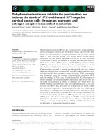
Figure 1 Expression of miRNAs in gastric cancer cell lines (AGS and BCG) and 3 randomly selected normal gastric samples (GN1-GN3).
RT-PCR was carried out to determine the expression of microRNAs. Expression of miR-204 was the most significantly down regulated compared
to the normal gastric samples (A). The average fold change of miRNAs calculated by qRT-PCR results are presented in the table (B).
was remarkably down-regulated in 24 primary tumors
stratified based on clinical progression that subsequently
metastasized when compared to the primary tumors
that did not recur (Figure 2B). These data indicate that
down-regulation of miR-204 may be related to the onset of gastric cancer metastasis. Notably, we found that
miR-204 down-regulation was significantly associated
with up-regulation of SIRT1 mRNA levels in gastric
cancer specimens (Table 2). N/T ratios were classified
as high based on highest textiles. MiR-204 and SIRT1
expressions were then dichotomized as high and low.
Therefore, SIRT1 may be the target of miR-204 in gastric
cancer cells.
Interaction of miR-204 with the 3’UTR of SIRT1 mRNA
The results presented so far demonstrate that the inactivation of miR-204 may be related to the up-regulation of
SIRT1 in gastric cancer cells. Down-regulation of SIRT1
in gastric cancer cells may occur through the binding
of miR-204 to the 3’UTR of SIRT1 mRNA. Target Scan
(release 5.1) predicted a single miRNA-responsive element
containing a conserved 8-mer exact seed match at positions
384–391 of SIRT1 3’-UTR as a miR-204 target (Figure 3A).
To investigate this potential interaction, wild-type SIRT1
3’-UTR as well as mutSIRT1 3’-UTR with mutated target
sites (A to G) were cloned into a pGL3 luciferase vector.
To examine the impact of miR-204 on SIRT1 3’-UTR
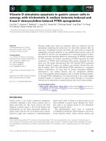
Figure 2 MiR-204 is down-regulated in gastric tumor tissues. (A) The analysis of the miR-204 expression level was performed in gastric
tumor tissues (n = 24) and matched normal tissues. The miR-204 level was significantly down-regulated in gastric cancer tissues compared with
that in matched normal tissues. (B) The gastric cancer samples were divided into two groups based on clinical progression. MiR-204 levels in the
metastasis group (n = 14) were lower than those in the no-metastasis group. Total RNA was subjected to RT-PCR to analyze the expression level
of miR-204 in each sample. U6 was used as a reference for miRNAs. Each sample was analyzed in triplicate. The standard curve method was used
to quantify the relative gene expression levels. **P < 0.01. *P < 0.05.
Zhang et al. BMC Cancer 2013, 13:290
/>
Page 5 of 9
Table 2 Correlation between miR-204 expression and
SIRT1 mRNA expression in 24 gastric carcinomas
miR-204 expression
Case number
N/T>7.5
N/T≤7.5
24
17
7
N/T≤2.5
18
15
3
N/T>2.5
6
2
4
Overall
SIRT1 mRNA expression
P
0.038*
*P<0.05.
activity, HEK293T cells were co-transfected with miR-204
precursor (Ambion) that restored miR-204 expression.
Figure 3B shows that miR-204 inhibits the luciferase activity
of the wild-type SIRT1 3’-UTR, but mutation of the miR204 miRNA-responsive element within the SIRT1 3’-UTR
abolishes miR-204 action, suggesting that miR-204 targets
one complementary sequence in the SIRT1 3’-UTR.
To determine whether miR-204 can affect endogenous
SIRT1 expression, we examined the effect of miR-204
activation on SIRT1 in AGS and BCG cells. Real-time
PCR and Western blots revealed significantly decreased
expression of both SIRT1 mRNA and protein in GCCs
transfected by miR-204 mimics (Figure 3C & D). These results indicate a negative role for miR-204 in the regulation
of SIRT1 expression.
Over-expression of miR-204 and down regulation of SIRT1
induces an mesenchymal -to- epithelial transition phenotype
We examined an in vitro model of EMT-like transformation and monitored alterations of SIRT1 and miR-204
expression. The immortalized cells were cultured in the
presence of 10% FBS and treated with 10 ng/ml human
TGF-β1 for 21 days. Treatment with TGF-β1 has been
shown to induce EMT-like transformation of epithelial
cells in many cell culture models (42). Figure 4A shows
that treatment with TGF-β1 leads immortalized HGC
cells (AGS and BCG cells) that undergo EMT-like transformation. This transformation is evidenced by loss of
cell-cell adhesion and alterations of morphology from
a round compact shape to a spindle shape. These
transformed cells were defined as HGC-T. To define
the role of miR-204 and SIRT1 in the progression of
cell metastasis in gastric cancer cells, we treated the AGS-T
and BGC-T cells with miR-204 mimics and SIRT1 SiRNA.
Up-regulation of miR-204 levels and down regulation
of SIRT1 was associated with the decrease of Vimentin
mRNA transcripts (Figure 4B & E). The increase of Ecadherin mRNA transcripts was detected in AGS-T cells
that overexpressed miR-204 or had SIRT1 knocked-down
(Figure 4C & F). These differences were also true at the
protein level, as shown for E-cadherin and Vimentin using
Western blot analysis (Figure 4D).
Over-expression of miR-204 and knock-down of SIRT1
inhibit gastric cancer cell metastasis
To study the physiological role of miR-204 and SIRT1 in
metastasis, the gastric cancer cell lines treated with
miR-204 mimics and SIRT1 SiRNA were analyzed. The
ability of SIRT1 and miR-204 to regulate cell migration
and cell invasion was investigated utilizing transwell
invasion assays. HGC-T cells transfected with either
SiRNA for SIRT1 (HGC-T/SiSIRT1) or miR-204 mimics
(HGC-T /miR-204) showed dramatically decreased cell
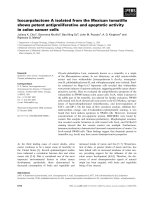
Figure 3 SIRT1 is a new target for regulation by miR-204. (A) illustration of SIRT1 3’UTR as well as the seed sequence of miR-204 showing the
computationally predicted target region on the 3’UTR of SIRT1 mRNA. (B) miR-204 decreases SIRT1 3’UTR luciferase activity. HEK293T cells were
transfected with 2μg of pGL3 luciferase vector containing either SIRT1 3’UTR or SIRT1 MUT 3’UTR (with an A-to-G mutation in miRNA-responsive
element). Cells were co-transfected along with 50 nM miR-204 precursor for 24 h and then lysed and assessed for luciferase activity. Mean-S.E.
(n =3). (C) SIRT1 mRNA levels in AGS and BGC transfected with 50 nM miR-204 precursor for 72 h. Mean-S.E. (n=3). (D) Western blotting of SIRT1
protein levels. SIRT1 levels in AGS and BGC transfected with 50 nM miR-204 precursor for 48 h (n=3). **P < 0.01. *P < 0.05.
Zhang et al. BMC Cancer 2013, 13:290
/>
Page 6 of 9
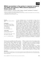
Figure 4 Transformation of gastric cancer cells is associated with altered SIRT1 and miR-204 expression. (A) An altered morphology is
associated with transformation. Representative images display morphological changes from HGC to HGC-T (treated with 10 ng/ml TGF-β1for 21
days). Scale bar ,100μm. Vimentin (B) and E-cadherin (C) mRNA levels in transformed cells transfected with 50 nM miR-204 precusor for 72h.
(D) Western bloting of E-cadherin and Vimentin protein levels. E-cadherin and Vimentin levels in HGC-T transfected with 50 nM miR-204 precusor or
SIRT1 SiRNA for 48h. Vimentin (E) and E-cadherin (F) mRNA levels in transformed cells transfected with 50 nM SIRT1 SiRNA for 72h. Representative
results from 3 independent experiments. Real-time RT-PCR results for mRNA levels were normalized to GAPDH mRNA. **P < 0.01. *P < 0.05.
invasion (Figure 5). These findings further confirmed a
functional role for SIRT1 in promoting EMT-like
transformation of gastric cancer epithelial cells as well
as establishing an inhibitory role for miR-204 in this
process. These data suggest that miR-204 negatively
regulates and SIRT1 positively regulates gastric cancer
cell metastasis.
Restoration of miR-204 and down-regulation of SIRT1
mediate suppression of anoikis resistance
Given the known role of anoikis resistance association
with EMT, we investigated the effect of miR-204 and
SIRT1 on anoikis by performing FACS analysis of Annexin
V/PI stained cells. In these assays, the cells were placed on
poly-HEMA coated plates, which prevent them from adhering. The cells were forced to float in suspension until
harvested for analysis. In the cell apoptosis analysis,
miR-204 restoration and consequent SIRT1 knock-down
resulted in increased cells positive for Annexin V/FITC
staining, indicating an increase in apoptosis in these
samples (Figure 6). Thus, restoration of miR-204 and
knock-down of SIRT1 decreased anoikis resistance as
indicated by a decrease in the viability of suspended
cells and a concurrent increase in apoptosis.
Down-regulation of miR-204 promotes cancer cell
invasion by activating LKB1 through SIRT1
LKB1 has a particularly tight link with EMT and anoikis.
Therefore, we investigated the role of miR-204 in regulating
LKB1. Western blot analysis of GC cells treated with
miR-204 showed that miR-204 overexpression significantly
increased LKB1 accumulation (Figure 7A). GC cells were
transfected with SIRT1 SiRNA. Figure 7B shows that
SIRT1 knockdown cells had a significant increase in LKB1
expression. AGS and BGC cells were co-transfected with
either the LKB1 SiRNA or the negative control, along with
either the miR-204 mimic or the microRNA control. Cells
transfected with miR-204 mimics along with the negative
control showed significantly decreased cell invasion ability
compared with the two LKB1 SiRNA transfected groups
(*P<0.05). LKB1 protein expression was negatively correlated with the cell invasion ability (Figure 7C). These data
suggest that decreased expression of miR-204 might promotes cell metastasis by inactivating LKB1.
Discussion
Class III histone deacetylase SIRT1 blocks senescence and
apoptosis and promotes cell growth and angiogenesis,
making it a critical regulator of tumor initiation, prognosis
Zhang et al. BMC Cancer 2013, 13:290
/>
Page 7 of 9
Figure 5 MiR-204 promotes gastric cancer cell invasion. (A) miR-204 was involved in AGS and BCG cell migration. The profiles and the
images are representative of at least three independent experiments. (B) SIRT1 SiRNA inhibited AGS and BCG cell migration. The profiles and the
images are representative of at least three independent experiments. N.C, negative control. *P < 0.05.
and drug resistance. Our previous studies have suggested
that the up-regulation of SIRT1 is related to lymph node
metastasis in gastric cancer [12]. The underlying mechanism by which this occurs is still unclear. There are 34
miRNAs are predicted to target the 3’UTR of SIRT1,
which is 1.7 kb. We evaluated and analyzed the expression
of these miRNAs that are conserved across various species
in normal gastric mucosa tissue, gastric cancer specimens,
and 2 gastric cancer cell lines. The results showed that reduced expression of miR-204 frequently occurred in gastric cancer tissues and was related with the up-regulation
of SIRT1. We also verified this result in 24 gastric cancer
tissues and found that the decreased expression of
miR-204 was related with cancer metastasis.
miRNAs are involved in several important biological
events such as tumorigenesis and cancer metastasis.
miRNAs are known to act as regulators in gastric cancer
cell growth and to regulate gastric cancer metastasis. Deregulation of some miRNAs, including miR-101, miR-107,
miR-221, and miR-222, has been observed in gastric cancer [13-15]. miR-101 was down-regulated in gastric cancer
tissues. The ectopic expression of miR-101 significantly
inhibited cellular proliferation, migration, and invasion of
gastric cancer cells by targeting EZH2, Cox-2, Mcl-1, and
Figure 6 MiR-204 increases sensitivity to anoikis. AGS (A) and BGC (B) cells were transfected with miRNA constructs and plated on polyHEMA coated plates. Cells were collected for apoptosis analysis by FACS analysis of Annexin V/PI stained cells. Columns mean of three biological
replicates, bars, standard deviation of the mean. ANOVA * P < 0.05.
Zhang et al. BMC Cancer 2013, 13:290
/>
Figure 7 MiR-204 activates LKB1 by repressing SIRT1expression.
(A) Western blot analysis showed the level of the endogenous LKB1
protein in AGS and BCG cells that were transfected with SIRT1 SiRNA.
(B) Western blot analysis showed the level of endogenous LKB1
protein in AGS and BCG cells that were transfected with miR-204.
(C) Inhibition of LKB1 could sabotage the miR-204 induced-cancer cell
migration reduction. The profiles are representative of at least three
independent experiments. N.C, negative control. *P < 0.05, **P<0.01.
Fos. miR-107 is frequently up-regulated in gastric cancers,
and its overexpression is significantly associated with gastric cancer metastasis. Here, we demonstrated miR-204,
another miRNA specific to gastric cancer metastasis, and
its specific target, SIRT1.
Epithelial–mesenchymal transition (EMT) consists of a
rapid and often reversible change of cell phenotype [16].
Epithelial cells loosen cell–cell adhesion structures, including adherens junctions and desmosomes, to modulate
their polarity and rearrange their cytoskeleton. Specifically,
intermediate filaments typically switch from cytokeratins
to Vimentin [17,18]. Cells become isolated, motile and resistant to apoptosis. Many genes and pathways have been
implicated in inducing EMT in tumor cells. Typically,
these pathways are also active in other processes, including
cell proliferation, apoptosis and differentiation during early
developmental stages, tissue morphogenesis and wound
healing. Their specific role during human tumor progression is usually not well understood [19]. Our previous analysis of the clinical characteristics indicated that SIRT1
expression was significantly associated with tumor stage
Page 8 of 9
and the presence of metastasis, which further indicated
that SIRT1 acts as a tumor promoter and facilitates the
infiltration of gastric cancer. The oncogenic epithelialto-mesenchymal transition (EMT) is thought to play
an important role in tumor progression. Our current
results suggest that miR-204 down-regulation and SIRT1
restoration can induce EMT in GC cells.
There is a tight correlation between anoikis resistance
and oncogenic EMT [20-22]. A common hallmark of
EMT is the breakdown of E-cadherin expression or function [23], which suffices to circumvent anoikis in some contexts. For example, the targeted knockout of the E-cadherin
gene in a mouse mammary tumor model or the stable
knockdown of E-cadherin in a mammary epithelial cell line
confers anoikis resistance [24]. This finding implies that
EMT-promoting transcription factors such as ZEB1/2,
Snail1/2 and Twist can block anoikis both by directly
regulating apoptosis control genes and by suppressing
E-cadherin expression. Here, we discuss the mechanism
by which E-cadherin suppression triggers signaling events
that control other apoptosis regulatory genes [25]. This
study also investigated whether the miR-204-SIRT1
pathway was involved in anoikis resistance and metastasis
promotion in GC cells. We demonstrate that miR-204
down-regulation and SIRT1 overexpression both can induce anoikis resistance in GC cells.
LKB1 was identified originally as the tumor suppressor
gene on human chromosome 19p13. LKB1 inactivation
triggers EMT in lung cancer cells through the induction
of ZEB1 [26]. Cheng et al. reported that LKB1 was an essential upstream regulator of p53-mediated anoikis [27].
Recent studies have revealed that many proteins, such as
SIRT1, are involved in the regulation of LKB1 [28]. Overexpression of LKB1 promoted cellular senescence and retarded endothelial proliferation, which could be blocked
by increasing SIRT1 levels. Knocking down of SIRT1 induced senescence and elevated the protein levels of LKB1.
SIRT1 antagonized LKB1 activation through promoting
deacetylation, ubiquitination and proteasome-mediated
degradation of LKB1. Our data show that over-expression
of miR-204 increased LKB1 expression in GCCs, while
down-regulation of SIRT1 can also restore LKB1 expression in GCCs. LKB1 down-regulation could promote cancer cells invasion even when miR-204 was upregulated. As
a result, miR-204 may modulate LKB1 by interacting with
SIRT1. These data suggest that reduction of miR-204 promotes EMT by inactivating LKB1, and SIRT1 might be
the direct target of miR-204 in the LKB1 pathway.
Conclusion
In conclusion, our data demonstrate that the downregulation of miR-204 promotes gastric cancer cell metastasis by activating the SIRT1-LKB1 pathway. Therefore,
we show that miR-204 is an important regulator in gastric
Zhang et al. BMC Cancer 2013, 13:290
/>
cancer metastasis and suggest the potential application of
miR-204 in gastric cancer therapy.
Competing interests
The authors declare that they have no competing interests.
Authors’ contributions
LZ was responsible for planning the study. XW carried out the molecular
genetic studies and involved in all steps of the data analysis and manuscript
writing. PC provided laboratory support and helped to draft the manuscript.
All authors read and approved the final manuscript.
Page 9 of 9
14.
15.
16.
17.
Acknowledgements
The authors wish to thank Hong Fan for clinical, laboratory and logistic
support.
Financial support
This work was supported by grants from the natural science foundation of
Jiangsu Province (BK2012750), and the Natural Science Foundation of China
(81101856).
Author details
1
Department of Pathology, Southeast University, Zhongda Hospital, Nanjing
210009, P R China. 2Department of Pathology, Southeast University, Nanjing
210009, P R China.
Received: 22 February 2013 Accepted: 5 June 2013
Published: 14 June 2013
References
1. Wang F, Sun GP, Zou YF, Hao JQ, Zhong F, Ren WJ: MicroRNAs as promising
biomarkers for gastric cancer. Cancer Biomark 2011, 11:259–267.
2. Yin Y, Li J, Chen S, Zhou T, Si J: MicroRNAs as Diagnostic Biomarkers in
Gastric Cancer. Int J Mol Sci 2012, 13:12544–12555.
3. Zheng L, Pu J, Qi T, Qi M, Li D, Xiang X, Huang K, Tong Q: MicroRNA-145
targets v-ets erythroblast sis virus E26 oncogene homolog 1 to suppress
the invasion, metastasis and angiogenesis of gastric cancer cells.
Mol Cancer Res 2013, 11:182–193.
4. Thiery JP, Sleeman JP: Complex networks orchestrate epithelial-mesenchymal
transitions. Nat Rev Mol Cell Biol 2006, 7:131–142.
5. Voulgari A, Pintzas A: Epithelial-mesenchymal transition in cancer
metastasis: mechanisms, markers and strategies to overcome drug
resistance in the clinic. Biochim Biophys Acta 2009, 1796:75–90.
6. Du C, Zhang C, Hassan S, Biswas MH, Balaji KC: Protein kinase D1
suppresses epithelial-to-mesenchymal transition through
phosphorylation of snail. Cancer Res 2010, 70:7810–7819.
7. Adam L, Zhong M, Choi W, Qi W, Nicoloso M, Arora A, Calin G, Wang H,
Siefker-Radtke A, McConkey D, et al: MiR-200 expression regulates
epithelial-to-mesenchymal transition in bladder cancer cells and reverses
resistance to epidermal growth factor receptor therapy. Clin Cancer Res
2009, 15:5060–5072.
8. Saito Y, Suzuki H, Tsugawa H, Nakagawa I, Matsuzaki J, Kanai Y, Hibi T:
Chromatin remodeling at Alu repeats by epigenetic treatment activates
silenced microRNA-512-5p with down regulation of Mcl-1 in human
gastric cancer cells. Oncogene 2009, 28:2738–2744.
9. Mazar J, DeYoung K, Khaitan D, Meister E, Almodovar A, Goydos J, Ray A,
Perera RJ: The regulation of miRNA-211 expression and its role in
melanoma cell invasiveness. PLoS One 2010, 5:e13779.
10. Tie J, Pan Y, Zhao L, Wu K, Liu J, Sun S, Guo X, Wang B, Gang Y, Zhang Y,
et al: MiR-218 inhibits invasion and metastasis of gastric cancer by
targeting the Robo1 receptor. PLoS Genet 2010, 6:e1000879.
11. Wang X, Huang G, Mei S, Qian J, Ji J, Zhang J: Over-expression of C/EBP-alpha
induces apoptosis in cultured rat hepatic stellate cells depending on p53
and peroxisome proliferator-activated receptor-gamma. Biochem Biophys Res
Commun 2009, 380:286–291.
12. Feng AN, Zhang LH, Fan XS, Huang Q, Ye Q, Wu HY, Yang J: Expression of
SIRT1 in gastric cardiac cancer and its clinicopathologic significance.
Int J Surg Pathol 2011, 19:743–750.
13. Chun-Zhi Z, Lei H, An-Ling Z, Yan-Chao F, Xiao Y, Guang-Xiu W, Zhi-Fan J,
Pei-Yu P, Qing-Yu Z, Chun-Sheng K: MicroRNA-221 and microRNA-222
18.
19.
20.
21.
22.
23.
24.
25.
26.
27.
28.
regulate gastric carcinoma cell proliferation and radio resistance by
targeting PTEN. BMC Cancer 2010, 10:367.
Wang HJ, Ruan HJ, He XJ, Ma YY, Jiang XT, Xia YJ, Ye ZY, Tao HQ:
MicroRNA-101 is down-regulated in gastric cancer and involved in cell
migration and invasion. Eur J Cancer 2010, 46:2295–2303.
Li X, Zhang Y, Shi Y, Dong G, Liang J, Han Y, Wang X, Zhao Q, Ding J, Wu K,
Fan D: MicroRNA-107, an oncogene microRNA that regulates tumour
invasion and metastasis by targeting DICER1 in gastric cancer. J Cell Mol
Med 2011, 15:1887–1895.
Polyak K, Weinberg RA: Transitions between epithelial and mesenchymal
states: acquisition of malignant and stem cell traits. Nat Rev Cancer 2009,
9:265–273.
Brabletz S, Brabletz T: The ZEB/miR-200 feedback loop–a motor of cellular
plasticity in development and cancer? EMBO Rep 2010, 11:670–677.
Sanchez-Tillo E, Siles L, de-Barrios O, Cuatrecasas M, Vaquero EC, Castells A,
Postigo A: Expanding roles of ZEB factors in tumorigenesis and tumor
progression. Am J Cancer Res 2010, 1:897–912.
Sanchez-Tillo E, de-Barrios O, Siles L, Cuatrecasas M, Castells A, Postigo A:
Beta-catenin/TCF4 complex induces the epithelial-to-mesenchymal
transition (EMT)-activator ZEB1 to regulate tumor invasiveness. Proc Natl
Acad Sci USA 2011, 108:19204–19209.
Savagner P: The epithelial-mesenchymal transition (EMT) phenomenon.
Ann Oncol 2010, 21(Suppl 7):vii89–vii92.
Klymkowsky MW, Savagner P: Epithelial-mesenchymal transition: a cancer
researcher's conceptual friend and foe. Am J Pathol 2009, 174:1588–1593.
Lee JM, Dedhar S, Kalluri R, Thompson EW: The epithelial-mesenchymal
transition: new insights in signaling, development, and disease. J Cell Biol
2006, 172:973–981.
Zavadil J, Haley J, Kalluri R, Muthuswamy SK, Thompson E:
Epithelial-mesenchymal transition. Cancer Res 2008, 68:9574–9577.
Onder TT, Gupta PB, Mani SA, Yang J, Lander ES, Weinberg RA: Loss of
E-cadherin promotes metastasis via multiple downstream transcriptional
pathways. Cancer Res 2008, 68:3645–3654.
Kurrey NK, Jalgaonkar SP, Joglekar AV, Ghanate AD, Chaskar PD, Doiphode
RY, Bapat SA: Snail and slug mediate radioresistance and
chemoresistance by antagonizing p53-mediated apoptosis and
acquiring a stem-like phenotype in ovarian cancer cells. Stem Cells 2009,
27:2059–2068.
Roy BC, Kohno T, Iwakawa R, Moriguchi T, Kiyono T, Morishita K,
Sanchez-Cespedes M, Akiyama T, Yokota J: Involvement of LKB1 in
epithelial-mesenchymal transition (EMT) of human lung cancer cells.
Lung Cancer 2010, 70:136–145.
Cheng H, Liu P, Wang ZC, Zou L, Santiago S, Garbitt V, Gjoerup OV, Iglehart
JD, Miron A, Richardson AL, et al: SIK1 couples LKB1 to p53-dependent
anoikis and suppresses metastasis. Sci Signal 2009, 2:ra35.
Zheng Z, Chen H, Li J, Li T, Zheng B, Zheng Y, Jin H, He Y, Gu Q, Xu X:
Sirtuin 1-mediated cellular metabolic memory of high glucose via the
LKB1/AMPK/ROS pathway and therapeutic effects of metformin. Diabetes
2012, 61:217–228.
doi:10.1186/1471-2407-13-290
Cite this article as: Zhang et al.: MiR-204 down regulates SIRT1 and
reverts SIRT1-induced epithelial-mesenchymal transition, anoikis
resistance and invasion in gastric cancer cells. BMC Cancer 2013 13:290.
Submit your next manuscript to BioMed Central
and take full advantage of:
• Convenient online submission
• Thorough peer review
• No space constraints or color figure charges
• Immediate publication on acceptance
• Inclusion in PubMed, CAS, Scopus and Google Scholar
• Research which is freely available for redistribution
Submit your manuscript at
www.biomedcentral.com/submit