Prognostic value of C-reactive protein and neutrophil-to-lymphocyte ratio in patients with hepatocellular carcinoma
Bạn đang xem bản rút gọn của tài liệu. Xem và tải ngay bản đầy đủ của tài liệu tại đây (556.99 KB, 9 trang )
Oh et al. BMC Cancer 2013, 13:78
/>
RESEARCH ARTICLE
Open Access
Prognostic value of C-reactive protein and
neutrophil-to-lymphocyte ratio in patients with
hepatocellular carcinoma
Byong Sun Oh1, Jeong Won Jang1,5*, Jung Hyun Kwon1, Chan Ran You1, Kyu Won Chung1, Chul Seung Kay2,
Hyun Suk Jung3 and Seungok Lee4
Abstract
Background: Accumulating evidence indicates that components of the systemic inflammatory response, such as
C-reactive protein (CRP) and neutrophil-to-lymphocyte ratio (NLR), have been associated with prognosis of various
cancers. We aimed to elucidate whether CRP and NLR could serve as potential surrogate markers for response and
survival in patients with hepatocellular carcinoma (HCC).
Methods: The study population consisted of 318 consecutive patients with HCC. CRP and NLR were measured at
baseline with follow-up measurements.
Results: With the mean follow-up of 13.9 months, the median survival time was 13.8 months. Child-Pugh class,
tumor size > 5 cm, tumor multiplicity, presence of portal vein thrombosis, α-fetoprotein > 200 ng/mL, CRP > 6.3 mg/
L and NLR > 2.3 were identified as independent factors for worse survival of HCC (all p < 0.05). Patients with
elevated CRP (> 6.3 mg/L) and elevated NLR (> 2.3) had a significantly shorter overall survival than those with low
CRP and low NLR (all p < 0.001). The combined use of CRP and NLR provided incremental prognostic information.
With significant inter-correlations, levels of CRP and NLR escalated with aggravating Child-Pugh class from A to C or
progressing tumor stage from I to IV. CRP and NLR on baseline and serial measurements were well predictive of
treatment response (p < 0.001).
Conclusions: CRP and NLR are independent indicators for survival in HCC patients, reflecting tumor burden and
hepatic reserve. Their role in predicting tumor response and survival is more enhanced when used in combination.
This study suggests that CRP and NLR are important prognostic biomarkers for HCC.
Keywords: Inflammation markers, Neutrophil-to-lymphocyte ratio, C-reactive protein, Hepatocellular carcinoma,
Survival
Background
Hepatocellular carcinoma (HCC) is the fifth most common
malignancy and the third leading cause of cancer-related
deaths worldwide [1]. Although there have been many
advances in the treatment of HCC, the outcome is still not
satisfactory [1,2]. Since a long lasting inflammatory process
like cirrhosis continually induces hepatocarcinogenesis,
* Correspondence:
1
Department of Internal Medicine, The Catholic University of Korea, Incheon
St. Mary’s Hospital, Incheon, Korea
5
Division of Hepatology, Department of Internal Medicine, College of
Medicine, The Catholic University of Korea, #222 Banpo-daero 22-gil,
Seocho-gu, Seoul 137-701, Korea
Full list of author information is available at the end of the article
there are limitations on curative therapy. Besides known
prognostic factors representing tumor status and liver
function [3], it is now clear that inflammation plays a significant role in tumor progression [4]. In this situation, Creactive protein (CRP) and neutrophil-to-lymphocyte ratio
(NLR), indicators of inflammation, have been suggested as
surrogate markers for a relationship between inflammation
and cancer [5].
CRP has been identified as a prognostic factor for HCC
[6-10] as well as other various malignancies, such as
gastrointestinal tumor, renal cell cancer and ovarian cancer [5,11]. It is an acute phase reactant, synthesized in the
liver [12], and regulated by proinflammatory cytokines,
© 2013 Oh et al.; licensee BioMed Central Ltd. This is an Open Access article distributed under the terms of the Creative
Commons Attribution License ( which permits unrestricted use, distribution, and
reproduction in any medium, provided the original work is properly cited.
Oh et al. BMC Cancer 2013, 13:78
/>
like interleukin (IL)-6, which plays an important role in
carcinogenesis [13]. The NLR has recently been evaluated
as a predictor of prognosis of HCC [14-16] as well as
other malignancies like colorectal cancer, gastric cancer,
breast cancer and ovarian cancer [5,17-20]. It has been
shown that high levels of NLR could predict a risk of recurrence and survival in patients with various malignancies.
HCC is unique among other cancers, in that its prognosis not only depends on the tumor characteristics but
also on the hepatic functional reserve. In this respect, it
seems to be more relevant to understand inflammatory
process in hepatocarcinogenesis and evaluate its effect
on the patient prognosis. To date, there have been no
comprehensive data on the relationship between the inflammatory markers and hepatic reserve and tumor status in HCC. In addition, the significance of both CRP
and NLR in HCC survival has not yet been explicitly
studied in one research.
In the present study, we therefore evaluated the clinical value of CRP as well as NLR measured at the same
point in time in clinical outcome in a large number of
patients with HCC. In addition, changes in CRP and
NLR after treatment were examined in relation to the
treatment response. The present findings show the prognostic utility of both CRP and NLR as a surrogate
marker for efficacy in treatment as well as HCC survival.
Page 2 of 9
University of Korea in accordance with the 1975 Declaration of Helsinki.
Methods
Laboratory data including CRP and NLR were obtained
from the study subjects prior to the initiation of treatment
and at each treatment cycle. All serum samples were
measured as fresh state. Serum CRP level was determined
as highly-sensitive CRP (hs-CRP) by immunoturbidimetric
assay using CRPH (C-Reactive Protein, High Sensitivity)
reagent (Beckman Coulter, Inc., Fullerton, CA USA; limit
of detection, 0.08 mg/L). The NLR was calculated by dividing the neutrophil count by the lymphocyte count. CRP
and NLR measurements were obtained without demonstrable infection. To evaluate the relationship between
changes in CRP levels and NLR and tumor response, serial
measurements of the CRP levels and NLR were carried
out 3 to 4 months after treatment. Based on the modified
Response Evaluation Criteria In Solid Tumors (RECIST)
criteria [22], patients with complete response and partial
response after treatment were grouped together as
responders, whereas patients with stable disease and progressive disease were defined as non-responders.
Statistical analysis
Methods
Patients
A database of all 318 patients with newly diagnosed
HCC between January 2007 and December 2010 at
Incheon St. Mary’s hospital, Incheon, South Korea was
prospectively collected and retrospectively analyzed. The
diagnosis of HCC was made based on histological evidence or elevated serum α-fetoprotein (AFP) levels
(> 200 ng/mL) with typical radiological findings (arterial
enhancement with portal washout) [21]. The staging of
tumor was determined according to the TNM classification of Malignant Tumors/International Union Against
Cancer (UICC) classification system, which is widely
used in Korea [21]. Our main treatment modality for
HCC was transarterial chemotherapy (TAC)-based locoregional therapy. The chemotherapeutic regimen used
for TAC was intra-arterial chemotherapy using doxorubicin (50 mg) for patients who had multifocal tumors
≤ 10 cm or a combination of epirubicin (50 mg) and cisplatin (60 mg) for patients with Child-Pugh class A who
had large tumors (> 10 cm) with or without portal vein
thrombosis (PVT). Patients with PVT or extrahepatic
metastasis were considered for radiotherapy in addition
to TAC. No patients treated with loco-regional therapy
were given glucocorticoids. Each patient provided
informed consent to participate in the study. This study
was approved by the Ethics Committees of The Catholic
SPSS version 18 (SPSS Inc., Chicago, IL, USA) and
MedCalc version 11.6.1 (MedCalc Software Inc.,
Mariakerke, Belgium) were used to analyze the data. The
cut-off values of CRP and NLR were determined using
receiver operating characteristic (ROC) curve analysis.
The optimal cut-off levels for CRP and NLR were
established at 6.3 mg/L and 2.3, respectively, and these
cutoff values were used to categorize the high and low
CRP or NLR groups. Univariate analysis was performed
to assess significant differences in clinical characteristics.
A multivariate analysis was performed by Cox regression
for variables significant on univariate analysis. Overall
survival was calculated from the date of diagnosis to the
date of death or last follow-up. For patients undergoing
liver transplantation, follow-up was censored at the time
of transplantation. To compare overall survival rates
according to CRP or NLR, the data were analyzed by the
Kaplan-Meier method, and the differences in survival
between groups were compared by the log-rank test.
Correlations between the inflammatory markers and
Child-Pugh class and tumor stage were assessed using
Spearman correlation coefficients. Serial changes in CRP
levels and NLR were evaluated using repeated measures
ANOVA. Each continuous variable was appropriately
categorized in order to achieve the largest statistical
power. A p value of less than 0.05 was considered to be
statistically significant.
Oh et al. BMC Cancer 2013, 13:78
/>
Results
Baseline characteristics
A total of 318 patients were consecutively enrolled in this
study. Table 1 shows baseline characteristics of the entire
study population. Patient age ranged from 32 to 89 years
(median: 58 years) and 240 (75.5%) of the patients were
males. At diagnosis, 200 (62.9%) patients were classified into
Child-Pugh class A; 91 (28.6%) patients into Child-Pugh
class B; 27 (8.5%) patients into Child-Pugh class C. The median tumor size was 5.5 (0.8–26.5) cm and the number of
patients with solitary tumor was 144 (45.3%). Each case of
presence of PVT and extrahepatic metastasis was 107
(33.6%) and 62 (19.5%), respectively. Distribution of tumor
stage after UICC (I/II/III/IVa/IVb) classification in our
patients was as follows: 41 (12.9%)/76 (23.8%)/85 (26.7%)/64
(20.1%)/52 (16.4%), respectively. The median levels of serum
AFP, CRP and NLR were 92.3 (1.6–2,753,500) ng/mL, 4.7
(0.1–343.6) mg/L and 2.6 (0.6–49.9), respectively. The
allocated treatments for the 318 patients were surgical resection (n = 41), liver transplantation (n = 10), TAC-based locoregional therapy (n = 221), and supportive care (n = 46).
Factors affecting prognosis of HCC in the whole study
population
To identify factors for HCC survival, 12 potential variables
of interest were analyzed, as listed in Table 2. Of these,
elevated ALT, Child-Pugh class, tumor size, tumor multiplicity, presence of PVT, presence of metastasis, elevated
AFP, high CRP and high NLR were significantly associated
with poorer survival. With multivariate analysis using a
Cox regression model, Child-Pugh class (p < 0.001; Hazard
ratio [HR] 1.711; 95% confidence interval [CI] 1.377–
2.125), tumor size > 5 cm (p = 0.003; HR 1.778; 95% CI
1.209–2.615), tumor multiplicity (p = 0.035; HR 1.391;
95% CI 1.023–1.892), presence of PVT (p = 0.001; HR
1.827; 95% CI 1.284–2.598), AFP > 200 ng/mL (p = 0.001;
HR 1.734; 95% CI 1.248–2.407), CRP > 6.3 mg/L
(p = 0.027; HR 1.519; 95% CI 1.049–2.199) and NLR > 2.3
(p = 0.009; HR 1.601; 95% CI 1.124–2.280) were identified
as independent poor prognostic factors for HCC (Table 2).
When we included the combination of high CRP and high
NLR as a variable into the analysis, the combination of
CRP and NLR (p < 0.001; HR 1.905; 95% CI 1.345–2.697)
together with Child-Pugh class (p < 0.001; HR 1.806; 95%
CI 1.464–2.228), tumor size > 5 cm (p = 0.002; HR 1.858;
95% CI 1.258–2.743), presence of PVT (p < 0.001; HR
1.893; 95% CI 1.329–2.697) and AFP > 200 ng/mL
(p < 0.001; HR 1.821; 95% CI 1.324–2.504) were identified
as independent factors for worse survival instead of CRP
or NLR alone.
Overall survival according to CRP and NLR level
During the mean follow-up period of 13.9 months, 202
(63.5%) of the patients died. The median survival time
Page 3 of 9
was 13.8 months. Since CRP and NLR played a significant role in predicting the HCC survival, we evaluated
the differences in survival according to the low versus
high CRP and NLR levels among the entire group. As
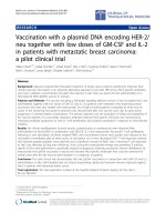
depicted in Figure 1A, the median survival of patients
with elevated CRP (> 6.3 mg/L) was 6.0 months, which
was significantly shorter than 26.9 months for patients
with a low level of CRP (log-rank test, p < 0.001). Likewise, the survival in the elevated NLR (> 2.3) group was
significantly worse than that in the low NLR group, with
the median survival times of 7.9 versus 32.5 months
(log-rank test, p < 0.001; Figure 1B). The statistical
differences for survival by the levels of CRP and NLR
were still maintained in the patients receiving treatments
as well as the entire patients (data not shown). An additional analysis was done to examine the synergistic effect of a combined use of CRP and NLR on the patient
outcome. When the Kaplan-Meier survival curves were
plotted over time, there was a stepwise increase in the
overall survival rate from both high CRP (> 6.3 mg/L)
and NLR (> 2.3) levels, one of them and to both low
CRP (≤ 6.3 mg/L) and NLR (≤ 2.3) levels, indicating the
benefit of the combined use of the two inflammatory
markers (Figure 1C).
Correlation of CRP and NLR with Child-Pugh class and
tumor stage
The relationship of CRP and NLR with Child-Pugh class
and tumor characteristics, which were the two major
determinants of the prognosis of patients with HCC,
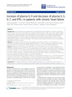
were evaluated. There was a significant correlation between CRP concentrations and Child-Pugh class or
tumor stage (r = 0.311, p < 0.001; r = 0.475, p < 0.001, respectively). As shown in Figures 2A and 2B, the level of
CRP tended to increase as liver disease progressed from
Child-Pugh class A to C as well as tumor stage from I to
IV. Likewise, there was also a significant correlation between NLR and Child-Pugh class or tumor stage
(r = 0.306, p < 0.001; r = 0.358, p < 0.001, respectively).
Figure 2C and Figure 2D illustrate the escalating values
of NLR with aggravating Child-Pugh class from A to C
or progressing tumor stage from I to IV. When analyzed
regarding the relationship between the CRP levels and
NLR, there was a significant positive inter-correlation
between CRP and NLR levels (r = 0.570, p < 0.001;
Figure 2E).
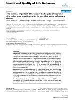
Tumor response according to CRP and NLR level
When baseline levels of CRP and NLR were analyzed for
tumor response, there were more non-responders in the
group with serum CRP > 6.3 mg/L and NLR > 2.3,
whereas there were more good responders in the group
with both baseline serum CRP ≤ 6.3 mg/L and NLR ≤ 2.3
Characteristics
Total patients (n = 318)
Patients grouped by CRP level (n = 318)
CRP ≤ 6.3 mg/L (n = 171)
Sex (male:female)
Age (years)
Cause (HBV/HCV/Alcohol/Others,
%)
Platelet counts (×103/mm3)
ALT (IU/L)
Total bilirubin (mg/dL)
Albumin (g/dL)
Prothrombin time (%)
Child-Pugh class (A/ B/ C, %)
Tumor size (cm)
CRP > 6.3 mg/L (n = 147)
Patients grouped by NLR level (n = 318)
NLR ≤ 2.3 (n = 129)
NLR > 2.3 (n = 189)
240:78
122:49
118:29
93:36
147:42
58 (32–89)
59 (33–80)
58 (32–89)
58 (33–80)
59 (32–89)
119 (81.0)/ 7 (4.8)/ 13 (8.8)/
8 (5.4)
100 (77.5)/ 14 (10.9)/ 8 (6.2)/
7 (5.4)
142 (75.1)/ 19 (10.1)/ 17 (9.0)/
11 (5.8)
242 (76.1)/ 33 (10.4)/ 25 (7.9) / 123 (71.9)/ 26 (15.2)/ 12 (7.0)/
18 (5.7)
10 (5.8)
139.6 ± 86.5
121.1 ± 63.9
161.1 ± 103.1
121.8 ± 64.9
151.7 ± 96.9
39.0 (1.0–2,430.0)
35.0 (1.0–2,430.0)
42.0 (6.0–503.0)
39.0 (1.0–2,430.0)
39.0 (6.0–503.0)
1.0 (0.2–26.9)
0.9 (0.2–9.4)
1.2 (0.2–26.9)
0.9 (0.2–7.8)
1.1 (0.2–26.9)
3.5 ± 0.6
3.7 ± 0.4
3.4 ± 0.5
3.6 ± 0.6
3.5 ± 0.6
79.4 ± 18.0
81.3 ± 17.6
77.1 ± 18.2
80.4 ± 18.4
78.6 ± 17.7
200 (62.9)/ 91 (28.6)/ 27 (8.5)
128 (74.9)/ 36 (21.1)/ 7 (4.1)
72 (49.0)/ 55 (37.4)/ 20
(13.6)
99 (76.7)/ 26 (20.2)/ 4 (3.1)
101 (53.4)/ 65 (34.4)/ 23 (12.2)
5.5(0.8–26.5)
3.0 (0.8–18.6)
10.0 (1.0–26.5)
3.0 (0.8–17.4)
7.9 (1.3–26.5)
Tumor number, solitary (%)
144 (45.3)
93 (54.4)
51 (34.7)
74 (57.4)
70 (37.0)
Presence of PVT (%)
107 (33.6)
24 (14.0)
83 (56.5)
23 (17.8)
84 (44.4)
Presence of metastasis (%)
62 (19.5)
14 (8.2)
48 (32.7)
11 (8.5)
51 (27.0)
Tumor stage (I/ II/ III/ IVa/ IVb, %)
41 (12.9)/ 76 (23.8)/ 85 (26.7)/ 34 (19.9)/ 57 (33.3)/ 51 (29.8)/
64 (20.1)/ 52 (16.4)
17 (9.9)/ 12 (7.0)
AFP (ng/mL)
92.3 (1.6–2,753,500.0)
CRP (mg/L)
NLR
7 (4.8)/ 19 (12.9)/ 34 (23.1)/
47 (32.0)/ 40 (27.2)
Oh et al. BMC Cancer 2013, 13:78
/>
Table 1 Baseline patient characteristics according to total patients, CRP and NLR level
30 (23.3)/ 40 (31.0)/ 35 (27.1)/ 11 (5.8)/ 36 (19.0)/ 50 (26.5)/ 46 (24.3)/
18 (14.0)/ 6 (4.7)
46 (24.3)
28.9 (1.6–331,580.0)
350.0 (2.2–2,753,500.0)
32.3 (1.7–331,580.0)
207.1 (1.6–2,753,500.0)
4.7 (0.1–343.6)
1.2 (0.1–6.3)
24.9 (6.4–343.6)
1.7 (0.1–64.8)
14.3 (0.2–343.6)
2.6 (0.6–49.9)
1.8 (0.6–13.0)
4.0 (0.7–49.9)
1.5 (0.6–2.2)
3.8 (2.3–49.9)
CRP, C-reactive protein; NLR, neutrophil-to-lymphocyte ratio; HBV, hepatitis B virus; HCV, hepatitis C virus; ALT, alanine aminotransferase; PVT, portal vein thrombosis; AFP, α-fetoprotein.
Page 4 of 9
Oh et al. BMC Cancer 2013, 13:78
/>
Page 5 of 9
Table 2 Univariate and multivariate analysis of prognostic factors of overall survival by Cox regression model
Univariate
Multivariate
HR (95% CI)
P value
Sex (male)
0.842 (0.604–1.173)
0.309
HR (95% CI)
P value
Age > 60 (years)
0.777 (0.586–1.031)
0.080
Cause (viral)
0.978 (0.652–1.469)
0.916
ALT > 40 (IU/L)
1.511 (1.146–1.992)
0.003
1.333 (0.985–1.804)
0.062
Child-Pugh class
1.803 (1.480–2.198)
< 0.001
1.711 (1.377–2.125)
< 0.001
Tumor size > 5 (cm)
3.548 (2.622–4.801)
< 0.001
1.778 (1.209–2.615)
0.003
Tumor multiplicity (≥ 2)
2.023 (1.510–2.711)
< 0.001
1.391 (1.023–1.892)
0.035
Presence of PVT
4.197 (3.141–5.609)
< 0.001
1.827 (1.284–2.598)
0.001
Presence of metastasis
3.002 (2.178–4.139)
< 0.001
1.242 (0.868–1.779)
0.236
AFP > 200 (ng/mL)
2.783 (2.099–3.690)
< 0.001
1.734 (1.248–2.407)
0.001
CRP > 6.3 (mg/L)
3.923 (2.929–5.255)
< 0.001
1.519 (1.049–2.199)
0.027
NLR > 2.3
3.050 (2.223–4.185)
< 0.001
1.601 (1.124–2.280)
0.009
HR, hazard ratio; CI, confidence interval; ALT, alanine aminotransferase; PVT, portal vein thrombosis; AFP, α-fetoprotein; CRP, C-reactive protein; NLR,
neutrophil-to-lymphocyte ratio.
(p < 0.001; Figure 3A). We next examined whether the
changes in CRP concentrations or NLR serially
measured before and after therapy could predict tumor
response. At follow-up, compared with responders,
patients with progressive disease showed an increase in
levels of CRP and NLR. In contrast, responders exhibited
a decrease in serial measurement of serum CRP and
NLR (Figures 3B and 3C). There were statistically significant differences in the CRP and NLR changes between
responders and those with progressive disease (all
p < 0.001, repeated measures ANOVA).
Discussion
The present study focused on the predictive value of CRP
and NLR in the outcome of patients with HCC. From the
analysis of a large cohort, we found that elevated CRP and
NLR independently predicted worse survival in patients with
HCC. In this study, CRP and NLR were utilized as prognostic indicators of HCC which appeared to be more evident
when used in combination. This is probably due to the significant synergistic effect of the two inflammatory markers.
Moreover, these markers correlated well with tumor response, as evidenced by serial measurements. The significant
prognostic role of CRP and NLR is supported by the
evidence that the levels of both markers displayed a linear
relationship with the progressing stage of tumor and ChildPugh classification, known as the two key prognostic factors
for HCC.
There have been not yet clear explanation how
elevated CRP and NLR would be responsible for tumor
progression. Some studies have compiled the possible
roles of CRP in cancer development as follows: i) antiapoptotic activity and tumorigenic potency by over expression of IL-6, ii) T cell impairment, iii) resistance to
chemotherapy, and iv) increased levels of serum
angiogenic factors [23]. The levels of CRP have been inversely related to tumor-infiltrating CD4+ T-lymphocytes
within the tumor microenvironment, which in turn carriers
a poor prognosis [24]. Because not only normal hepatocytes
but also hepatoma cells can produce serum CRP [25], it
could be regarded that elevated CRP is related to hepatic
tumor burden, and this has supporting evidence [7].
Although measured easily, NLR is more complicated
due to its special feature as a combined factor of inflammation and host immune reaction. Increased counts of
neutrophil can provide an adequate environment for
tumor growth and even metastasis via angiogenesis. One
study showed that neutrophils enhance tumor invasion
via paracrine regulation mediated by neutrophil-derived
hepatocyte growth factor [26]. The accumulated IL-17
-recruited neutrophils into peritumoral stroma of HCC
were the major source of matrix metalloproteinase-9,
which stimulates proangiogenic activity in HCC [27].
Circulating vascular endothelial growth factor, a key
proangiogenic factor, is contained in granulocyte, particularly in the neutrophils [28]. Neutrophil can contribute to cancer metastasis via promoting motility of
cancer cells and adhesion to hepatic sinusoids [29,30].
Since neutrophil could also suppress T cell activation
through the production of arginase, nitric oxide and reactive oxygen species [31], it induces depletion of
lymphocyte-mediated immune response. Weaker immune reaction due to relative lymphocytopenia and
elevated NLR could explain for depletion of tumorinfiltrating lymphocytes that are independently predictive of cancer-specific survival [32].
With multivariate analysis, the present study revealed
Child-Pugh class, tumor size, tumor multiplicity,
Oh et al. BMC Cancer 2013, 13:78
/>
Page 6 of 9
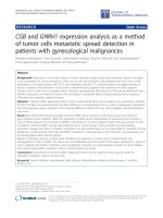
Figure 1 Kaplan-Meier curves for overall survival probability according to (A) CRP, (B) NLR and (C) a combination of CRP and NLR.
(A) Patients with CRP > 6.3 mg/L (dotted line) had a significantly shorter overall survival than those with CRP ≤ 6.3 mg/L (solid line) (median 6.0
vs. 26.9 months, respectively; p < 0.001). (B) Likewise, survival among patients with NLR > 2.3 (dotted line) was shorter than those with NLR ≤ 2.3
(solid line) (median 7.9 vs. 32.5 months, respectively; p < 0.001). (C) There is a significantly longer survival in the group with both CRP ≤ 6.3 mg/L
and NLR ≤ 2.3 (solid line, 39.2 months) than the group with either CRP > 6.3 mg/L or NLR > 2.3 (dashed line, 14.6 months) or than the group with
both CRP > 6.3 mg/L and NLR > 2.3 (dotted line, 4.6 months).
presence of PVT, AFP, CRP and NLR as independent
factors predictive of outcome of HCC. A recent study
evaluating CRP and NLR in transplant recipients with
HCC showed that CRP did not affect overall patient
survival, but NLR did with statistical significance [33].
The discrepancy could be due to some differences in
assays measuring CRP and the study population. Our
study employed a high-sensitivity CRP with cut-off value
6.3 mg/L, whereas the study detected CRP with a
conventional sensitivity with cut-off value 1 mg/dL
(= 10 mg/L). Additionally, our study recruited patients
with various stages of HCC undergoing different treatment modalities, which is a distinction to the study
recruiting patients with limited stages of HCC undergoing transplantation, which could result in no more
lasting inflammation. There is no consensus on the cutoff value for NLR. It has been therefore set empirically
between 2.42 and 5 in studies [5,14-20]. In this analysis,
the cut-off value of 2.3 for NLR offered the most significant association with the patient outcomes. Further
Oh et al. BMC Cancer 2013, 13:78
/>
Page 7 of 9
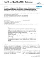
Figure 2 Correlations between (A) CRP and Child-Pugh class, (B) CRP and tumor stage, (C) NLR and Child-Pugh class, (D) NLR and
tumor stage, and (E) CRP and NLR. The levels of CRP and NLR tends to escalate with aggravating Child-Pugh class from A to C (A, r = 0.311,
p < 0.001; C, r = 0.306, p < 0.001) or progressing tumor stage from I to IV (B, r = 0.475, p < 0.001; D, r = 0.358, p < 0.001). (E) There is a significant
positive inter-correlation between CRP and NLR (r = 0.570, p < 0.001).
studies are needed to determine the optimal cut-off
point of CRP and NLR in predicting prognosis in
patients with HCC.
In our results, it is interesting to note that an elevated
CRP level was strongly associated with an elevated NLR.
It seems to be due to some relationship between these
two markers in terms of host inflammation and immune
reaction, as evidenced by the observations that high CRP
levels associated with inflammation are inversely related
to low-level tumor lymphocyte infiltration, which may
contribute to an elevated NLR [24]. Although they had
modest individual associations on the prognosis of the
patients, the significance was quite stronger when used
in combination. It is of note that both CRP and NLR
levels were significantly related to tumor burden and
underlying hepatic reserve, known as the two key
determinants of HCC survival, showing a linear positive
relationship with tumor stages or Child-Pugh classes.
Oh et al. BMC Cancer 2013, 13:78
/>
Page 8 of 9
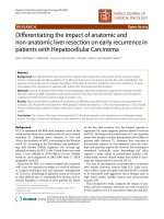
Figure 3 Tumor response according to baseline levels of serum (A) CRP and NLR and changes in (B) CRP and (C) NLR levels after
therapy. Tumor response was better in the group with both baseline serum CRP ≤ 6.3 mg/L and NLR ≤ 2.3 than the other groups (p < 0.001;
Figure 3A). At follow-up, patients with progressive disease showed an increase in levels of CRP and NLR, whereas responders exhibited a decrease
in the levels (repeated measures ANOVA, all p < 0.001; Figure 3B and 3C). CR, complete response; PR, partial response; SD, stable disease; PD,
progressive disease.
Thus, their role as prognostic indicators seems to be
synergistically enhanced with combined use of them, as
presented in our survival analysis.
Moreover, our results showed that patients with
elevated CRP and NLR had worse treatment response,
while those with a low-level of CRP and NLR had more
favorable response. Consistent with survival analysis,
there were significant differences in tumor response on
the combined use of serum CRP level and NLR. Serial
measurements of CRP and NLR exhibited a reduction in
CRP and NLR among responders as well as their rise in
progressive disease during treatment. Thus, it is
expected that the outcome of treatment response before
and during management would be predictable to some
extent with the levels of CRP and NLR.
The current study has some limitations. There is some
heterogeneity in treatment used for HCC, and the majority of the patients received TAC-based loco-regional
therapy, with limited number of surgical cases, which
could have affected the outcome. However, the prognostic effect of those inflammatory markers was still apparent when analyzed specifically in selected patients
receiving loco-regional therapies. Although there are
many other conditions affecting the levels of CRP and
NLR, like an infectious state and autoimmune disease,
the possibility may be minimized because most of those
markers were indeed tested prior to treatment, without
evidence of serious infection. Rather, the results of our
study could be reliable, because we described the largest
cohort to date of consecutive patients and provided
comprehensive data from serial measurements of CRP
and NLR simultaneously, which have not been explicitly
evaluated thus far.
Conclusions
In summary, this study demonstrated that CRP and NLR
are not only markers of inflammation, but also independent prognostic indicators for HCC, reflecting tumor
burden and hepatic reserve. Their role as a surrogate
marker for tumor response and survival is more
enhanced when used in combination. Further studies are
needed to confirm and update a more detailed clinical
relevance and biological mechanisms of CRP and NLR.
Abbreviations
HCC: Hepatocellular carcinoma; HBV: Hepatitis B virus; HCV: Hepatitis C virus;
CRP: C-reactive protein; IL: Interleukin; NLR: Neutrophil-to-lymphocyte ratio;
PVT: Portal vein thrombosis; AFP: α-fetoprotein.
Competing interests
The authors declare that they have no competing interests.
Authors’ contributions
BSO and JWJ participated in the design of the study, performed the
statistical analysis and interpretation of data, and drafted the manuscript.
JHK, CRY and KWC recruited patients to the study. CSK is a radiation
oncologist who contributed to treat the patients. HSJ is an interventional
radiologist who contributed to treat the patients. SL is a clinical pathologist
who participated in the study design. All authors read and approved the
final manuscript.
Authors’ information
Jeong Won Jang, Division of Hepatology, Department of Internal Medicine,
College of Medicine, The Catholic University of Korea, #222 Banpo-daero
22-gil, Seocho-gu, Seoul 137-701, Korea, Telephone: +82-32-280-5866, Fax:
+82-32-280-5987.
Oh et al. BMC Cancer 2013, 13:78
/>
Author details
1
Department of Internal Medicine, The Catholic University of Korea, Incheon
St. Mary’s Hospital, Incheon, Korea. 2Department of Radiation Oncology, The
Catholic University of Korea, Incheon St. Mary’s Hospital, Incheon, Korea.
3
Department of Radiology, The Catholic University of Korea, Incheon St.
Mary’s Hospital, Incheon, Korea. 4Department of Laboratory Medicine, The
Catholic University of Korea, Incheon St. Mary’s Hospital, Incheon, Korea.
5
Division of Hepatology, Department of Internal Medicine, College of
Medicine, The Catholic University of Korea, #222 Banpo-daero 22-gil,
Seocho-gu, Seoul 137-701, Korea.
Page 9 of 9
20.
21.
22.
Received: 5 April 2012 Accepted: 30 January 2013
Published: 15 February 2013
23.
References
1. Llovet JM, Burroughs A, Bruix J: Hepatocellular carcinoma. The Lancet 2003,
362(9399):1907–1917.
2. Bruix J, Sherman M: Management of hepatocellular carcinoma. Hepatology
2005, 42(5):1208–1236.
3. Tandon P, Garcia-Tsao G: Prognostic indicators in hepatocellular
carcinoma: a systematic review of 72 studies. Liver Int 2009, 29(4):502–510.
4. Colotta F, Allavena P, Sica A, Garlanda C, Mantovani A: Cancer-related
inflammation, the seventh hallmark of cancer: links to genetic instability.
Carcinogenesis 2009, 30(7):1073–1081.
5. Roxburgh CS, McMillan DC: Role of systemic inflammatory response in
predicting survival in patients with primary operable cancer. Future Oncol
2010, 6(1):149–163.
6. Morris-Stiff G, Gomez D, Prasad KR: C-reactive protein in liver cancer
surgery. Eur J Surg Oncol 2008, 34(7):727–729.
7. Hashimoto K, Ikeda Y, Korenaga D, Tanoue K, Hamatake M, Kawasaki K,
Yamaoka T, Iwatani Y, Akazawa K, Takenaka K: The impact of preoperative
serum C-reactive protein on the prognosis of patients with
hepatocellular carcinoma. Cancer 2005, 103(9):1856–1864.
8. Lin ZY, Wang LY, Yu ML, Chen SC, Chuang WL, Hsieh MY, Tsai JF, Chang
WY: Role of serum C-reactive protein as a marker of hepatocellular
carcinoma in patients with cirrhosis. J Gastroenterol Hepatol 2000,
15(4):417–421.
9. Nagaoka S, Yoshida T, Akiyoshi J, Akiba J, Torimura T, Adachi H, Kurogi J,
Tajiri N, Inoue K, Niizeki T, et al: Serum C-reactive protein levels predict
survival in hepatocellular carcinoma. Liver Int 2007, 27(8):1091–1097.
10. Jang JW, Oh BS, Kwon JH, You CR, Chung KW, Kay CS, Jung HS: Serum
interleukin-6 and C-reactive protein as a prognostic indicator in
hepatocellular carcinoma. Cytokine 2012, 60(3):686–693.
11. Mahmoud FA, Rivera NI: The role of C-reactive protein as a prognostic
indicator in advanced cancer. Curr Oncol Rep 2002, 4(3):250–255.
12. Hurlimann J, Thorbecke GJ, Hochwald GM: The liver as the site of Creactive protein formation. J Exp Med 1966, 123(2):365–378.
13. Porta C, De Amici M, Quaglini S, Paglino C, Tagliani F, Boncimino A, Moratti
R, Corazza GR: Circulating interleukin-6 as a tumor marker for
hepatocellular carcinoma. Ann Oncol 2008, 19(2):353–358.
14. Chen TM, Lin CC, Huang PT, Wen CF: Neutrophil-to-lymphocyte ratio
associated with mortality in early hepatocellular carcinoma patients after
radiofrequency ablation. J Gastroenterol Hepatol 2012, 27(3):553–561.
15. Gomez D, Farid S, Malik HZ, Young AL, Toogood GJ, Lodge JP, Prasad KR:
Preoperative neutrophil-to-lymphocyte ratio as a prognostic predictor
after curative resection for hepatocellular carcinoma. World J Surg 2008,
32(8):1757–1762.
16. Huang ZL, Luo J, Chen MS, Li JQ, Shi M: Blood neutrophil-to-lymphocyte
ratio predicts survival in patients with unresectable hepatocellular
carcinoma undergoing transarterial chemoembolization. J Vasc Interv
Radiol 2011, 22(5):702–709.
17. Cho H, Hur HW, Kim SW, Kim SH, Kim JH, Kim YT, Lee K: Pre-treatment
neutrophil to lymphocyte ratio is elevated in epithelial ovarian cancer
and predicts survival after treatment. Cancer Immunol Immunother 2009,
58(1):15–23.
18. Azab B, Bhatt VR, Phookan J, Murukutla S, Kohn N, Terjanian T, Widmann
WD: Usefulness of the Neutrophil-to-Lymphocyte Ratio in Predicting
Short- and Long-Term Mortality in Breast Cancer Patients. Ann Surg Oncol
2012, 19(1):217–224.
19. Ding PR, An X, Zhang RX, Fang YJ, Li LR, Chen G, Wu XJ, Lu ZH, Lin JZ,
Kong LH, et al: Elevated preoperative neutrophil to lymphocyte ratio
24.
25.
26.
27.
28.
29.
30.
31.
32.
33.
predicts risk of recurrence following curative resection for stage IIA
colon cancer. Int J Colorectal Dis 2010, 25(12):1427–1433.
Jung MR, Park YK, Jeong O, Seon JW, Ryu SY, Kim DY, Kim YJ: Elevated
preoperative neutrophil to lymphocyte ratio predicts poor survival
following resection in late stage gastric cancer. J Surg Oncol 2011,
104(5):504–510.
Korean Liver Cancer Study Group and National Cancer Center K: Practice
guidelines for management of hepatocellular carcinoma 2009. Korean J
Hepatol 2009, 15(3):391–423.
Eisenhauer EA, Therasse P, Bogaerts J, Schwartz LH, Sargent D, Ford R,
Dancey J, Arbuck S, Gwyther S, Mooney M, et al: New response evaluation
criteria in solid tumours: revised RECIST guideline (version 1.1).
Eur J Cancer 2009, 45(2):228–247.
Shimada H, Nabeya Y, Okazumi S, Matsubara H, Shiratori T, Aoki T, Sugaya
M, Miyazawa Y, Hayashi H, Miyazaki S, et al: Elevation of preoperative
serum C-reactive protein level is related to poor prognosis in
esophageal squamous cell carcinoma. J Surg Oncol 2003, 83(4):248–252.
Canna K, McArdle PA, McMillan DC, McNicol AM, Smith GW, McKee RF,
McArdle CS: The relationship between tumour T-lymphocyte infiltration,
the systemic inflammatory response and survival in patients undergoing
curative resection for colorectal cancer. Br J Cancer 2005, 92(4):651–654.
Goldman ND, Liu TY: Biosynthesis of human C-reactive protein in
cultured hepatoma cells is induced by a monocyte factor(s) other than
interleukin-1. J Biol Chem 1987, 262(5):2363–2368.
Imai Y, Kubota Y, Yamamoto S, Tsuji K, Shimatani M, Shibatani N, Takamido
S, Matsushita M, Okazaki K: Neutrophils enhance invasion activity of
human cholangiocellular carcinoma and hepatocellular carcinoma cells:
an in vitro study. J Gastroenterol Hepatol 2005, 20(2):287–293.
Kuang DM, Zhao Q, Wu Y, Peng C, Wang J, Xu Z, Yin XY, Zheng L:
Peritumoral neutrophils link inflammatory response to disease
progression by fostering angiogenesis in hepatocellular carcinoma.
J Hepatol 2011, 54(5):948–955.
Kusumanto YH, Dam WA, Hospers GA, Meijer C, Mulder NH: Platelets and
granulocytes, in particular the neutrophils, form important
compartments for circulating vascular endothelial growth factor.
Angiogenesis 2003, 6(4):283–287.
Wu Y, Zhao Q, Peng C, Sun L, Li XF, Kuang DM: Neutrophils promote
motility of cancer cells via a hyaluronan-mediated TLR4/PI3K activation
loop. J Pathol 2011, 225(3):438–447.
McDonald B, Spicer J, Giannais B, Fallavollita L, Brodt P, Ferri LE: Systemic
inflammation increases cancer cell adhesion to hepatic sinusoids by
neutrophil mediated mechanisms. Int J Cancer 2009, 125(6):1298–1305.
Muller I, Munder M, Kropf P, Hansch GM: Polymorphonuclear neutrophils
and T lymphocytes: strange bedfellows or brothers in arms? Trends
Immunol 2009, 30(11):522–530.
Ohtani H: Focus on TILs: prognostic significance of tumor infiltrating
lymphocytes in human colorectal cancer. Cancer Immun 2007, 7:4.
Bertuzzo VR, Cescon M, Ravaioli M, Grazi GL, Ercolani G, Del Gaudio M,
Cucchetti A, D’Errico-Grigioni A, Golfieri R, Pinna AD: Analysis of factors
affecting recurrence of hepatocellular carcinoma after liver
transplantation with a special focus on inflammation markers.
Transplantation 2011, 91(11):1279–1285.
doi:10.1186/1471-2407-13-78
Cite this article as: Oh et al.: Prognostic value of C-reactive protein and
neutrophil-to-lymphocyte ratio in patients with hepatocellular
carcinoma. BMC Cancer 2013 13:78.