Báo cáo y học: "Comparison of osteogenic potentials of human rat BMP4 and BMP6 gene therapy using [E1-] and [E1-,E2b-] adenoviral vectors"
Bạn đang xem bản rút gọn của tài liệu. Xem và tải ngay bản đầy đủ của tài liệu tại đây (742.3 KB, 9 trang )
Int. J. Med. Sci. 2006, 3
97
International Journal of Medical Sciences
ISSN 1449-1907 www.medsci.org 2006 3(3):97-105
©2006 Ivyspring International Publisher. All rights reserved
Research paper
Comparison of osteogenic potentials of human rat BMP4 and BMP6 gene
therapy using [E1-] and [E1-,E2b-] adenoviral vectors
Hongwei Li
1
, Jin Zhong Li
1
, Debra D. Pittman
2
, Andy Amalfitano
3
,
Gerald R. Hankins
1
and Gregory A. Helm
1 4
1
Departments of Neurological Surgery, University of Virginia Health System, Charlottesville, Virginia 22908, USA;
2
Genetics Institute, Andover, Massachusetts 01810, USA;
3
Departments of Pediatrics and Human Genetics, Duke University Medical Center, Durham, North Carolina 27710, USA;
4
Departments of Biomedical Engineering, University of Virginia Health System, Charlottesville, Virginia 22908, USA
Corresponding address: Jin Zhong Li, D.V.M. Ph.D., Department of Neurological Surgery, University of Virginia Health
System, P. O. Box 800212, Charlottesville, Virginia 22908, USA
Received: 2006.05.02; Accepted: 2006.05.31; Published: 2006.06.01
Osteogenic potentials of some recombinant human bone morphogenetic protein (BMP) first-generation
adenoviral vectors (ADhBMPs) are significantly limited in immunocompetent animals. It is unclear what role
expression of viral proteins and foreign proteins transduced by adenoviral vectors play in the host immune
response and in ectopic bone formation. In this study two sets of experiments were designed and performed.
First, rat BMP6 cDNA were amplified, sequenced, and recombined in first-generation adenoviral vector
(ADrBMP6). A comparison of human and rat BMP6 adenoviral vectors demonstrated identical osteogenic
activities in both immunodeficient and immunocompetent rats. Second, the activities of recombinant human
BMP6 in E1- (ADhBMP6) and [E1-,E2b-] ( [E1-,E2b-]ADGFP&hBMP6, and [E1-,E2b-]ADhBMP6) adenoviral
vectors were compared in both in vitro and in vivo models. Similar activities of these two generations of BMP
adenoviral vectors were found in all models. These results indicate that the amount of viral gene expression and
the source of the BMP cDNA are not major factors in the interruption of osteogenic potentials of recombinant
BMP6 adenoviral vectors in immunocompetent animals.
1. INTRODUCTION
Gene therapy provides a novel method to repair
damaged bone by using bone morphogenetic protein
(BMP). BMP gene vectors can be divided into DNA,
viral, and cell vectors [10, 19]. Among the BMP gene
therapy vectors, human BMP adenoviral vectors
(ADhBMPs) are very commonly used and have
displayed strong osteogenic potentials in
immunodeficient animals [1, 25]. Nevertheless, the
functions of ADhBMPs have proved to be significantly
limited in immunocompetent animals [1, 25, 35, 38].
All five human BMP adenoviral vectors (ADhBMPs 2,
4, 6, 7, and 9) have been shown to induce large
volumes of ectopic bone formation in athymic nude
(AN) rats. Bone volumes induced by these BMP
vectors were greatest when ADhBMPs 4, 6, and 9 were
used, followed by ADhBMP2 and, finally, by
ADhBMP7. The osteogenic potentials of ADhBMPs 2,
4, and 7, however, were not shown in
immunocompetent animals. In addition, bone
volumes induced by ADhBMP6 were significantly
smaller in immunocompetent animals than in
immunodeficient animals. In contrast, bone formation
induced by ADhBMP9 was similar in AN and
immunocompetent rats [25]. These results may be
related to different BMP signal transduction pathways
and the host immune response.
The BMP family includes more than 30 members
[9, 46]. According to previous studies, BMPs combine
with type 1 (Alk2, Alk3, and Alk6) and type 2 (BR2,
ActR2, and ActR2B) receptors, activate the Smad and
p38/MAPK signal transduction pathways, and, finally,
activate transcription of bone formation factors [36,
44]. BMP2 and BMP4 combine with Alk3 and use
Smad1, Smad5, or Smad8 to transduce signals [4, 31].
BMP6 and BMP7 may strongly combine with Alk2
and weakly combine with Alk3 and Alk6. Their
signals are mainly transferred with Smad5 and,
possibly, with Smad1 but not with Smad8 [11, 13].
Compared with other BMPs, BMP9 uses a different
type of receptor and signal transduction pathway, the
details of which are not yet clear [30, 40]. The
functional performances of various BMP adenoviral
vectors may reflect different mechanisms in vivo.
The host immune response to an adenovirus and
its infected cells may be a primary cause of this
functional limitation. The antigenicity of adenoviral
vectors, which induce the host immune response,
consist of directly injected viral particles and viral
genome–expressed proteins that include viral proteins
and foreign transgene-encoded proteins [27, 42]. It is
unclear what roles expression of viral proteins and
foreign proteins transduced by adenoviral vectors
play in the host immune response and ectopic bone
formation. The adenoviral genome can be divided into
four early regions: E1, E2, E3, and E4 [6]. Adenoviral
vectors in which the E1 and E3 regions have been
deleted have commonly been used in the field of gene
therapy and are called first-generation adenoviral
vectors, that is, replication-defective adenoviruses.
Int. J. Med. Sci. 2006, 3
98
Unfortunately, because of the presence of E1-like
factors in many cell types, vectors with the E1 deletion
may still express a certain amount of viral gene
products in vivo. The expression of viral proteins in
infected target cells may increase the host immune
response and further interrupt the expression of the
foreign gene [12]. To limit the productions of
potentially immunogenic viral proteins, investigators
have pursued the construction of adenoviral vectors in
which more or even all viral coding genes have been
deleted. Helper-dependent adenoviral vectors in
which all viral protein–coding DNA sequences have
been deleted have been developed [34, 45]. This
modification reportedly has reduced toxicity and
prolonged gene expression in some experiments [5,
33]. Nevertheless, we constructed a recombinant
BMP9 helper-dependent adenoviral vector and found
that the osteogenic potentials of this vector were not
significantly different from those of the corresponding
recombinant BMP9 first-generation vector (ADhBMP9)
in immunodeficient and immunocompetent rats [23].
Because there are different signal transduction
pathways among the BMPs, the complex procedure
needed to produce the helper-dependent vector and
the possible roles of viral gene products in the process
of bone formation need to be clarified further. An
adenoviral vector with deletions of E1, E3, and the
polymerase and terminal protein ( [E1-,E2b-]AD, also
known as the second-generation adenoviral vector)
was selected in the following study. This class of
modified adenovirus vector has several potential
benefits including the following: 1) clonal
preparations that do not require a helper virus for
growth; 2) theoretically, decreased frequency of
replication-competent adenovirus generation; 3)
increased carrying capacity; 4) rapid scale up of
production; and 5) decreased potential for eliciting an
immunogenic response in vivo [20]. On the other hand,
the sources of BMP cDNAs may also cause the
functional limitation of ADhBMPs in
immunocompetent animals. We selected rat BMP4
and BMP6 to help us answer these questions.
In the present study, two different sets of
experiments were designed and performed. In the first,
rat BMP6 cDNA from adult Sprague–Dawley (SD) rats
was cloned and sequenced. A recombinant first-
generation adenovirus ADrBMP6 was constructed and
compared with ADhBMP6 in the process of bone
formation. In the second set of experiments, human
BMP6 cDNA was constructed to form a second-
generation human BMP adenoviral vector ( [E1-,E2b-
]ADhBMP). The viral vectors [E1-,E2b-]ADhBMP6,
and [E1-,E2b-]ADGFP&BMP6 which include the green
fluorescent protein [GFP] were tested in in vitro and in
vivo models. The roles of the viral genome and the
sources of BMP cDNA in the process of bone
formation were determined in this study.
2. MATERIALS AND METHODS
Cloning and identification of rat BMP4 and BMP6 cDNA
coding sequences
Total RNA was prepared from the spleen of a 2-
month-old SD rat by using an RNeasy Mini Kit
(Qiagen, Valencia, CA) according to the
manufacturer’s instructions. Rat BMP4 and BMP6
cDNAs containing full coding sequences were
generated by performing an RT-PCR with the OneStep
RT-PCR Kit (Qiagen). The designs of the PCR primers
were based on a known rat BMP4 sequence and a
partial rat BMP6 sequence in which mouse and
human BMP6 sequences were inserted in places in
which the rat sequence was not known [17, 22, 39].
For BMP4, the upstream primer was 5´–CCA CC
A
TGA TTC CTG GTA ACC GAA TGC TG–3´, to which
CCACC was added to give the amplified product a
typical Kozak consensus sequence around the initiator
methionine; the downstream primer was 5´–CTC AGC
GGC ATC CGC ACC CCT C–3´. For BMP6, the
upstream primer was 5´–TTAGAT CTC CAC C
AT
GCC CGG GCT GGG G–3´, and the downstream
primer was 5´–AGA ATC ACA GCC CCT GCA A–3´.
Single PCR products of the expected size (1.2 kb for
BMP4 and 1.5 kb for BMP6) were purified by
performing agarose gel electrophoresis and cloned
into the EcoRV site of pShuttleCMV after having been
blunted with T4 DNA polymerase. The purified rat
BMP4 and BMP6 PCR products and the recombinant
plasmids of pShuttle-rBMP4 and pShuttle-rBMP6
were prepared and sequenced (both strands) to ensure
that the rat BMP inserts were correct.
Construction of rat BMP4 and BMP6 recombinant
adenoviruses
The AdEasy Vector System [18] was used for
construction of the rat BMP4 and BMP6 adenoviral
vectors. The linear pShuttle-rBMPs and the pAdEasy 1
plasmid were cotransformed into the competent
Escherichia coli strain BJ5183 to obtain the BMP viral
DNA plasmid. Briefly, 1 µg of linearized recombinant
transfer vector pShuttle-rBMP (5 µl) and 1.0 µl of
pAdEasy-1 vector (100 ng/µl) were added to 200 µl of
competent BJ5183 cells. The next procedure followed
methods outlined in previous reports [18]. The
recombinant clones were identified by using a PCR.
The pAdeasy-1 contains the human adenovirus
type 5 genome with E1 and E3 deletions. The
recombinant adenoviral plasmids, pADrBMP4 and
pADrBMP6, were cleaved with PacI to expose their
inverted terminal repeats and transfected into 293A
cells to produce viral particles. The recombinant
viruses were identified by performing PCR, RT-PCR,
Southern blot analysis, immunocytochemical staining,
and Western blot analysis. The recombinant
ADrBMP4 and ADrBMP6 were purified through two
cesium chloride gradients, after which the purified
virus was desalted by dialysis at 4°C against 10 mM
Tris–hydrochloric acid buffer with 10% glycerol and
stored in aliquots in liquid nitrogen. The titer of the
virus preparations was determined by measuring the
Int. J. Med. Sci. 2006, 3
99
spectrophotometric absorbance at 260 nm and by
performing a plaque assay.
The AdEasy1ΔpolΔpTp
small
Vector System ( [E1-
,E2b-]AD) [3] was used for construction of human
BMP second-generation adenoviral vectors ( [E1-,E2b-
]ADhBMP6, [E1-,E2b-]ADGFP&hBMP4, and [E1-,E2b-
]ADGFP&hBMP6), which lack polymerase and
terminal protein [E2b] as well as the E1 and E3 genes.
Human BMP4 cDNA was inserted into the shuttle
vector pTrackCMV, and a recombinant adenoviral
vector was made that encoded both GFP and human
BMP4 ( [E1-,E2b-]ADGFP&hBMP4). Human BMP6
cDNA was inserted into the shuttle vectors
pShuttleCMV and pTrackCMV, and recombinant
adenoviral vectors were made that encode human
BMP6 with GFP ( [E1-,E2b-]ADGFP&hBMP6) and
without GFP ( [E1-,E2b-]ADhBMP6), as described
earlier for the AdEasy Vector System. The viruses
were produced in C7 cells, which are stably
transformed with adenoviral E1 and E2b genes [2].
The viral titer was measured by determining the
spectrophotometric absorbance at 260 nm.
Southern blot analysis of genomic DNA of recombinant
adenovirus
Viral DNA was isolated from 293A or C7 cells
that had been transduced with recombinant
adenovirus. One hundred nanograms of each DNA
was digested with HindIII, BstXI (BMP4 and BMP6),
or BglII plus EcoRV (BMP6); electrophoretically
separated in a 0.8% agarose gel; and transferred onto a
nylon membrane. The membranes were baked at 80°C
for 30 min and probed with the pAdEasy1 plasmid,
BMP4 cDNA fragment, or BMP6 cDNA fragment,
each of which was labeled with digoxigenin by using
the DIG-Chem-Link Labeling and Detection Set
(Roche Diagnostics Corp., Indianapolis, IN). Detection
of DIG-labeled nucleic acids was performed using the
DIG Luminescent Detection Kit (Roche).
Western blot detection of BMP4 and BMP6
The pShuttle-rBMP4 and pShuttle-rBMP6
plasmids were transfected into 293A cells with
Lipofectamine 2000 reagent (Invitrogen, Carlsbad,
CA). African green monkey kidney cells (Vero cells,
No. CCL-81; American Type Culture Collection
[ATCC], Manassas, VA), which have a higher
potential to produce BMPs than 293A or C2C12 cells,
were infected with ADrBMP4, ADhBMP4 [25], [E1-
,E2b-]ADGFP&hBMP4, ADrBMP6, ADhBMP6 [25],
[E1-,E2b-]ADhBMP6, [E1-,E2b-]ADGFP&hBMP6, or
ADNULL (2 × 10
8
particles per well of a 24-well plate)
in Opti-MEMI medium without fetal bovine serum.
After transfection, the 293A cells or infected Vero cells
were incubated for 48 hrs at 37°C. The transfected cells
and media were harvested, treated in LDS sample
buffer (Invitrogen) at 70°C for 10 min with reducing
reagent (Invitrogen Corp., Carlsbad, CA),
electrophoresed on a NuPAGE 10% Bis-Tris System
(Invitrogen), and transferred to a PVDF membrane
(Invitrogen). Recombinant human BMP4 and BMP6
(R&D Systems, Minneapolis, MN) were used as
standard proteins. The blots were reacted with
monoclonal mouse BMP4 or BMP6 antibody
(Chemicon, Temecula, CA) at 1 μg/ml or with mouse
β-actin antibody (Sigma Chemical Co, St. Louis, MO)
at a 1:5000 dilution. The Novex Chemiluminescent
Western Blotting Immunodetection System
(Invitrogen) was used.
Three concentrations of
standard BMPs were used to make the standard
curves. We calculated the sample BMP concentrations
based on the results of film scanning by using the
Personal Densitometer SI (Amersham Biosciences,
Piscataway, NJ).
Rat BMP4 and BMP6 biological activity assays.
Mouse C2C12 myoblastic cells (No. CRL-1772;
ATCC) were used in bioassays to determine the
biological activity of the BMPs. We chose the C2C12
cell line for this experiment not only because increased
ALP activity in these cells is dependent on stimulation
by BMPs, but also because the C2C12 cell has a lower
background of ALP and constitutes a defined cell line
that is much more convenient to use than human
mesenchymal stem cells. Cells in 48-well plates that
had reached 80% to 90% confluence were infected
with ADrBMP4, ADhBMP4, ADrBMP6, ADhBMP6, or
ADNULL at concentrations of 3 × 10
8
, 1.5 × 10
8
, 7.5 ×
10
7
, and 3.8 × 10
7
particles per well. Seven days later,
the cells were stained to measure ALP by using the
Sigma Diagnostics ALP Kit (Sigma Diagnostics, Inc., St.
Louis, MO).
In vivo ectopic bone formation in rats
We studied ectopic bone formation in 2-month-
old male rats. Eight AN rats and 20 SD rats were used
for the study of BMP4, and 16 AN rats and 21 SD rats
for the study of BMP6. Animal protocols were
approved by the University of Virginia Animal Use
and Research Committee and conformed to National
Institutes of Health guidelines. The rats were
anesthetized with a mixture of ketamine and xylazine,
and in each animal the thigh was prepared in a sterile
fashion. Using a 19-gauge guide needle, the skin 1 cm
above the knee joint was punctured and the needle
advanced 1 cm proximally. Through this needle, a
Hamilton microsyringe was inserted and 50 μl of viral
solution (2.8 × 10
10
, 5 × 10
10
, or 1.4 × 10
11
particles) was
injected.
For BMP4, the AN rats were separated into two
groups (four rats per group). Animals in the first
group received an injection of ADrBMP4 in one thigh
and an injection of ADNULL in the other. Animals in
the other group received an injection of [E1-,E2b-
]ADGFP&hBMP4 in one thigh and an injection of
ADhBMP4 in the other. Each injection contained 5 ×
10
10
particles. The SD rats were separated into four
groups (5 animals in each group). These animals
received bilateral injections (5 × 10
10
particles in one
thigh and 1 × 10
10
particles in the other) of ADNULL,
ADhBMP4, ADrBMP4, or [E1-,E2b-] ADGFP&hBMP4.
For BMP6, the AN rats were separated into four
groups (four rats per group). Animals in three groups
received an injection of 2.8 × 10
10
particles of
Int. J. Med. Sci. 2006, 3
100
ADNULL in one thigh and an injection of 2.8 × 10
10
particles of either ADhBMP6, [E1-,E2b-
]ADGFP&hBMP6, or [E1-,E2b-]ADhBMP6 in the other
thigh. Animals in the remaining group were injected
in one thigh with 1.4 × 10
11
of ADrBMP6 particles and
in the other thigh with 2.8 × 10
10
particles of ADrBMP6.
The SD rats were separated into five groups. Only one
vector was injected into each SD rat. Animals in the
first four groups (four rats per group) received
bilateral injections of either ADNULL, ADhBMP6, [E1-
,E2b-]ADGFP&hBMP6, or [E1-,E2b-]ADhBMP6 (2.8 ×
10
10
particles per thigh). The other five rats received
bilateral injections of ADrBMP6 (2.8 × 10
10
particles in
one thigh and 1.4 × 10
11
particles in the other thigh).
On Day 35, the rats were euthanized and scanned
using CT. Axial CT images (1-mm collimation and 1-
mm table increment) were obtained using the
standard algorithm with 130 kV, 100 mA, a 2-second
scan time, and a 40-mm image size. The three-
dimensional reconstruction was performed using a
Voxel Q workstation. Serum samples were collected
on Days 0 and 35.
Detection of antibodies to adenovirus
Ninety-six-well Nunc Maxisorb plates (Nunc,
Inc., Roskilde, Demark) were coated with 100 μl of
purified adenovirus (5 × 10
9
particles AdCMV-
βgal/ml) in phosphate-buffered saline (PBS) overnight
at 4°C, washed four times in PBS containing 0.05%
Tween-20, and blocked in PBS supplemented with 1%
bovine serum albumin for 1 hr at 37°C. Appropriately
diluted serum samples were added to antigen-coated
plates and incubated overnight at 4°C. Plates were
washed four times in PBS–0.05% Tween-20 and
incubated with anti–rat IgG (H+L) ALP conjugate
(1:2500 dilution, Promega, Madison, WI) for 2 hrs at
37°C. The plates were washed in the manner
described earlier, and ρ-nitrophenyl phosphate (ρNPP)
substrate (Invitrogen) was added. Optical densities
were recorded at 410 nm on an OPTImax tunable
microplate reader (Molecular Devices Corp.,
Sunnyvale, CA).
Detection of antibodies to BMP4 and BMP6
Microtiter plates (96-well, Nunc) were coated
with the purified human BMP4 and BMP6 (R&D
Systems, Minneapolis, MN), followed by blocking, as
described earlier. Rat sera were added in serial
dilutions. Captured antibodies were detected as
described previously.
3. RESULTS
Cloning, sequencing, and identification of rat BMP4 and
BMP6 cDNAs
Rat BMP4 and BMP6 cDNA fragments
containing complete coding regions were amplified by
RT-PCR from the total spleen RNA of an adult SD rat.
Afterward the fragments were directly cloned into
pShuttleCMV, which is a transfer vector used with the
CMV promoter for the pAdEasy Vector System.
For rat BMP4 cDNA, five recombinant plasmids
were selected and transfected into 293A cells. The
expression of BMP4 was determined by performing
immunocytochemical staining and Western blot
analysis. Three of the five clones produced and
secreted BMP4, but the amounts of BMP4 expressed
by these clones varied; the other two clones could not
produce BMP4 (Fig. 1). The three clones with BMP4
expression were sequenced and their sequences were
compared with the BMP4 coding region sequence
published in GenBank. Only three consecutive base
pairs in the propeptide coding region differed from
the rat BMP4 sequence described by GenBank, but
these base pairs were the same as those found in
murine and human BMP4. One clone that
demonstrated a high BMP4 expression was used to
construct the rat BMP4 recombinant adenoviral vector.
The complete rat BMP4 cDNA sequence was
submitted to GenBank (accession No. AY184241).
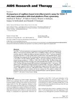
Figure 1 Western blot detection of rat BMP4 or BMP6
in conditioned media. The conditioned media were
prepared from 293A cells transfected with rat BMP4 or
BMP6 cDNA recombinant clones. Detection was
accomplished using BMP4 antibody (upper panel) and
BMP6 antibody (lower panel). Lane 1, pShuttleCMV;
Lanes 2–6, rat BMP4 or BMP6 recombinant clones; Lane 7,
pShuttle-hBMP4 or pShuttle-hBMP6.
For rat BMP6 cDNA, five recombinant plasmids
(Nos. 1, 2, 7, 8, and 23] and the purified PCR product
were sequenced. The clones differed from each other
and from the partial sequence of the rat BMP6 gene
published in GenBank. Only clone No. 8 could
produce and secrete BMP6, as observed using
immunocytochemical staining and Western blot
analysis (Fig. 1). Based on a comparison of the BMP6
sequences of the five clones and the purified PCR
product, the first complete rat BMP6 cDNA sequence
was obtained and submitted to GenBank (accession
No. AY184240
). For the derived amino acid sequence,
amino acids 301 through 506 are identical to those
derived from the previously reported partial sequence
for the Lewis rat [39] (GenBank accession No. X58830
),
and amino acids 77 through 280 are identical to those
derived from the previously reported partial sequence
for the Wistar rat [22] (GenBank accession No. U66298
)
except for amino acid 146, which in our sequence is
valine as opposed to alanine in the Wistar rat. The
remaining amino acids, 1 through 76 and 281 through
300, are identical to those found in the mouse [8, 16,
17, 28] (GenBank accession No. NM_007556
). To
Int. J. Med. Sci. 2006, 3
101
ensure that the sequence of rat BMP6 cDNA was
correct, BMP6 cDNA from another SD rat was
amplified, cloned, and sequenced. The findings
confirmed that the BMP6 sequences from the two rats
were identical. Genomic DNA containing the codon
for the polymorphic amino acid was amplified from
four additional SD rats and sequenced. The results of
the genomic sequencing also verified the presence of
valine at position 146 in the SD rats.
In each of the five rat BMP6 clones one or several
amino acids were mutated, even though clone No. 8
could produce and secrete BMP6. A rat BMP6
recombinant clone was generated by ligation at the
EcoNI site, between the upstream segment of clone No.
8 and the downstream segment of clone No. 2, to
obtain a complete clone with no mutated amino acid.
Construction and identification of ADrBMP4 and
ADrBMP6
The AdEasy system was used to construct
ADrBMP4 and ADrBMP6. The viral DNA of
ADrBMP4 and ADrBMP6 were identified by
performing a Southern blot analysis. Figure 2 shows
the Southern blot for BMP6. The viral DNA contained
the correct rat BMP cDNA inserts, and the expected
DNA fragments were generated by using several
restriction endonucleases. The titers of the purified
viral solutions were 1.0 × 10
12
particles/ml or 2.1 ×
10
10
plaque forming units (PFU)/ml for ADrBMP4 and
2.7 × 10
12
particles/ml or 3.3 ×10
10
PFU/ml for
ADrBMP6.
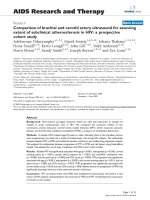
Figure 2 Identification of recombinant BMP6
adenoviruses by Southern Blot analysis. A. pAdEasy1
probe. B. BMP6 cDNA fragment probe. DNA samples were
digested by HindIII (Lanes 1, 2 and 3), BstXI (Lanes 4, 5,
and 6), and BglII plus EcoRV (Lanes 7, 8, and 9). M, 1-kb
DNA ladder. Lanes 1, 4, and 7, 293A cells; Lanes 2, 5, and
8, ADrBMP6 DNA; Lanes 3, 6, and 9, ADhBMP6 DNA.
This finding indicates that the construction of rat BMP6
recombinant adenovirus is correct.
BMP4 and BMP6 expression by corresponding
adenoviral vectors in Vero cells
Protein expression by the BMP4 vectors is
demonstrated in Fig. 3A and B, which depicts the
results of a Western blot analysis of lysed Vero cells
transduced with ADNULL, ADhBMP4, or ADrBMP4.
The blot was tested with BMP4 antibody (Fig. 3A) and
with β-actin antibody (Fig. 3B). The ADrBMP4-
transduced cells produced mature protein that
exhibited the same electrophoretic mobility as
ADhBMP4 on a reduced gel. There was no significant
difference in the amount of BMP4 expressed by
ADrBMP4- and ADhBMP4-transduced Vero cells.
Protein expression by the BMP6 vectors is
demonstrated in Fig. 3C, which shows a Western blot
of lysed Vero cells transduced with ADNULL,
ADhBMP6, or ADrBMP6. The blot was probed with
an antibody to BMP6 (Fig. 3C) and with β-actin
antibody (Fig. 3D). The ADrBMP6-transduced cells
produced mature protein that exhibited the same
electrophoretic mobility as ADhBMP6 on a reduced
gel. Also there was no significant difference in the
amounts of expressed BMP6 protein between
ADrBMP6- and ADhBMP6-transduced Vero cells,
which were 8.1 μg/ml and 7.9 μg/ml, respectively.
After a denaturing gel electrophoresis under reducing
conditions and a Western blot analysis, a 19-kD
protein reacted with the BMP6 antibody. The staining
differed from that of standard human BMP6, which
was purchased from R&D Systems (Minneapolis, MN)
and displayed two bands at 18 kD and 23 kD. This
difference may be due to dissimilar cell types, which
may produce different numbers of glycosylation sites.
Alkaline phosphatase activity induced by BMP
adenoviral vectors in C2C12 cells
To evaluate the biological function of rat and
human BMP4 and BMP6, C2C12 cells transduced with
BMP adenoviral vectors were stained to demonstrate
alkaline
phosphatase
(ALP) activity,
which is an
important
indicator of
BMP activity.
The
ADrBMP4-
induced ALP
expression in
C2C12 cells
was similar to
that observed
following
ADhBMP4
treatment
(data not
shown);
however, at
the same number of viral particles, the ALP expression
induced by ADrBMP6 in C2C12 cells was significantly