Control of Cell Proliferation and Growth byMyc Proteins
Bạn đang xem bản rút gọn của tài liệu. Xem và tải ngay bản đầy đủ của tài liệu tại đây (296.26 KB, 14 trang )
Results Probl Cell Differ (42)
P. Kaldis: Cell Cycle Regulation
DOI 10.1007/004/Published online: 24 February 2006
© Springer-Verlag Berlin Heidelberg 2006
Control of Cell Proliferation and Growth by Myc Proteins
Sandra Bernard · Martin Eilers (✉)
Institute for Molecular Biology and Tumor Research, University of Marburg,
35033 Marburg, Germany
Abstract Myc proteins act as signal transducers that alter cell proliferation in dependence
on signals from the extracellular environment. In normal cells, the expression of MYC
genes is therefore under tight control by growth factor dependent signals. The enormous
interest in the function of these proteins is motivated by the observation that the close
control of MYC expression is disrupted in a large percentage of human tumors, leading to
deregulated expression of Myc proteins. A large body of evidence shows that this dereg-
ulation is a major driving force of human tumorigenesis; in cells with deregulated Myc,
proliferation often takes place in the complete absence of external stimuli. We will dis-
cuss current models to understand Myc function and also potential avenues to selectively
interfere with the proliferation of Myc-transformed cells.
1
Introduction
MYC genes form a small multigene family; the family has attracted enormous
attention, since the enhanced expression of one of its three members (MYC,
MYCL or MYCN) contributes to multiple human tumors. Deregulation of ex-
pression can occur through diverse mechanisms, only some of which involve
the mutation of MYC genes themselves. More frequently, mutations occur in
human tumors in pathways that control the expression of MYC genes or that
control the function of the encoded proteins. The full spectrum of such mu-
tations remains to be elucidated, since the regulation of any member of the
MYC gene family is complex. As a result, the precise percentage of human
tumors in which MYC genes are activated and/or deregulated remains a mat-
ter of some debate; it is possible that the ‘Myc pathway’, as suggested for
the E2F pathway, needs to be deregulated for any human tumor to emerge.
Alternatively, there is evidence that at least some cells proliferate in a Myc-
independent manner and therefore it is possible that tumors derived from
such cells do not have an ‘activated’ MYC gene.
Exogenously introduced MYC genes that are expressed under the con-
trol of a strong promoter (to mimic the activation of MYC genes seen in
human tumors) elicit a stereotype response in most cells into which they
are introduced: they promote cell proliferation even in the absence of mi-
togenic signals, they often promote cell growth and they almost invari-
330 S. Bernard · M. Eilers
ably promote apoptosis or at least sensitize cells to apoptotic stimuli. Ker-
atinocytes are one of only a few examples of cells that respond differently
to Myc: in skin, proliferation is tightly linked to adhesion to the basal lam-
ina and Myc disrupts this adhesion; therefore, keratinocytes respond to Myc
with premature differentiation and arrest of proliferation (Gandarillas and
Watt 1997).
The frequent activation of MYC genes in tumorigenesis has suggested that
Myc proteins might also have an important function in normal prolifera-
tion and growth and this suggestion has by now been tested in numerous
experimental systems: Rat1 fibroblasts, in which both alleles of c-myc have
been deleted, show a reduced rate of proliferation and cell growth (Mateyak
et al. 1997; Mateyak et al. 1999). Similarly, mouse embryo fibroblasts that
carry a floxed allele of c-myc arrest upon cre-mediated excision of c-myc (de
Alboran et al. 2001). Importantly, deletion of the mnt gene, which encodes
a member of the Mad family of antagonists of Myc, restores proliferation to
c-myc deleted cells, suggesting that the balance of expression of members of
theMyc/Max/Madnetworkofproteins(seeFig.1)maydictatetheresponse
to loss of Myc (Walker et al. 2005).
In mice, the effects of deletion of the c-myc or n-myc genes in vivo are
not uniform. Deletion of c-myc in mice results in embryonic lethality and
certain cell types do not proliferate (de Alboran et al. 2001; Trumpp et al.
2001). In the hematopoietic lineage, stem cells continue to proliferate upon
genetic ablation of c-myc, whereas differentiated (lineage positive) cells ar-
rest (Wilson et al. 2004). While these data show that c-myc is not required
for proliferation of hematopoietic stem cells, such cells also express n-myc
and therefore it is possible that n-myc provides essential functions of Myc
proteins in these cells. Similarly, postnatal proliferation of hepatocytes in the
liver does not require c-Myc, but again the potential compensation by other
myc family members is not completely clear (Baena et al. 2005). In the small
intestine, deletion of c-myc leads to a transient failure to form normal num-
bers of crypts in the small intestine, but in long-term experiments the mice
maintain a normal epithelium in the absence of c-Myc activity and without
apparent compensation by N-Myc or L-Myc (Bettess et al. 2005). Both findings
argue that at least some cell types can proliferate in the absence of functional
Myc. There are also clear examples for a strict requirement for Myc func-
tion in cell proliferation: for example, deletion of n-myc in neuronal precursor
cells leads to a dramatic loss of proliferative capacity of such cells (Knoepfler
et al. 2002). As long as questions of redundancy between different members
of the myc gene family and compensation by loss of antagonists such as mnt
are not fully resolved, it seems to us that no definitive answer is possible as
to whether Myc function is generally required for cell proliferation and cell
growth or whether its essential role in proliferation is restricted to specific
cell types.
Control of Cell Proliferation and Growth by Myc Proteins 331
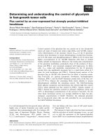
Fig. 1 Transcriptional regulatory complexes formed by Myc proteins and their co-factors.
For details, see text. Almost certainly, Myc forms more than one repressive complex, so
the Myc/Miz1 complex should be viewed as one well-understood example of a group of
similar complexes
332 S. Bernard · M. Eilers
2
Mechanisms of Myc Action
Mycproteinsactatleastinpartastranscriptionfactorsthatactivateandre-
press large groups of genes (see Fig. 2). They activate transcription as part of
a binary complex together with an obligate partner protein, Max; the complex
binds to a specific sequence, termed E-Box (CACGTG or related sequences),
which is found in all genes that are activated by Myc. Interestingly, Myc
proteins not only regulate protein-coding genes that are transcribed by poly-
merase II.
There are at least three exceptions: first, there is evidence that microRNAs
can be target genes that are activated by Myc: for example, a microRNA tar-
geting E2F1 is induced by Myc (O’Donnell et al. 2005). Since the e2f 1gene
isatthesametimeatargetfortranscriptionalupregulationbyMyc,thedata
suggest that there may be a fine tuning of expression of Myc target genes and
proteins (Baudino et al. 2003). In the case of E2F1, there is evidence both that
it mediates proliferation downstream of Myc (in B-lymphocytes) and that it
mediates Myc-dependent apoptosis, so the microRNA-mediated regulation of
E2F1 protein levels may ensure the correct balance between the two (Baudino
et al. 2003; Leone et al. 2001).
The second exception are ribosomal RNA genes. Myc activates transcrip-
tion through direct binding to canonical binding sites in the rDNA promoter
(Arabi et al. 2005; Grandori et al. 2005; Poortinga et al. 2004). rDNA genes are
transcribed by polymerase I in the nucleolus, arguing that at least a fraction
of Myc proteins are localized in this compartment. Indeed, immunofluores-
cence experiments show that Myc can be found in the nucleolus, in particular
after proteasome inhibition. The latter finding also suggests that Myc can be
degraded in the nucleolus and at least one E3 ligase that targets Myc, Fbw7γ ,
is specifically localized in this compartment (Welcker et al. 2004a,b; Yada et al.
2004).
The third exception are several tRNA genes, demonstrating that Myc can
also stimulate polymerase III-dependent transcription (Gomez-Roman et al.
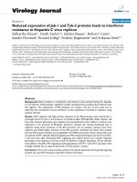
Fig. 2 Genetic targets of Myc in cell proliferation and their proposed functions
Control of Cell Proliferation and Growth by Myc Proteins 333
2003). There is clear evidence that this effect is direct and not mediated
by activation of protein-coding genes that regulate polymerase III function;
however, the mechanism of activation has not been clarified.
Myc proteins also act as transcriptional repressors; all current models
suggest that this is mediated by protein/protein interactions with other tran-
scription factors. One such factor is the Myc-interacting zinc finger protein,
Miz1: through interaction with Miz1 (and likely additional proteins) Myc is
recruited to non-canonical sites in the genome (Schneider et al. 1997; Seoane
et al. 2002; Seoane et al. 2001; Staller et al. 2001; Wanzel et al. 2003). Free
Miz1 acts as a transcriptional activator; in contrast, the Myc/Miz1 complex re-
presses transcription from the same binding sites. Interestingly, at least one
other oncogene, Bcl6, uses Miz1 as a ‘platform’ to repress transcription, sug-
gesting that both Bcl6 and Myc may repress a common set of target genes
(Phan et al. 2005).
Mechanistically, many questions remain open. Myc proteins interact with
a number of potential co-activator and co-repressor proteins and, for some
of them, functions in Myc-dependent activation and repression have been
demonstrated.SomeoftheseareshowninFig.1.Potentially,thebest-
understood interaction is that with TRRAP, since Myc recruits two distinct
histone acetylases, Gcn5 and Tip60, to Myc/Max target sites in vivo through
interaction with TRRAP (Bouchard et al. 2001; Frank et al. 2003; McMahon
et al. 1998; McMahon et al. 2000). TRRAP binds to a highly conserved do-
main in the amino-terminus of Myc proteins (MycboxII), and Myc controls
acetylation of its target genes in a MycboxII-dependent manner. Mutations
in this domain impair both activation and repression of many, but not all,
genes by Myc, and abolish transformation by Myc (Nikiforov et al. 2002). To-
gether, the data strongly support the view that TRRAP-dependent stimulation
of local histone acetylation is a key function in transcriptional activation by
Myc. The precise role of most other interactions is less clear, mainly because
genetic analyses are missing or have yielded unexpected results. For example,
while mutations in Tip48 or Tip49 (which also bind to MycboxII) geneti-
cally interact with mutations in Myc in Drosophila, the pattern of genes that
are regulated by either mutation show only very little overlap (Bellosta et al.
2005). Clearly, more work will be required to achieve a clear picture of which
protein interactions of Myc contribute to which aspects of its transcriptional
regulatory functions and how those link its biological properties.
This is most certainly true for an exciting link that emerges between ubi-
quitination of Myc and its transcriptional properties. There are two aspects
to this link: first, phosphorylation of threonine 58 (T58) by GSK3 stimulates
recognition by Fbw7γ , an SCF-type E3 ligase complex (Welcker et al. 2004b;
Yada et al. 2004). T58 mutations are frequently found in human lymphomas,
suggesting that this is one mechanism selecting for enhanced levels of Myc
in human tumors. Surprisingly, however, these mutations also show an al-
tered gene-regulatory behavior in that they fail to repress p21
Cip1
,atargetof