Mineralization of Natural Hydroxyapatite for High Efficiency of Pb2+ion Removal in Aqueous Solution
Bạn đang xem bản rút gọn của tài liệu. Xem và tải ngay bản đầy đủ của tài liệu tại đây (716.97 KB, 8 trang )
<span class='text_page_counter'>(1)</span><div class='page_container' data-page=1>
50
Original Article
Mineralization of Natural Hydroxyapatite for High Efficiency
of Pb
2+ion Removal in Aqueous Solution
Bui Xuan Vuong
<i>Faculty of Pedagogy in Natural Sciences, Sai Gon University, </i>
<i>273 An Duong Vuong street, District 5, Ho Chi Minh City, Vietnam </i>
Received 14 February 2019
Revised 22 March 2019; Accepted 22 March 2019
<b>Abstract: The mineralized hydroxyapatite (m-HA) was prepared by soaking natural hydroxyapatite </b>
(n-HA) extracted from pig bone in the simulated body fluid (SBF) for 3 days. The m-HA was much
better in comparison with the n-HA for removing Pb2+<sub> ions from aqueous solution. After 4 hours of </sub>
adsorption experiments, m-HA material eliminated almost 100% of lead ions while n-HA removes
only 65.4%. The adsorption isotherm study was effectuated for the m-HA. The experimental data
was fitted for both Langmuir and Freundlich models in which the Langmuir model was more suitable
due to the higher value of R2<sub> coefficient. The maximum adsorption capacity (Q</sub>
m) of Pb2+ ions on
the m-HA was calculated from the Langmuir isotherm equation, which was the high value of 574.1
(mg/L). The mechanism of lead ion removal for m-HA was determined by XRD analysis. The
obtained result highlighted the ion exchange between the m-HA and the Pb2+<b><sub> ions. </sub></b>
<i>Keywords: Natural hydroxyapatite (n-HA), mineralized hydroxyapatite (m-HA), Pb</i>2+<sub>, SBF, removal. </sub>
<b>1. Introduction </b>
In recent years, water pollution by heavy
metal elements due to fast industrialization is a
serious problem and harm to public health [1-2].
In particular, Pb2+<sub>ions are typical pollutants </sub>
because of their high toxicity even at low
concentrations in water environment. Lead
poisoning can cause irreversible damage and
________
<sub>Corresponding author. </sub>
<i> Email address: </i>
</div>
<span class='text_page_counter'>(2)</span><div class='page_container' data-page=2>
Hydroxyapatite (HA - Ca10(PO4)6(OH)2) is
the main inorganic component in natural bone. It
has been proved as an adsorptive material for
treatment of lead ion contamination [6-8].
Several studies have performed to separate
natural HA (n-HA) from animal bones for the
purpose of adsorbing lead ions in water
environment. T. Kaludjerovic et al [9] have
studied the Pb2+<sub> sorption and it’s kinetic by using </sub>
natural HA extracted from Lisina ore. J. Cha et
al [10] have investigated the adsorption of Pb2+
onto HA powder synthesized from waste cow
bone. Three-dimensional natural HA has been
developed by R. Zhu et al [11], the microspheres
of obtained HA can remove some heavy metal
ions including of Pb2+<sub> ions. </sub>
Keep up with the trend of lead treatment
according to the above studies, a special type of
HA material was developed in this study, which
is the mineralized HA (m-HA). The mineralization
process was performed by soaking n-HA in
Simulated Body Fluid (SBF) with the aim of
modifying the surface structure of adsorptive
material, thereby increasing the efficiency of
lead ion removal in aqueous solution.
<b>2. Materials and methods </b>
<i>2.1. Preparation of natural HA (n-HA) </i>
Natural hydroxyapatite was extracted from
pig bone by using a typical thermal treatment
without using any chemicals. The separated
process of n-HA is briefly described as follows:
Firstly, pig bone was boiled for 6 hours to
remove fats and impurities. Next, the cleaned
bone was heated at 300 o<sub>C for 2 hours to burn off </sub>
some organic compounds. The bone of this step
is black color due to some char appeared.
Finally, the black sample was hated at 750 o<sub>C for </sub>
6 hours to remove the remaining char and convert
into ceramic material. The obtained sample was
crushed to achieve the fine white powder.
<i>2.2. Preparation of mineralized HA (m-HA) </i>
The simulated body fluid (SBF) solution,
used for mineralization process, was synthesized
according to Kokubo’s method [12]. The SBF
solution has an ionic composition similar to that
of human blood plasma. The mineralization of
n-HA was performed by soaking 500 mg of natural
HA powder in 1000 mL of SBF solution for 3
days at room temperature. During this time, the
mixture was continuously stirred by using a
magnetic mixer. After the end of immersing
time, the powder was collected and rinsed with
distilled water, then dried at 100 o<sub>C for 24 hours. </sub>
<b>The resulting powder is called m-HA. </b>
<i>2.3. Lead ion adsorption experiments </i>
To compare the efficiency of Pb2+<sub> ion </sub>
removal from aqueous solution of n-HA and
m-HA materials, the adsorption experiments were
performed according to the reference of the
previous study [13]. The Pb2+<sub> ion solutions with </sub>
concentration of 100 ppm and pH of 4.7 were
obtained by dissolving Pb(NO3)2 salt in
deionized water. The experiments of lead ion
removal were carried out by immersion of 50 mg
of each powder samples in 100 mL of Pb2+<sub> ion </sub>
solution. The mixtures were stirred with speed of
100 rpm at room temperature for different
contact times. At the end of experimental
periods, the mixtures were filtered to separate
into two parts (liquid and powder). The liquid
parts were taken immediately to measure
remaining lead contents. The powder samples
were rinsed three times with deionized water,
dried at 100 o<sub>C and further served for </sub>
characterization. The percentages of lead ions
removed by n-HA and m-HA materials were
calculated by following equation:
𝑅𝑒𝑚𝑜𝑣𝑎𝑙 (%) =𝐶0− 𝐶𝑓
𝐶0 . 100 (𝑒𝑞1)
<i>Where, C0 and Cf </i> <i>are initial and final </i>
<i>concentrations (mg/L - ppm) of lead ions in </i>
<i>solution, respectively. </i>
<i>2.4. Physic-chemical characterizations </i>
</div>
<span class='text_page_counter'>(3)</span><div class='page_container' data-page=3>
after lead ion adsorption. The XRD data were
measured with a scanning speed of 1°/min. Field
emission scanning electron microscopy
(FE-SEM) – an innovation technique, was served to
observe surface morphology of synthetic HA
and mineralized HA. Lead ion concentrations in
water environment were investigated by using
inductively coupled plasma – mass spectrometry
(ICP-MS) following to EPA method 200.8,
revision 5.4.
<b>3. Results and discussion </b>
<i>3.1. Characterization of natural HA (n-HA) </i>
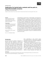
The XRD diagram of n-HA was compared
with JCPDS PDF no. 09-432 standard HA card
[14]. All characteristic peaks of HA were
identified and no strange peaks were presented
as seen in Fig. 1. The clear and sharp peaks
confirmed the purity and high crystallinity of
n-HA material extracted from pig bone by using a
typical thermal process in this study.
Fig. 1. XRD diagram of n-HA extracted from pig bone.
<b> </b>
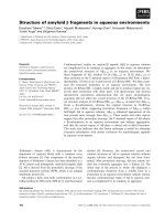
Fig. 2. FE-SEM images of n-HA at different magnifications.
The FE-SEM micrographs at 20.000 and
50.000 magnifications clearly show the particles,
rods, scales and porous holes in the structure of
n-HA (Fig. 2). The porous characteristic is an
important factor of adsorption material,
especially for physical adsorption property. The
result obtained by FE-SEM is quite similar to the
one reported in the reference [15], in which the
authors have extracted the HA material from
bovine bone.
<i>3.2. Characterization of mineralized HA (m-HA) </i>
</div>
<span class='text_page_counter'>(4)</span><div class='page_container' data-page=4>
observed. The FE-SEM images of m-HA clearly
indicated the newly mineral layer consisting of
homogeneous crystalline scales covered on the
surface and interwoven into the pores of n-HA
after 3 days of immersing in SBF as seen in Fig.
4. The combination of two results XRD and
FE-SEM confirmed the formation of newly HA
crystal layer after mineralization process.
Fig. 3. XRD diagram of mineralized HA in SBF
solution.
<b> </b>
Fig. 4. FE-SEM images of m-HA at different magnifications.
<i>3.3. Lead ion adsorption in aqueous solution of </i>
<i>n-HA and m-HA </i>
Fig. 5 and Fig. 6 present the behaviors of
lead ion adsorption of n-HA and m-HA
respectively as a function of times. For n-HA,
lead removal rapidly reached 46.3% after 1 hour
of adsorption. This was followed by a slight
increase until 4 hours before achieving a
saturated period of adsorption from 4 to 8 hours.
In the study of Y. Zhou [13], n-HA was
separated from pig bone by a different thermal
processing which showed only 28.7% of lead ion
adsorption within1 hour in the same conditions
of initial concentration of lead ion solution and
amount of adsorbent. Therefore, n-HA extracted
in this work showed higher levels of lead
removal than previous study.
</div>
<span class='text_page_counter'>(5)</span><div class='page_container' data-page=5>
Fig. 6. Lead ion removal efficiency of m-HA as a
function of times.
For m-HA, a very effective removal of lead
ions was recorded in comparison with n-HA.
The percentage of lead ion removal was reached
95.56% after 1 hour of adsorption. This value
was almost constant when the contact time
increased to 2 hours. Therefore, it is possible to
consider that the equilibrium adsorption time is
1 hour. The maximum value of lead ion removal
was recorded as 99.67% after 4 hours of
adsorption. Then, the efficiency of lead removal
represented a slight decrease. At 24 hours of
contact time, the percentage of lead removal was
96.86%. Thus, the m-HA expressed the highly
efficiency in removing lead ions in aqueous
solution. The surface modification of m-HA
after mineralized process of n-HA can be an
important factor to capture Pb2+<sub> ions. The m-HA </sub>
consists of a newly layer of HA crystals which
provides more sites for physical adsorption.
<i>3.4. Adsorption isotherm studies </i>
From the obtained results in the section 3.3,
the time of adsorption equilibrium for m-HA is
chosen as 1 hour. The Pb2+<sub> ion adsorption </sub>
isotherms for m-HA were investigated with a
series of experiments by immersing the powder
samples (each 50 mg) in 100 mL of Pb2+<sub> solution </sub>
with pH of 4.7 at different initial concentrations
of 50, 100, 150, 200, 250 and 300 mg/L for 1
hour. The obtained data was examined using the
two well-known isotherm models as follows:
Langmuir isotherm model:
𝐶<sub>𝑒</sub>
𝑄<sub>𝑒</sub>=
𝐶<sub>𝑒</sub>
𝑄<sub>𝑚</sub>+
1
𝐾<sub>𝐿</sub>. 𝑄<sub>𝑚</sub> (𝑒𝑞2)
Freundlich isotherm models:
𝐿𝑛𝑄<sub>𝑒</sub>= 𝐿𝑛𝐾<sub>𝐹</sub>+ 1
𝑛. 𝐿𝑛𝐶𝑒 (𝑒𝑞3)
<i>Here, Ce (mg/L) and Qe (mg/g), respectively, </i>
<i>represents the concentration and adsorption </i>
<i>capacity at the equilibrium; The Qm (mg/g) is the </i>
<i>maximum adsorption capacity; KL and KF are </i>
<i>the Langmuir and Freundlich constants, </i>
<i>respectively; n is the Freundlich coefficient. </i>
The Qe values were calculated as following
equation:
𝑄<sub>𝑒</sub>=(𝐶𝑜− 𝐶𝑒). 𝑉
</div>
<span class='text_page_counter'>(6)</span><div class='page_container' data-page=6>
Tab. 1. The calculated values for Langmuir and
Freundlich models
<b>Co</b>
<b>(mg/L) </b>
<b>Ce</b>
<b>(mg/L) </b>
<b>LnCe </b> <b>Qe</b>
<b>(mg/g) </b>
<b>LnQe </b> <b>Ce/Qe</b>
<b>(g/L) </b>
50 1.8 0.588 96.4 4.568 0.019
100 4.4 1.480 191.2 5.253 0.023
150 10.2 2.322 279.6 5.633 0.036
200 23.3 3.148 353.4 5.867 0.079
250 34.3 3.535 431.4 6.070 0.080
300 51.2 3.940 497.6 6.210 0.103
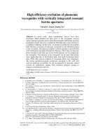
Fig. 7. Langmuir adsorption isotherm for Pb2+
adsorption on m-HA.
Fig. 8. Freundlich adsorption isotherm for Pb2+
adsorption on m-HA.
Tab. 2. The experimental constants in Langmuir and
Freundlich models
<b>Langmuir </b> <b>Freundlich </b>
<b>Qm </b> <b>KL </b> <b><sub>R</sub>2 </b> <b><sub>n </sub></b> <b><sub>K</sub><sub>F </sub></b> <b><sub>R</sub>2 </b>
574.
1
0.09
6
0.9802
1
2.14
9
84.
4
0.9629
3
<i>3.5. Identification of type of lead ion adsorption </i>
<i>by XRD analysis </i>
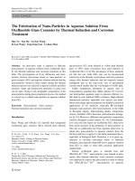
The above results confirmed the efficiency
of lead ion adsorption by using m-HA. To
identify the types of lead ion adsorption, the
XRD diagrams of n-HA, m-HA and m-HA after
lead adsorption for 1 hour were coupled as
presented in Fig. 9. According to the literatures
[16-19], different phases were determined on
m-HA diffraction pattern after adsorption
processing. All characteristic peaks of HA material
were found but they were slightly shifted to the
right side. This result is attributed to the
exchange of lead ions in aqueous solution with
m-HA material according to the following reaction:
Ca10(PO4)6(OH)2 + xPb2+↔ Ca10-
xPbx(PO4)6(OH)2 +xCa2+ (eq5)
The appearance of Pb(NO3)2 phase can be
assigned to sign of the physical adsorption of
m-HA. When m-HA soaked in Pb(NO3)2 solution,
Pb2+<sub> and NO</sub>
3- ions adhere to the surface or
infiltrate in the pores of absorbent. These ions
recombine to make lead nitrate salt when the
material sample dried after adsorption processing.
In addition, the presence of PbO and PbCO3
phases can be explained by chemical reactions
that occur during the experiment. The phase of
PbCO3 may be due to the combination of Pb2+
cations and CO32- anions produced by dissolving
CO2 in atmosphere into the aqueous solution.
Pb2+<sub> + CO</sub>
32- → PbCO3 (eq6)
The phase of PbO can be generated by
Pb(NO3)2 decomposition when drying absorbent
sample after adsorption experiment.
</div>
<span class='text_page_counter'>(7)</span><div class='page_container' data-page=7>
Fig. 9. XRD identification of phases on m-HA after
<i>lead ion adsorption. </i>
<b>4. Conclusion </b>
The natural hydroxyapatite (n-HA) was
successfully extracted from pig bone by using a
thermal processing. The mineralized HA
(m-HA) was achieved by soaking n-HA powder in
Simulated Body Fluid (SBF) for 3 days.
Experiments of lead ion adsorption were
effectuated for both n-HA and m-HA. The
obtained results showed that the m-HA
expressed a high efficiency of lead ion removal
in comparison with n-HA. The lead ion removal
percentage reached 95.56% for m-HA after only
1 hour of experiment while this value was only
46.3% for n-HA in the same contact time. The
maximum efficiency of lead ion adsorption for
m-HA was almost 100% after 4 hours. The
adsorption process of Pb2+<sub> ions on the m-HA </sub>
follows both Langmuir and Freundlich models.
However, the Langmuir model is more suitable
due to the higher value of R2<sub> coefficient. The </sub>
maximum adsorption capacity (Qm) of m-HA is
much higher than that of n-HA without
mineralization process. The mechanism of lead
ion removal for m-HA was investigated to clearly
define the ion exchange of absorbent material.
So, m-HA was proven to be a potential adsorbent
for lead ion removal compared to n-HA.
<b>References </b>
[1] S.A. Alrumman, A.F. El-kott, M.A. Kehsk,
Water pollution: Source and treatment, Am. J.
Environ. Eng. 6 (2016) 88-98. https://doi:
10.5923/j.ajee.20160603.02.
[2] F. Fu, Q. Wang, Removal of heavy metal ions
from waste waters: A review, J. Environ. Manag.
<i>92 (2011) 407-418. </i>
<i>jenvman.2010.11.011. </i>
[3] W. Zhang, F.H. Wang, P.L. Wang, Facile
synthesis of hydroxyapatite/yeast biomass
composites and their adsorption behaviors for
lead (II), J. Coll. Inter. Sci. 477 (2016) 181-190.
https: doi: 10.1016/j.jcis.2016.05.050.
[4] I. Ali, New generation adsorbents for water
treatment, Chem. Rev. 112 (2012) 5073-5091.
[5] B. Kizilkaya, A.A. Tekınay, Utilization to
remove Pb(II) ions from aqueous environments
using waste fish bones by ion exchange, J. Chem.
204 (2014) 1-12.
2014/739273.
[6] F. Wang, Y. Guo, H. Wang, Facile preparation
of hydroxyapatite with a three dimensional
architecture and potential applicationin water
treatment, Cryst. Eng. Comm. 13 (2011)
5634-5637. https://DOI:10.1039/C1CE05485F.
[7] A.R. Ibrahim, Y. Zhou, X. Li, Synthesis of
rod-like hydroxyapatite with high surface area and
pore volume from eggshells for effective
adsorption of aqueous Pb2+<sub>, Mater. Res. Bull. 62 </sub>
(2015) 132-141.
materresbull.2014.11.023.
[8] L.C. Palmer, C.J. Newcomb, S.R. Kaltz, E.D.
Spoerke, S.I. Stupp, Biomimetic systems for
hydroxyapatite mineralization inspired by bone
and enamel, Chem. Rev. 108 (2008) 4754-4783.
https://doi:10.1021/cr8004422.
[9] T. Kaludjerovic, S. Raicevic, Aqueous Pb
sorption by synthetic and natural apatite:
kinetics, equilibrium and thermodynamic
studies, Chem. Eng. J. 160 (2010) 503-510.
[10] J. Cha, M. Cui, M. Jang, S.H. Cho, D.H. Moon,
J. Khim, Kinetic and mechanism studies of the
adsorption of lead onto waste cow bone powder
(WCBP) surfaces, Environ. Geochem. Health.
33 (2011) 81-89. https://doi:
10.1007/s10653-010-9357-z.
</div>
<span class='text_page_counter'>(8)</span><div class='page_container' data-page=8>
Inor. Chem. 16 (2012) 2665-2668. https://doi.
org/10.1002/ejic.201101038.
[12] T. Kokubo, H. Takadama, How useful is SBF in
predicting in vivo bone bioactivity, Biomat. 27
(2006) 2907-2915.
biomaterials.2006.01.017.
[13] Y. Zhou, D. Chang, J. Chang, Preparation of
nano-structured pig bone hydroxyapatite for
high-efficiency adsorption of Pb2+<sub> from aqueous </sub>
solution, App. Ceram. Tech. 14 (2017)
1125-1133.
[14] JCPDS PDF card no. 09-432.
[15] A.M. Nasser et al, Extraction of pure natural
hydroxyapatite from the bovine bones bio waste
by three different methods, J. Mater. Process.
Tech. 209 (2009) 3408-3415.
10.1016/j.jmatprotec.2008.07.040.
[16] S.M. Mousa, N.S. Ammar, H.A. Ibrahim,
Removal of lead ions using hydroxyapatite
nano-material prepared from phosphogypsum
waste, J. Saudi. Chem. Soc. 20 (2016) 357-365.
[17] D. Wang et al, Facile fabrication of Pb(NO3)2/C
as advanced anode material and its lithium
storage mechanism, Electrochim. Acta. 120
(2014) 110-121.
<b>electacta.2013.12.080. </b>
[18] J. Wang et al, Spray pyrolyzed PbO/C
nanocomposites as anode for lithium-ion
batteries, J. Electrochem. Soc. 153 (2006)
<b>787-793. https://doi: 10.1149/1.2172570. </b>
</div>
<!--links-->
<a href=' /><a href=' /> Risk Assessment of Fenthion Oxide Derivatives in Aqueous Environment
- 7
- 416
- 0